The Digestive System Functions mechanical and chemical breakdown

The Digestive System

Functions: mechanical and chemical breakdown of food absorption of nutrients Consists of alimentary canal and accessory organs

Villi increase surface area for absorption of nutrients.


Mixing Movements Contractions mix food with digestive juices Propelling Movements Peristalsis - pushes food down the tube

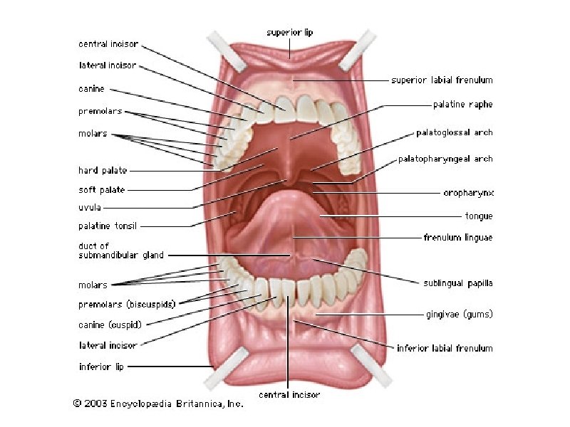
Anatomy of the Mouth - begins digestion by chewing and mixing with saliva Tongue - moves food, connects to floor of mouth via FRENULUM; has papillae Palate - forms roof of oral cavity (hard and soft); Uvula - back of the mouth

Palatine Tonsils - part of the immune system Tonsillitis is an inflammation of the tonsils and will often, but not necessarily, cause a sore throat and fever. In chronic cases tonsillectomy may be indicated.
 vs Secondary teeth")
Teeth Primary (baby) vs Secondary teeth

Teeth INCISORS CUSPIDS BICUSPIDS 1 st, 2 nd, 3 rd Molars CANINES

Anatomy of a Tooth

Tooth Decay

ROOT CANAL


Salivary Glands produce AMYLASE this enzyme breaks down starch into sugars Mucus cells also produce mucus for lubrication during swallowing 1. Parotid Gland 2. Submandibular Gland 3. Sublingual Gland

Esophagus esophageal hiatus is where it penetrates the diaphragm cardiac sphincter at entrance to stomach

STOMACH MUSCLES: Longitudinal, Circular, Oblique FUNDUS ESOPHAGUS CARDIAC REGION Longitudinal muscles LESSER CURVATURE PYLORIC SPHINCTER Circular muscles DUODENUM Oblique muscles BODY of stomach GREATER CURVATURE RUGAE
 ● Pyloric")
Stomach Regions ● Cardiac ● Fundic ● Body (greater and lesser curvature) ● Pyloric

Stomach Lining Mucus prevents stomach from digesting itself, small openings called gastric pits contain glands Glands secrete gastric juices to breakdown food PEPSIN - most important digestive enzyme for breaking down food

Chyme - paste, after food has been broken down, released then into the duodenum via the pyloric sphincter valve Rugae - folds within stomach

PANCREAS - secretes insulin which breaks down sugars Pancreatic Juice also breaks down fat Chemicals empty into the DUODENUM

Liver 1 large right lobe | 1 smaller left lobe

BILIARY SYSTEM Consists of liver, gall bladder, ducts Functions to create bile used in digestion (5) Cystic Duct (gall bladder) (4) Hepatic Duct (liver) Join to form the (6)COMMON BILE DUCT Empties into the duodenum

Liver Functions 1. blood glucose levels 2. breakdown of lipids and fats 3. protein metabolism 4. stores vitamins 5. recycles RBCs 6. removes toxins 7. secretes bile Bile – yellowish-green liquid aids in digestion, breakdown of fat

Small Intestine Main function: absorption of nutrients 1. Duodenum 2. Jejunum 3. Ileum MESENTERY - supports coils of small intestine and contains blood vessels
 a \"curtain-like\" membrane that covers the intestines, stores fat and lays")
Greater Omentum (peritoneum) a "curtain-like" membrane that covers the intestines, stores fat and lays like a drape

Greater Omentum

Intestinal villi - increase surface area to absorb nutrients, connect to vessels

Large Intestine 1. Cecum = start of large intestine, has an attached appendix 2. Colon = 4 sections, ascending / transverse / descending / sigmoid 3. Rectum – stores waste before it is expelled from the body 4. Anus -muscular sphincter which controls the exit of waste

Function of Large Intestine Secretes mucus, reabsorbs water, contains bacteria to aid in digestion (intestinal flora) Mass Movements (defecation) - removes undigested food The main job is WATER REABSORPTION. .


Types 1– 2 indicate constipation, with 3 and 4 being the ideal stools (especially the latter), as they are easy to defecate while not containing any excess liquid, and 5, 6 and 7 tending towards diarrhea.

Dysentery or Diarrhea Failure to reabsorb water in the large intestine, which leads to watery stool. Dehydration can lead to death.

Appendicitis

Hernia intestines poke through abdominal muscles
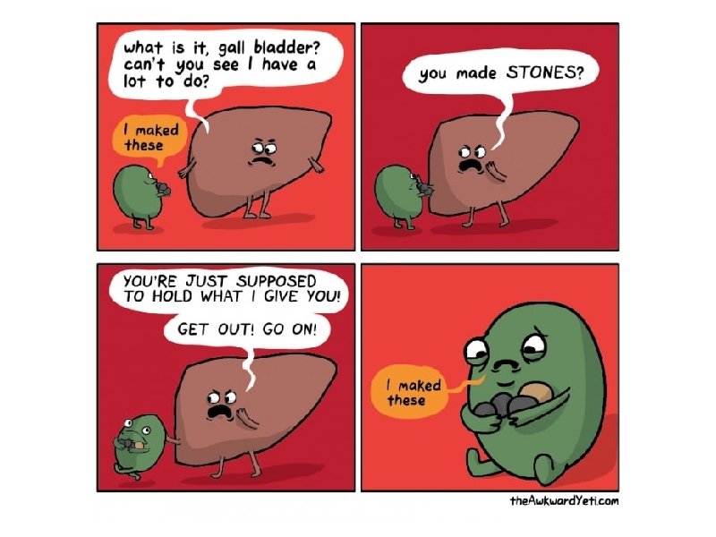
 Gallstones are made from cholesterol and other things found in the bile.")
Gallstones (Cholelithiasis) Gallstones are made from cholesterol and other things found in the bile. They can be smaller than a grain of sand or as large as a golf ball.

Gallstones within the gall bladder

Obesity - BMI is 30 or more Gastric Bypass Lap Band
- Slides: 39