The Cytoskeleton Section 4 6 Learning Objectives Contrast

The Cytoskeleton Section 4. 6

Learning Objectives �Contrast the structure & functions of the cytoskeleton with the membrane-bound organelles �Relate the structure of cilia & flagella to their functions

Cytoskeleton �Cytoskeleton = system of connected protein filaments inside the cell • Is always changing �Functions include: • Providing strength • Creating cell shape • Allowing cells to move �Filaments include: • Microtubules – the thickest filament • Microfilaments – the thinnest filament • Intermediate filaments – in-between

Microtubules �Straight, hollow tubes �Have thickest diameter �Made longer by adding pairs of tubulin subunits (dimers) �Easily broken down & reassembled

Functions of Microtubules �Provides rigidity & shape in areas of the cell �Holds organelles in place �Acts as tracks or roads for organelles to move around the cell (ex. Vesicles) �Guide the movement of chromosomes during cell division �The main structural component of cilia & flagella

Centrosome �Centrosome = area in non-dividing cell where microtubules are organized anchored • In animal cells it is also the area where centrioles are found

Centrioles �Organelles found only in animal cells • Barrel-shaped • Perpendicular to each other (90º) �Function is not certain • May play a role in microtubule organization or creation of cilia and flagella

Cilia and Flagella �Flagella = long whip-like projections used for movement �Cilia = short hair-like projections used to move liquids and particles across the cell’s surface • Also function in cell signaling

Cilia & Flagella Structure �Made of microtubules wrapped in the plasma membrane � 9 + 2 Arrangement • Ring of 9 microtubule pairs • Two microtubules in the center of the ring �Connected to a basal body • Has 9 sets of 3 microtubules arranged in a cylinder

Motor Proteins �Motor proteins = Proteins that move cell parts when given energy from ATP �Drag cell parts along microtubule & microfilament tracks �Also help to move flagella & cilia • Bend the microtubules to create movement

Microfilaments �Also called actin filaments • Made of chains of the beadlike protein actin �Looks like a twisted double chain �Are flexible

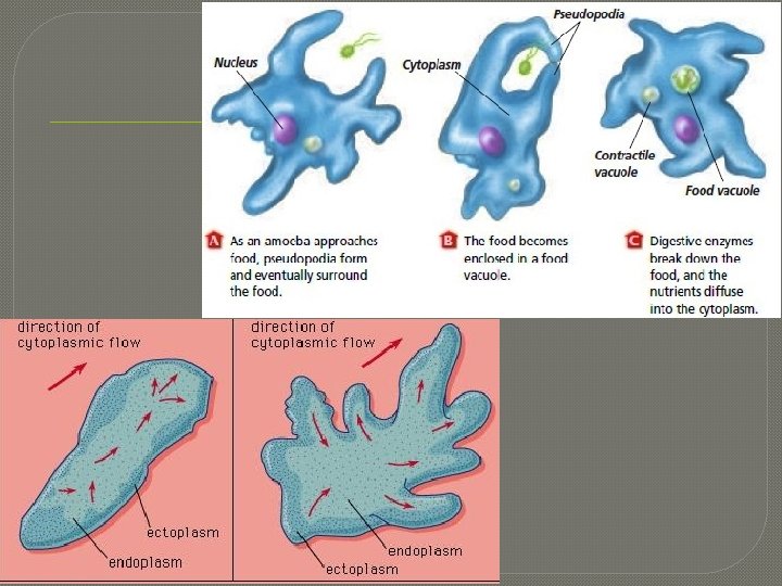
Functions of Microfilaments �Helps support the cell’s shape • Found just inside the plasma membrane �Works with other filaments to make cells contract muscle • Works with myosin filaments �Helps the cell change shape & move • Pseudopodia = “false feet” that help some cells move �Form when microfilaments & microtubules push the plasma membrane outward

Intermediate Filaments �Have a ropelike structure �Made of many fibrous proteins wound together �Are the most stable (strongest) filament �Only found in some animal groups

Functions of Intermediate Filaments �Reinforce cell shape • Prevent the cell from stretching excessively in response to outside forces �Anchors some organelles so they don’t float around • Ex. The nucleus is held in place by a cage of intermediate filaments
- Slides: 15