The Crdiovascular system II Dr Nabil khouri Conducting

The Crdio-vascular system II Dr. Nabil, khouri

Conducting System of Heart

Conduction System of the Heart • SA node: sinoatrial node. The pacemaker. – Specialized cardiac muscle cells. – Generate spontaneous action potentials (autorhythmic tissue). – Action potentials pass to atrial muscle cells and to the AV node • AV node: atrioventricular node. – Action potentials conducted more slowly here than in any other part of system. – Ensures ventricles receive signal to contract after atria have contracted • AV bundle: passes through hole in cardiac skeleton to reach interventricular septum • Right and left bundle branches: extend beneath endocardium to apices of right and left ventricles • Purkinje fibers: – Large diameter cardiac muscle cells with few myofibrils. – Many gap junctions. – Conduct action potential to ventricular muscle cells (myocardium)

Autonomic Innervation of the Heart Release Norepinephrine Release Acetylcholine

Great Vessels of the Heart

Coronary Circulation
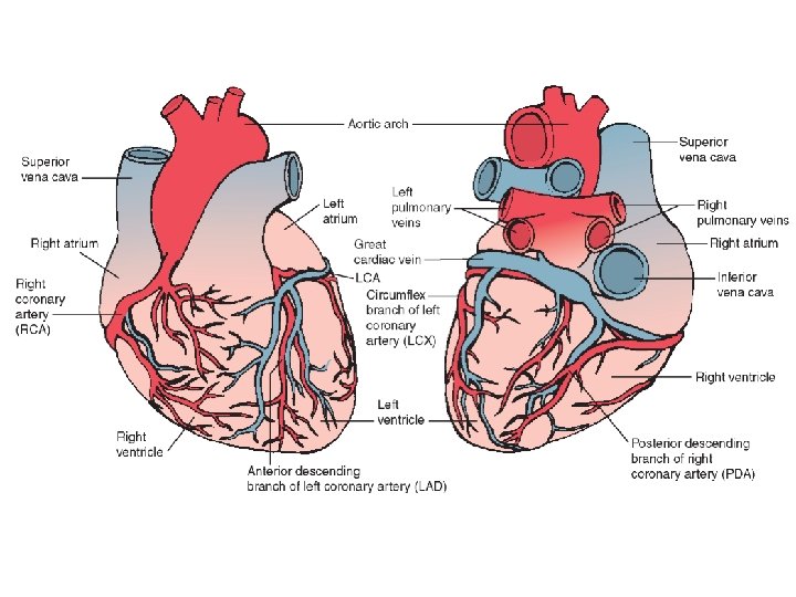

Coronary Veins

Arteries to the head and neck

Arteries of the Brain

Arteries of the Upper Limbs and Thorax

Abdominal Arteries
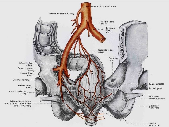

Arteries to the uterus and vagina
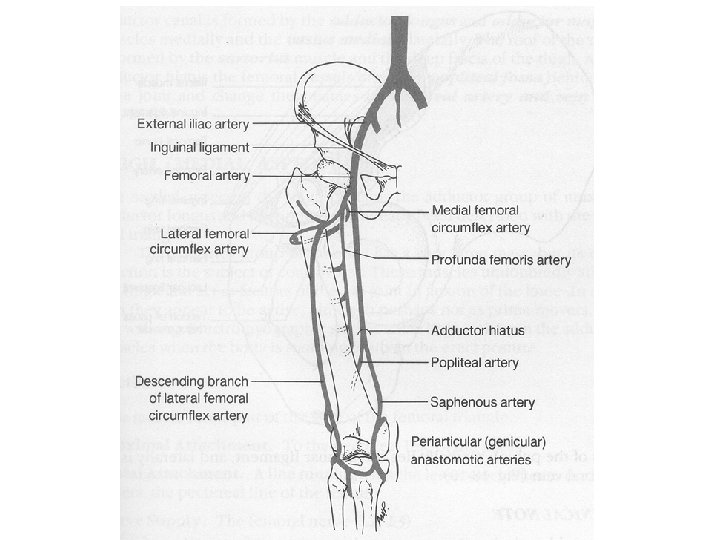

Arteries of the Lower Limbs

Systemic Circulation

VENOUS DISTRIBUTION Rt EXTERNAL JUGULAR V Lft BRACHIOCEPHALIC V Rt SUBCLAVIAN V Lft SUBCLAVIAN V Rt BRACHIOCEPHALIC V Since there is no leftward arch to the SVC, two short brachiocephalic veins are needed - Rt & Lft SUPERIOR VENA CAVA INFERIOR VENA CAVA Lft COMMON ILIAC V Lft EXTERNAL ILIAC V Iliac pattern is repeated on the right Lft INTERNAL ILIAC V

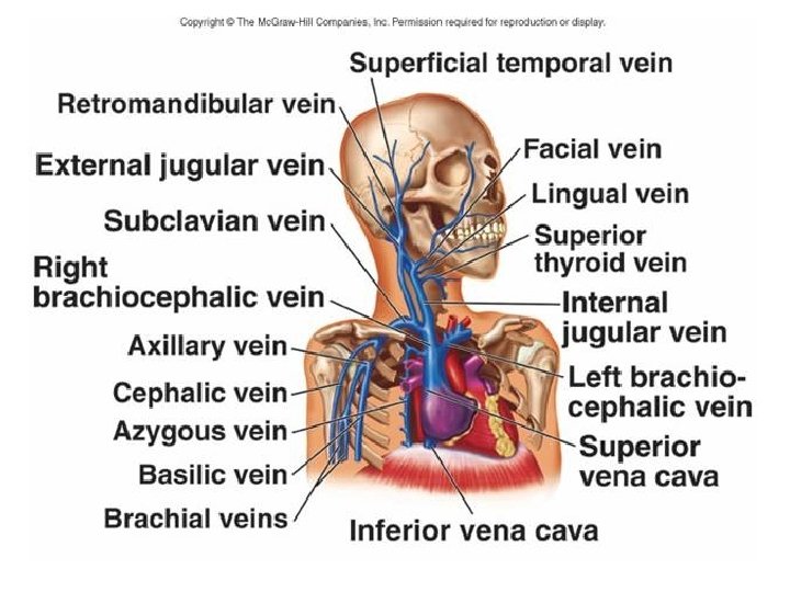
Veins of the Head and Neck

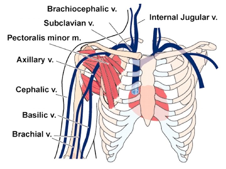
• • • Superficial veins Dorsal venous arch Cephalic vein Basilic vein Axillary Subclavian Veins of the Upper limbs

Veins of the lower limbs

Inferior Vena cava

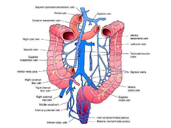
Portal System
- Slides: 26