The Cranial Nerves Honors Anatomy Physiology for copying


- Slides: 28

The Cranial Nerves Honors Anatomy & Physiology for copying

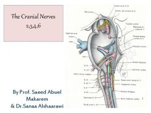
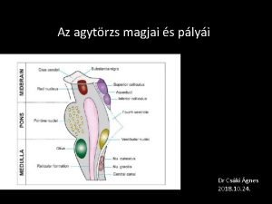
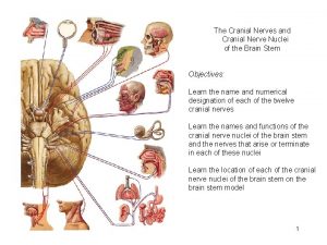
CRANIAL NERVES • 12 -pair • named “cranial” because each passes thru a foramina of the cranium • part of PNS • each with roman numeral (order from anterior posterior in which nerves arise from base of brain) & a name that indicates nerve distribution

CRANIAL NERVES • classified as: 1. sensory 2. motor 3. mixed (sensory & motor)

Cranial Nerve I: Olfactory • olfact = to smell • sensory • olfactory epithelium on superior surface of nasal cavity just inferior to cribiform plate of ethmoid bone • olfactory receptors are bipolar neurons – each: single odor-sensitive dendrite – their unmyelinated axons join above plate form rt or lt olfactory nerves

Course of Olfactory Nerve • olfactory nerves end in pair of olfactory bulbs: masses of gray matter resting just above cribiform plate where they synapse with next neurons in olfactory pathway

Course of Olfactory Nerve • axons of these neurons make up the olfactory tracts posteriorly to primary olfaction center in temporal lobe

Cranial Nerve II: Optic Nerve • optic = eye • sensory • rods & cones in retina: receptors initiating visual signals & relay them bipolar cells optic ganglion neurons their axons join forming optic nerves • pass thru optic foramen optic chiasm: a cross-over of medial half of each eye to opposite side (lateral half does not cross

Optic Tracts • from optic chiasm optic tracts – most axons thalamus synapse with neurons whose axons primary visual area of occipital lobe – some axons synapse with motor neurons in midbrain extrinsic eye muscles

Cranial Nerve III: Oculomotor • oculo = eye • mixed, mainly motor • its motor nucleus in ventral part of midbrain • 2 branches pass thru superior orbital fissure

Oculomotor Nerve Extrinsic Muscles of Eye Superior Branch • axons innervate: 1. superior rectus 2. levator palpebrae superioris (upper eyelid) Inferior Branch • 1. 2. 3. axons innervate: medial rectus inferior oblique

Oculomotor Nerve • inferior branch also: – parasympathetic innervation to intrisic muscle of eye (smooth muscle) 1. ciliary muscle: adjusts lens for near/far vision 2. circular muscle of iris: contracts/relaxes in response to amt of light (pupils constrict/dilate)

Oculomotor Nerve: Sensory • proprioception: nonvisual perception of movements & positions of body

Cranial Nerve IV: Troclear Nerve • • trochle = pulley mixed, mainly motor smallest of the 12 cranial nerves only 1 that arises from posterior of midbrain

Cranial Nerve IV: Troclear Nerve • motor: • axons from nucleus in midbrain superior orbital fissure • innervates superior oblique muscle • sensory: proprioception in superior oblique

Trigeminal Nerve • largest of 12 cranial nerves • mixed: – sensory: ganglion in temporal bone – motor: neurons in pons

Cranial Nerve V: Trigeminal Nerve • tri: has 3 branches 1. Ophthalmic: sensory only: upper eyelids, eyes, lacrimal glands, upper nasal cavity, side of nose, forehead, anterior ½ of scalp 2. Maxillary: sensory only: mucosa of nose, palate, part of pharynx, upper teeth, upper lip, lower eyelids 3. Mandibular: sensory: anterior 2/3 of tongue (not taste), cheek, lower teeth motor: muscles of mastication

Cranial Nerve VI: Abducens Nerve • ab: away / ducens: to lead (nerve impulses causes abduction of eyeball) • mixed mainly motor • nucleus in pons (motor): innervates lateral rectus muscle • sensory: proprioception in lateral rectus

Cranial Nerve VII: Facial Nerve • mixed • sensory: – taste buds anterior 2/3 of tongue, proprioceptors in face & scalp • motor: – nucleus in pons – innervates muscles of facial expression + stylohyoid muscle & posterior belly of digastric muscle • parasympathetic: lacrimal glands, palatine glands, salivary glands: sublingual & sub-mandibular

Cranial Nerve VIII: Vestibulocochlear Nerve • vestibule: small cavity; cochlear: snail-like • mixed, mainly sensory • 2 branches 1. Vestibular: – equilibrium 2. Cochlear: – – hearing motor: hair cells of spiral organ

Cranial Nerve IX: Glossopharyngeal Nerve • glosso: tongue, pharyngeal: throat • Mixed • sensory: taste buds & somatic sensory receptors on posterior 1/3 tongue, proprioceptors in swallowing muscles, baroreceptors (stretch) in carotid sinus, chemoreceptors in carotid bodies • motor: from nuclei in medulla, exit thru jugular foramen, innervate stylopharyngeus muscle (elevates pharynx & larynx) • parasympathetic: motor: stimulate parotid gland to secrete saliva

Cranial Nerve X: Vagus Nerve • vagus: wanderer, vagrant • mixed • distributed from head abdomen

Vagus Nerve • sensory: skin of external ear taste buds in epiglottis & pharynx proprioceptors in muscles of neck & throat baroreceptors in arch of aorta & chemoreceptors in aortic bodies – visceral sensory receptors in most organs of thorax & abdominal cavities – –

Vagus Nerve • parasympathetic motor: – heart & lungs – glands in GI tract – smooth muscle of airways, esophagus, stomach, gall bladder, small intestine, most of large intestine

Cranial Nerve XI: Accessory Nerve • mixed • originates from both the brainstem & spinal cord • cranial root: – motor: from medulla thru jugular foramen – supplies voluntary muscles of pharynx, larynx, & soft palate • spinal root: – mixed, mainly motor – motor:

Cranial Nerve XI: Accessory Nerve • spinal root: – mixed, mainly motor – motor: neurons in anterior gray horn of C 1 – C 5 axons come together fpramen magnum jugular foramen – innervates sternocleidomastoid & trapezius muscles – sensory: proprioceptors in muscles it supplies

Cranial Nerve XII: Hypoglossal • hypo: below, glossal: tongue • mixed • sensory: proprioceptors in tongue muscles medulla • motor: nucleus in medulla hypoglossal canal muscles of the tongue (speech, swallowing)

Development of the Nervous System • begins developing in 3 rd wk from a thickening of ectoderm called the neural plate

Development of the Brain & Spinal Cord
 Exercise 17 gross anatomy of the brain and cranial nerves
 Cranial nerve 11
 Cranial nerves
 Cranial nerves
 Cranial nerves special senses
 First and second cranial nerves
 Mixed cranial nerve
 Cranial nerves labeled with roman numerals
 Cranial nerves ipsilateral
 Afferent cranial nerves
 Ix agyideg
 Oo oo oo to touch and feel cranial nerves
 Ventricles brain
 Olfactory nerve
 Mnemonic for cranial nerves
 Cranial nerves foramina
 Trochlear nerve decussation
 Figure 14-2 cranial nerves labeled
 Old opie occasionally tries
 Cranial labeled
 Cranial accessory nerve supplies
 Oh oh oh to touch and feel cranial nerves mnemonic dirty
 Cranial nerves
 Cranial nerves classification
 Brachioradialis reflex
 Nucleus solitarius cranial nerves
 Spinal nerves labeled
 Crossed extensor reflex
 Minor salivary glands
