The connective tissue ground substance It is the

The connective tissue ground substance: It is the material that fills the spaces between the cells and contains the fibers. It is composed of: Ø Interstitial tissue fluid, formed of plasma proteins of low molecular weight that escape through the capillary wall as a result of the hydrostatic pressure. Edema: is an increase in the quantity of the tissue fluid due to loss of the equilibrium between the tissue fluids entering and leaving the matrix of CT. Ø Glycosaminoglycans (GAG) • linear (unbranched) polysaccharides, e. g. heparan sulfate, chondroitin sulfate, keratan sulfate, hyaluronic acid • attract sodium & hold water • very hydrophilic due to abundant negative charges. Ø Adhesive glycoproteins e. g. fibronectin and laminin. They serve mainly as connective tissue glue that allows connective tissue cells to bind themselves to matrix elements.
")
• Proteoglycans, consist of a protein Function : core to which glycosaminoglycans (GAGs) are attached. The strand-like Ø The ground substance holds GAGs are large, negatively-charged large amounts of fluid and polysaccharides that extend from the functions as a medium through core protein like the fibers of a bottle which nutrients and other brush. GAGs are like chondroitin dissolved substances can diffuse sulfate and keratan sulfate. between the blood capillaries The proteoglycans tend to form huge proteoglycan aggregates with and the cells. hyaluronic acid that trap water, forming a Ø Its gel state serves to resist substance that varies from a fluid to a compression and to act as a viscous= Jelly like lubricant. Ø It also acts as a barrier to bacterial penetration. Some virulent bacteria can secrete the enzyme hyaluronidase that hydrolyzes the ground substance and facilitates bacterial invasion to CT.

Connective tissue fibers • The fibers of connective tissue provide support. They are embedded in connective tissue matrix. There are three types of CT fibers; v collagen fibers, v elastic fibers v reticular fibers. v elastic fibers vcollagen fibers, v reticular fibers.
 Characters: Collagen fibers are the most abundant CT fibers. They")
Collagen fibers= (white fibers) Characters: Collagen fibers are the most abundant CT fibers. They are the strongest and provide high tensile strength (that is the ability to resist longitudinal stress). Stress test shows that collagen fibers are stronger than steel fibers of the same size. In fresh state, collagen fibers have a glistening white appearance; they are therefore also called white fibers. Histological features: In longitudinal section, collagen fibers appear as cylindrical structures that run in wavy bundles The individual fibers do not branch while the bundles of fibers often do. They stain pink with H&E (eosinophilic), green with Masson’s trichrome stain. blue with Mallory’s stain and

Synthesis of collagen: • Procollagen, a precursor of collagen protein is formed inside the fibroblasts then it is released by exocytosis into the extracellular space. • Procollagen is cleaved to form collagen molecules which assemble spontaneously into collagen fibrils. • Collagen fibrils in turn are further assembled into collagen fibers which may be bundled together into the thick collagen bundles. Types of collagen: • More than 20 different types of collagen fibers are known. They differ by their molecular composition, morphologic features, distribution in tissues and functions. The major types of collagen are: • Type I collagen fibers in connective tissue proper, and in fibrocartilage and bone matrix. • Type II collagen fibrils in cartilage matrix (hyaline and elastic). • Type III collagen fibers form the reticular fibers. • Type IV in basement membrane. • Type VII in basement membrane.

Collagen Synthesis DISULFIDE BONDS ALDOL CONDENSATION
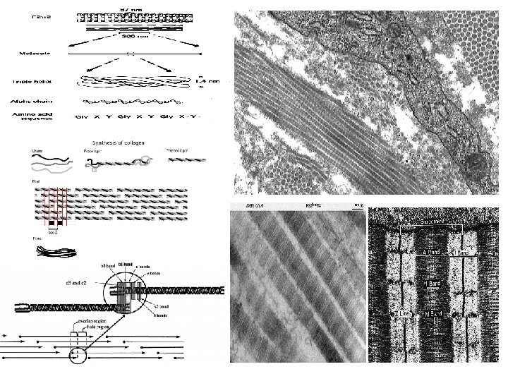

Assembly of collagen fiber bundles

H&E Collagen Fibers EM
 According to the chemical composition")
Major Collagen Fiber Types (out of at least 20) According to the chemical composition of collagen molecules Collagen Type Tissues Function Fibril-forming collagens (these are visible) I (most abundant) Skin, tendon, bone, dentin Resistance to tension II Cartilage, vitreous of eye Resistance to pressure III (reticulin) Skin, muscle, blood vessels, liver, etc. Structural framework and stability Network-forming collagens IV All basement membranes Support and filtration Anchoring filament collagens VII Epithelia Epidermis to basal lamina
 Fibers • Form a delicate supporting framework for highly cellular tissues (endocrine")
Reticular (Reticulin) Fibers • Form a delicate supporting framework for highly cellular tissues (endocrine glands, lymph nodes, liver, bone marrow, spleen, smooth muscle). • Composed mainly of Type III collagen, with a carbohydrate moiety that reduces Ag+ to metallic sliver = argyrophilic. • Special stain: silver impregnation to visualize. • Thinner than type I collagen (Type III fibrils are 30 -40 nm diameter; type I fibrils are ~200 nm diameter) • made by reticular cells (specialized fibroblasts) and vascular smooth muscle cells

Elastic fibers Characters: • These fibers contain protein, elastin that allows them to stretch and recoil like rubber bands. Because the fresh elastic fibers appear yellow, they are called the yellow fibers. Histological features: • Elastic fibers may exist in two different forms: • Individual long and thin fibers that branch in the extracellular matrix. • In the wall of large blood vessels they form fenestrated parallel sheets • They stain weakly with H&E. • Special staining with orcein stain gives a brick-red color to elastic fibers, while staining with V. VG stain gives them a dark violet color.

Network of elastin molecules can stretch and recoil like a rubber band
: can be stretched to one and one-half times")
Elastic Fibers Elastic fibers (yellow fibers): can be stretched to one and one-half times their length, but recoil to their initial length when released. Fresh elastic fibers appear yellow and are also called yellow fibers. Stain : H&E , Orcein , VVG

Fibers n n n n The most numerous White if in great number (white fibers) Strong and flexible Fibers do not branch but bundles can do Formed of collagen protein Stain pink with eosin Types of Collagen Fibres Yellow if in great number (Yellow fibers) Elastic and stretchable Fibers can branch and unit Formed of elastin protein Stained weakly by H&E Stain brick red by orcein Stain dark violet with V. V. G stain. Thin branching n Not stained by H&E n Stained dark brown with silver stain n Consist of type III collagen n Supportive function n

C. T. CELLS Fixed cells= resident q stable, long-lived cell e. g. 1. UDMC 2. Fibroblast , fibrocytes 3. Fat cell = adipocytes 4. Pigment cell Free cells = immigrating Transient = (wandering) cells. originate mainly in the bone marrow and circulate in the bloodstream. q motile, short-lived cells e. g. 1. Macrophages 2. Plasma cell 3. Mast cell 4. White blood cells= Leucocytes

Undifferentiated Mesenchymal Cell Histological features: q They are stellate cells with few processes. q They have euchromatic nuclei q with faint basophilic cytoplasm. Function: they are adult stem cells that can divide and differentiate into many types of CT cells. L. M.

Fibroblasts Fibrocytes • After they synthesize the matrix, they become quiescent and are called fibrocytes. They assume their Histological features: less active mode, indicated by the • By LM, fibroblast is a spindle-shaped suffix “cyte”. branching cell, with deeply basophilic cytoplasm and large euchromatic nucleus • are smaller cells with fewer processes than the fibroblasts. By with prominent nucleolus. LM, the cell has a small elongate, • By EM, its cytoplasm contains abundant heterochromatic nucleus and an rough endoplasmic reticulum and welleosinophilic cytoplasm. By EM, they developed Golgi complex. have fewer r. ER and small Golgi. they are the most common cells in CT. Function: • Fibroblasts, indicated by the suffix “blast” (means “forming”), are active protein-synthesizing cells that secrete the ground substance and the fibers of the matrix. Function: maintenance of the CT matrix. However, if the matrix is injured, they can easily return to their more active state (fibroblast) to repair and regenerate the matrix.

Fibroblasts the most common type §Origin : from UDMC § 2 types Young fibroblast= active §Large in size §Fusiform with processes § oval central paler nucleus §Basophilic cytoplasm= numerous r. ER Function : Fibrocytes mature (Fibrocytes) : inactive §Small in size §Fusiform smaller § oval central darker nucleus §acidophilic cytoplasm q Synthesize and secrete components of the ECM: fibers and ground substance. q Synthesize growth factors. q Rarely undergo cell division unless tissue is injured, which activates the quiescent cells. q Play a major role in the process of wound healing and respond to an injury by proliferating and enhanced fiber formation.

Fibroblasts Fibrocytes
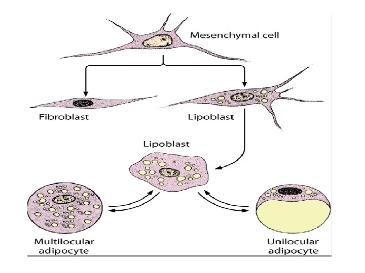

Fat Cell=Adipocytes= fixed cells Histological features: • They are large cells, spherical when single or polyhedral in shape when they are closely-grouped. • In unilocular adipocytes, the cytoplasm is occupied by a single large lipid droplet that pushes the cytoplasm to a thin peripheral rim with peripheral flattened nucleus giving the “signet-ring” appearance. • The multilocular adipocytes are polygonal and smaller than the unilocular adipocytes. Their cytoplasm contains a central rounded nucleus, numerous small lipid droplets and numerous mitochondria with abundant long cristae.

Adipocytes §Origin : UDMC §large, spherical or polyhedral • The flattened nucleus • The cytoplasm only forms a very narrow rim around a large central lipid droplet. • Single or several lipid droplets • Adipocytes are long-lived cells. Their number is determined by the number of preadipocytes generated during foetal and early postnatal development. EM Lipid storage/mobilisation is under: • nervous (sympathetic) hormonal (insulin) control. • Function : • Storage of lipid • Production of energy • endocrine function - they secrete the protein leptin which regulate appetite with feedback about the bodies fat reserves. Osmic acid
 Function : § gives")
Pigment Cells= § branched cells §Contain pigment granules: Melanin (melanocytes) Function : § gives the color of skin and iris of the eye fixed cells

Mast Cells §Origin: from haemopoetic stem cell in B. M Two types: q Connective tissue mast cells are found in skin (dermis) and peritoneal cavity; q mucosal mast cells are in the mucosa of the digestive and respiratory tracts. q Contain basophilic granules § (Metachromatic staining) when stained with toluidine blue, the granules bind the dye and change its color to red. Function : secretion of histamine and heparin

Mast Cells Histological features: • By LM, mast cell is a large CT cell. Its cytoplasm is full of basophilic granules that may obscure the nucleus. Its nucleus is rounded and central in position. • A distinctive staining feature of mast cells is "metachromasia" which means that certain basic stains give to their granules a color other than that of the dye itself e. g. toluidine blue stain gives a purple color instead of blue, due to the chemical composition of the secretory granules. • By EM, their cytoplasm contains numerous secretory granules. Function: they initiate allergic and local inflammatory responses by release (degranulation) of their granules which contain; the anticoagulant heparin and histamine which promotes increased vascular permeability and smooth muscles contraction.
: they include neutrophils, eosinophils, basophils, monocytes and lymphocytes.")
• White blood cells (leukocytes): they include neutrophils, eosinophils, basophils, monocytes and lymphocytes. • • Macrophages: they are derived from the monocytes that migrate from bloodstream into CT. • Histological features: they are large, irregular cells with eccentric kidney-shaped nucleus. The cytoplasm shows numerous lysosomes. • Function: • They are phagocytic cells; macrophages engulf a broad variety of foreign materials including bacteria, dead cells and dust particles. •

Macrophages= Histocytes Origin : From blood monocytes Three types: §Resident : resting §Elicited: moving to a stimulus §Activated: active in phagcytosis: -pseudopodea -Kidny shaped eccentric nucleus-large number of lysosomes Function : Phagocytosis

Origin : they are derived from B lymphocytes that enter the CT. Histological features: • By LM, they are large oval cells, with basophilic cytoplasm. The nucleus is spherical and eccentrically-placed. The chromatin of the nucleus is arranged giving the nucleus a cartwheel appearance. • The prominent juxtanuclear Golgi apparatus appears unstained "negative Golgi image" against the deeply-basophilic cytoplasm. Plasma Cells • By EM, the cytoplasm shows closely-packed cisternae of r. ER together with large juxtanuclear Golgi complex. Function: they are responsible for synthesis of antibodies against bacteria and foreign proteins penetrating into the CT.

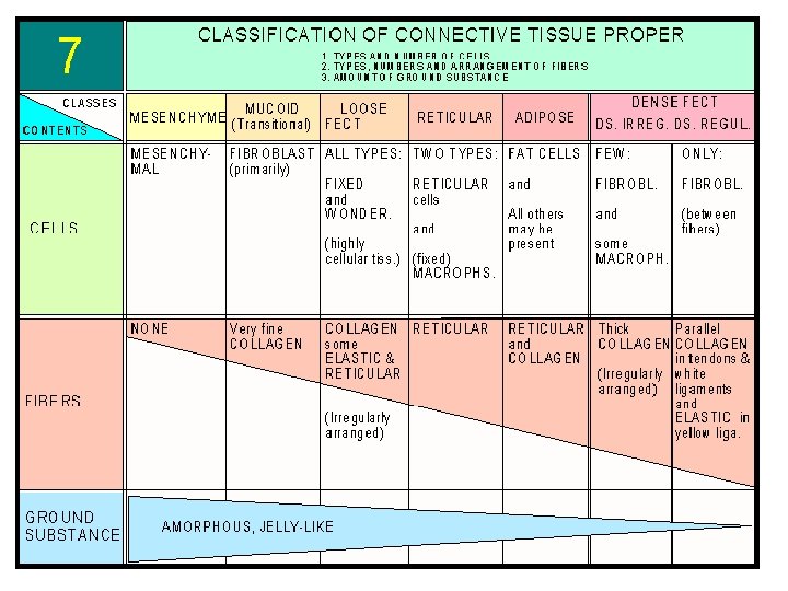
CLASSIFICATION OF CONNECTIVE TISSUE Classification depends on the I- Embryonic connective proportion of cells to fibers, and on tissue it includes: the arrangement, and the types of q. Mesenchymal CT. fibers. q. Mucoid CT. Three categories can be defined: II- Connective tissue proper: it includes: III- Specialized connective tissue; it q Loose areolar connective tissue. includes: q Dense irregular connective tissue q. Cartilage. q Dense regular connective tissue. q. Bone. q Elastic connective tissue. q. Blood. q Reticular connective tissue. q Adipose connection tissue.

Classification of C. T. Depend on ground substance Modified types v Blood =fluid v Cartilage= firm v Bone = hard q Embryonic C. T q Mucoid C. T. Jelly like C. T. Proper Ø Loose C. T. Ø Dense C. T.

Embryonic connective tissue Mesenchymal CT Mucoid CT Site: it is found in the umbilical cord and pulp of growing teeth. Site: it is found in embryo. Histological structure: it consists Histological structure: It consists of: q Abundant ground substance (Wharton's jelly) composed Ø Undifferentiated mainly of hyaluronic acid. It mesenchymal cells (UMCs) appears homogeneous and with their processes come in basophilic. contact with each other forming a network. q Spindle-shaped UMCs that are widely separated and fibroblasts. Ø A gel-like, amorphous ground substance. q Unapparent fine collagen fibers that have the same refractive Ø Scattered reticular fibers. index as the matrix.
")
Mucoid C. T. = Embryonic C. T • Mucoid connective tissue (or mucous tissue) is a type of connective tissue found during fetal development. • It is composed mainly of ground substance with few cells & fibers • It is most easily found as a component of Wharton's jelly. • Cells : UDMC, Fibroblasts • Fibers : present but not apparent collagen type II • Ground substance : Abundant Sites: • Mucous connective tissue forms the umbilical cord. • The vitreous of the eyeball is a similar tissue.

Connective tissue proper Loose: connective tissue is relatively cell rich, soft. It is also rich in vessels and nerves. Loose connective tissue may occur in some special variants: • LACT: connective tissue • Reticular: connective tissue • Adipose: tissue. Dense C. T. : connective tissues are completely dominated by fibres. They are subdivided according to the arrangement of the fibres in the tissue. Dense irregular: connective tissue the fibres do not show a clear orientation within the tissue but instead form a densely woven three-dimensional network (dermis). Dense Regular 1. White fibrous C. T. 2. Elastic C. T.

Connective tissue proper Loose areolar CT: Site: it is the most widely distributed connective tissue in the body. It binds body parts together while allowing them to move freely over one another. It contains many small blood vessels coursing through this tissue. • Loose CT is found in the following sites: It is present beneath the epithelium in all mucous membranes forming the lamina propria. It forms the papillary layer of dermis which attaches the skin epidermis to underlying structures. It surrounds glands, small blood vessels, and nerves.

Histological structure • All types of fibers; collagen, elastic and a small proportion of the reticular fibers. All types of connective tissue cells with predominance of fibroblasts and macrophages. Good amount of ground substance. Function: a. Supports and binds other tissues (by its fibers). b. Holds body fluids and provide nutrition (by its ground substance). c. Defends against infection (by its white blood cells, plasma cells, mast cells and macrophages).

Loose Areolar C. T.

Reticular CT: • Histological structure: Reticular fibers, forming a network. Reticular cells, these are the fibroblasts of reticular connective tissue, that synthesize the reticular fibers. • Site & function: reticular tissue is limited to certain sites. It forms the supporting stroma for: Hemopoietic tissue in the bone marrow. Lymphoid tissue in lymph nodes and spleen. Hepatocytes in liver. Liver (silver stain)

Adipose C. T. Unilocular adipose C. T. Yellow fat • • Unilocular fat cells C. T. fibers: collagenous F. rich in blood supply Carotenoids Sites: • Subcutaneous tissue • Around vital organs Multilocular adipose C. T. Brown fat • • Multilocular fat cells C. T. fibers – collagenous F. rich in blood supply Many blood vessels , numerous mitochondria , cytochrome pigment Sites: • Back & neck of newborne

Adipose C. T. Function: Storage of energy in the form of triglycerides. Subcutaneous adipose tissue shapes the body. Pads of fatty tissue in palms and soles act as shock absorber. Thermal insulation of the body; due to the poor heat conduction of adipose tissue. Fixation of the vital organs as heart and kidney, thus keeping them in position.
: • Closely-packed")
Dense regular C. T. White Fibrous C. T. Histological structure (Figure 84): • Closely-packed wavy bundles of collagen fibers running in the same direction and parallel to the direction of pull. • Rows of fibroblasts (tendon cells) with flattened nuclei aligned between the collagen bundles. • Little amount of ground substance. Sites & function: • Unlike areolar CT, this tissue is poorly vascularized. This type of tissue forms white flexible structures with great resistance to pulling forces wherever it is exerted in a single direction. It is found in: • Tendons, which attach muscles to bones. • Ligaments, which bind bones together at joints. § sclera of the eye

Dense Irregular CT: • Histological structure: Thick bundles of collagen fibers arranged irregularly (running in more than one plane). Little amount of ground substance with few fibroblasts. • Sites & function: this type of tissue forms sheets in body areas where tension is exerted from many different directions. It is found in the reticular layer of dermis of the skin. It forms the capsules of fibrous joints. It forms the capsules of body organs e. g. kidney, spleen, lymph nodes and liver.

Elastic C. T. • Histological structure: the elastic fibers predominate; they run in all directions, also they may form fenestrated membranes. • Site & function: this tissue is present where flexibility and elastic recoil are needed; it is found in: Elastic laminae of arteries. True vocal cords. Few ligaments in the body are very elastic such as ligamenta flava and ligamenta nuchae connecting adjacent vertebrae.

CONNECTIVE TISSUE • Connective tissues are the most abundant of the primary tissues. • The cells of the connective tissues are far apart, separated by an abundant amount of extracellular material, also called extracellular matrix Function : 1. Binding, support and packaging: 2. Protection, defense and repair: 3. Insulation: Fat cells or adipose tissue, is a connective tissue which not only cushions body organs but also insulates them and provides reserve energy fuel. 4. Transportation: Blood is a connective tissue and it carries and delivers oxygen and nutrient to tissues.

- Slides: 46