The Connective Tissue Dr Nabil Khouri MD Ms

ØThe Connective Tissue Dr. Nabil Khouri. MD, Ms. C, Ph. D

The Connective Tissue Ø Found throughout the body; most abundant and widely distributed in primary tissues l Connective tissue proper l Cartilage l Bone l Blood

Characteristics of Connective Tissue • CT Functions • Binding and support • Protection • Insulation • Transportation Ø Connective tissues have: l Mesenchyme as their common tissue of origin l Varying degrees of vascularity l Nonliving extracellular matrix, consisting of ground substance and fibers

Fundamental Cells Type Ø Ø Fibroblasts – connective tissue proper Chondroblasts – cartilage Osteoblasts – bone Hematopoietic stem cells – blood • White blood cells (WBCs) • plasma cells • Macrophages • mast cells

Connective Tissue cells Figure 4. 6

CT cells from Mesenchyme

Structural Elements of Connective Tissue • Ground substance: – unstructured material that fills the space between cells • Fibers: – collagen, elastic, or reticular • Cells: – fibroblasts, chondroblasts, osteoblasts, and hematopoietic stem cells
 fluid l Adhesion proteins: fibronectin and laminin l Proteoglycans: glycosaminoglycans")
Ground Substance Interstitial (tissue) fluid l Adhesion proteins: fibronectin and laminin l Proteoglycans: glycosaminoglycans (GAGs) l Functions: • As a molecular sieve through which nutrients diffuse between blood capillaries and cells Ø Fibers l Collagen – tough; provides high tensile strength l Elastic – long, thin fibers that allow for stretch l Reticular – branched collagenous fibers that form delicate networks Ø
 Collagen Elastin Mesothelial cell")
Loose CT in mesentery (areolar CT) Collagen Elastin Mesothelial cell

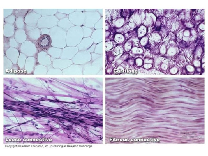
Adipose CT

Areolar CT

Reticular CT

Dense Regular Figure 4. 8 e

Dense irregular CT The only nuclei visible here are fibroblasts & fibrocytes NOTE that collagen fibers are wider than nuclei

Dense Irregular Fibrous CT Figure 4. 8 f

Dense irregular & regular CT NOTE fibrocytes in rows

Compare loose to dense in the dermis Loose CT Dense CT

Bones and Cartilages of the Human Body Figure 6. 1

Cartilage Ø Ø Ø Contains no blood vessels or nerves Surrounded by the perichondrium (dense irregular CT) that resists outward expansion Three types – hyaline, elastic, and fibro-cartilage

Connective Tissue: Cartilage Ø Hyaline cartilage l Amorphous, firm matrix with imperceptible network of collagen fibers l Chondrocytes lie in lacunae l Supports, reinforces, cushions, and resists compression l Forms the costal cartilage l Found in: embryonic skeleton, the end of long bones, nose, trachea, and larynx

Connective Tissue: 1. Hyaline Cartilage

Hyaline cartilage

Connective Tissue: 2. Elastic Cartilage Figure 4. 8 h

Elastic Cartilage

Fibro-cartilage Ø Highly compressed with great tensile strength Ø Contains collagen fibers Ø Found in menisci of the knee and in intervertebral discs

Connective Tissue: 3. Fibro-cartilage Figure 4. 8 i
")
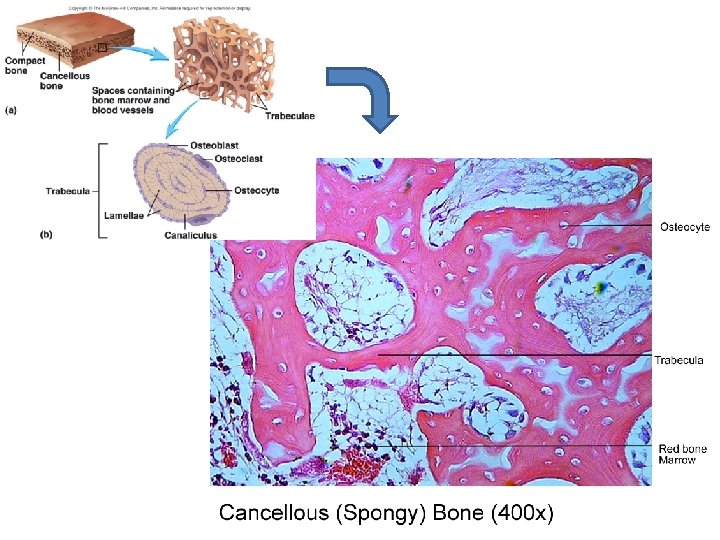
Bone tissue (osseous tissue)
")
Connective Tissue: Bone (Osseous Tissue)

Compact Bone 2 1 3 4

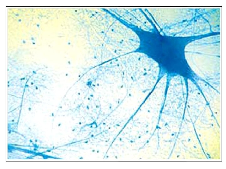
Nervous Tissue Figure 4. 10

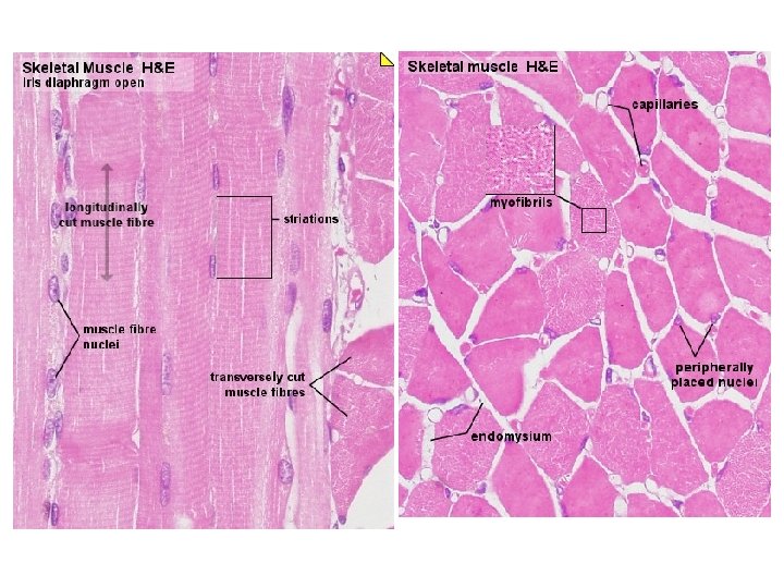
Muscle Connective tissue: 1. Skeletal Muscle

Skeletal muscle

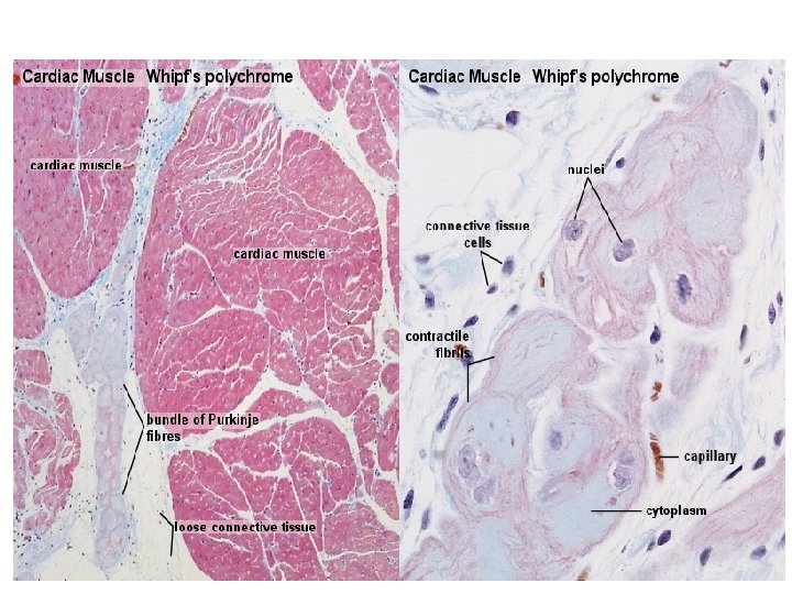
2. Cardiac Muscle Figure 4. 11 b
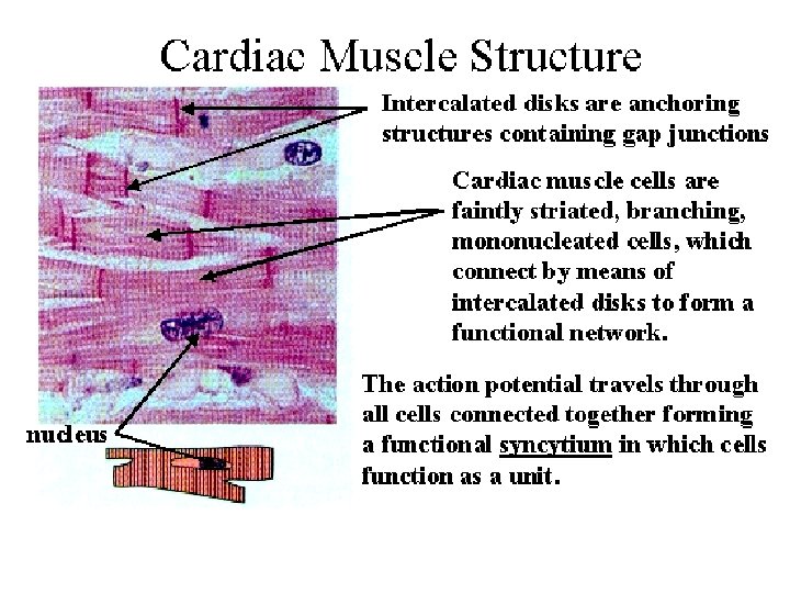

Cardiac muscle

3. Smooth Muscle Figure 4. 11 c

Smooth muscle

Cells derived from hematopoiesis
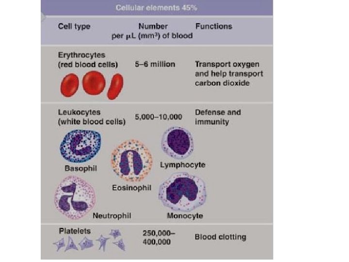

Connective Tissue: Blood Figure 4. 8 k

White blood cells found in CT Lymphocyte Eosinophil Neutrophil Monocyte
- Slides: 46