The complications of acute and chronic otitis media


 –")
 Pathology: – Occurs")


")


")


- Slides: 44
The complications of acute and chronic otitis media Dr. Abdulrahman Alsanosi Associate professor Otolaryngology consultant Otologist , Neurotologist &Skull Base Surgeon Director of cochlear implant program King Abdulaziz University Hospital& KFMC http: //faculty. ksu. edu. sa/alsanosi/default. aspx
What are the predisposing factors for developing complications ?
Predisposing factors • Virulent organisms. • Cholesteatoma and bone erosion. • Obstruction of drainage e. g. by a polyp. • Low resistance of the patient
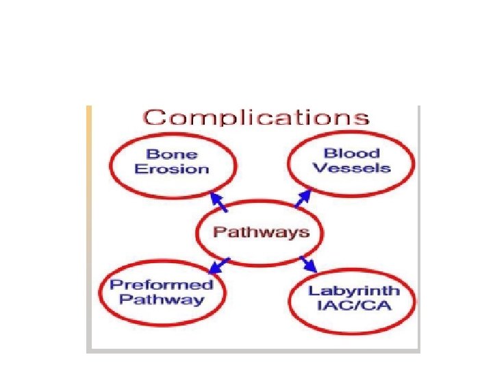
What are the pathways for spreading of infections beyond the ear?
Pathways of infection • The commonest way for extension of infection is by bone erosion due to a cholesteatoma. • Vascular extension (retrograde thrombophlebitis). • Extension along preformed pathways as – Congenital dehiscences, fracture lines, round window membrane, the labyrinth, – Dehiscences due to previous surgery
How do you classify the complications of otitis media ?
Classification • Intra-cranial complications • Intratemporal complications • Extra-cranial complications
Complications of otitis media Intratemporal Intracranial • Mastoiditis • Petrositis • Labyrinthitis • Facial paralysis • Labyrinthine fistula • Extradural abscess • Subdural abscess • Brain abscess • Meninigitis • Sinus thrombophilbitis Extracranial • Retropharyngeal abscess • Parapharyngeal abscess • Lymphadentitis
Intra-cranial complications
What are the intracranial complications? What is the commonest ? How does patient with possible intracranial complications present with ? What investigations to do to diagnose such complications ?
Intra-cranial complications Extradural Abscess: Definition: • Collection of pus against the dura of the • middle or posterior cranial fossa. • When pus collects against the walls of the • Extradural abscess is the commonest • intracranial complication of otitis media
Intra-cranial complications Extradural abscess: Clinical Picture – Persistent headache on the side of otitis media. – Pulsating discharge. – Fever – Asymptomatic (discovered during surgery) �� Diagnosis: – CT scans reveal the abscess as well as the middle ear pathology. �� Treatment: – Mastoidectomy and drainage of the abscess.
Intra-cranial complications Extradural abscess: Diagnosis – CT scans reveal the abscess as well as the middle ear pathology. �� Treatment: – Mastoidectomy and drainage of the abscess
Intra-cranial complications Subdural Abscess: Definition – Collection of pus between the dura and the arachnoid. – It’s a rare pathology Clinical picture: – Headache without signs of meningeal irritation – Convulsions – Focal neurological deficit (paralysis, loss of sensation, visual field defects)
Intra-cranial complications Subdural Abscess: Investigations – CT scan, MRI Treatment: – Drainage (neurosurgeons) – Systemic antibiotics – Mastoidectomy
Intra-cranial complications Meningitis Definition – Inflammation of meninges (pia & arachinoid) Pathology: – Occurs during acute exacerbation of chronic unsafe middle ear infection. – Two forms: • Circumscribed meningitis: no bacteria in CSF. • Generalized meningitis: bacteria are present in CSF
Intra-cranial complications Meningitis Clinical picture: – General symptoms and signs: • high fever, restlessness, irritability, • photophobia, and delirium. – Signs of meningeal irritation?
Intra-cranial complications Meningitis – Signs of meningeal irritation: • Neck rigidity. • Positive Kernig’s sign: difficulty to straighten the knee while the hip is flexed Positive Brudzinski’s sign: – passive flexion of one leg results in a similar movement on the opposite side or – if the neck is passively flexed, flexion occurs in the hips and knees
Intra-cranial complications Meningitis Diagnosis • – Lumbar puncture is diagnostic: Treatment: – Treatment of the complication itself and control of ear infection: • Specific antibiotics. • Antipyretics and supportive measures • Mastoidectomy to control the ear infection.
Intra-cranial complications Venous Sinus Thrombosis: Definition • Thrombophlebitis of the venous sinus. • Etiology: • It usually develops secondary to direct extension • from a perisinus abscess due to unsafe otitis media with cholesteatoma.
Intra-cranial complications
Intra-cranial complications Venous Sinus Thrombosis Clinical picture: – Signs of blood invasion: • (spiking) fever with rigors and chills • persistent fever (septicemia). – Positive Greissinger’s sign which is edema and tenderness over the area of the mastoid emissary vein. – Signs of increased intracranial pressure: headache, vomiting, and papilledema. – When the clot extends to the jugular vein, the vein will be felt in the neck as a tender cord.
Intra-cranial complications Venous Sinus Thrombosis: • • Diagnosis – CT scan with contrast – MRI, MRA, MRV – Angiography, venography – Blood cultures is positive during the febrile phase.
Intra-cranial complications Venous Sinus Thrombosis: Treatment – Medical: • Antibiotics and supportive treatment. • Anticoagulants – Surgical: • Mastoidectomy with exposure of the affected sinus and the intra-sinus abscess is drained.
Intra-cranial complications Brain Abscess: Definition • – Localized suppuration in the brain substance. • – It is most lethal complication of suppurative otitis media • Incidence: • – 50% is Otogenic brain abscess • – It is more common in males especially • between 10 – 30 years of age.
Intra-cranial complications Brain Abscess Pathology • – Site: Temporal lobe or • Less frequently, in the cerebellum. (more dangerous)
Intra-cranial complications Brain Abscess Diagnosis • – CT scans. • – MRI
Intra-cranial complications Brain Abscess Treatment Medical: • Systemic antibiotics. • Measure to decrease intracranial pressure. – Surgical: • Neurosurgical drainage of the abscess. • Appropriate mastoidectomy operation after subsidence of the acute stage.
What intratemporal bone complications do you know ? How does each present with? How do you manage each ?
Intratemporal complications Labyrinthine fistula • communication between middle and inner ear • It is caused by erosion of boney labyrinth due cholesteatoma • Lateral canal erosion is the most common location
Interatemporal complications Clinical picture : • Hearing loss • Attack of vertigo mostly during straining , sneezing and lifting heavy object • Positive fistula test
Interatemporal complications Labyrinthine fistula : Diagnosis • High index of suspicion • longstanding disease • fistula test • Ct scan of temporal bone Treatment : Mastoidectomy
Intratemporal complications Facial nerve paralysis: • Congenital or acquired dehiscence of nerve canal • It is possibly a result of the inflammatory response within the fallopian canal to the infection • Tympanic segment is the most commom site to be involved
Itratemporal complications Facial nerve paralysis Diagnosis • Clinical • May occur in acute or chronic ottis media • Ct scan Treatment
Intratemporal complications Facial nerve paralysis Treatment : -Acute otitis media (cortical mastoidectomy +ventilation tube) - chronic otitis media with cholestetoma ( mastoidecomy ± facial nerve decompresion )
MASTOIDITIS • • DEFINITION • It is the inflammation of mucosal lining of antrum and mastoid air cells system. Mastoiditis, per se, actually occurs with most infections of the middle ear. It is not considered a complication until bone destruction occurs
Intratemporal complication Mastoiditis : Pathology • Production of pus under tension • Hyperaemic decalcification • Osteoclastic resorption of bony walls
Clinical Features Symptoms: • Earache • Fever • Ear discharge Signs: • Mastoid tenderness • Sagging of posterosuperior meatal wall • TM perforation • Swelling over mastoid • Hearing loss
Investigations • • Blood CP • • CT scan temporal bones • • Ear swab for c/s
Differential Diagnosis • • Suppuration of mastoid lymph nodes • • Furunculosis of meatus • • Infected Sebaceous cyst
TREATMENT Medical treatment: − Hospitalize − Antibiotics − Analgesics Surgical treatment: −Myringotomy − Cortical mastoidectomy
Extracranial complications • Extension of infection to the neck • Bezold abscess ( extension of infection from mastoid to SCM)
Thanks