The Complement System Historical Background Pfeiffer Lysis of

The Complement System

Historical Background • Pfeiffer: - Lysis of Cholera bacilli - Demonstration of heat liability • Bordet: Confirmed the observations and called the substance Alexine • Ehrlich: Introduced the term Complement

Introduction • The complement system consists of a group of serum proteins that act in concert and in an orderly sequence to exert their effect • These proteins are not immunoglobulins and their concentrations in serum do not increase after immunization • Complement activation (fixation) leads to lysis of cells and to the generation of many powerful biologically active substances

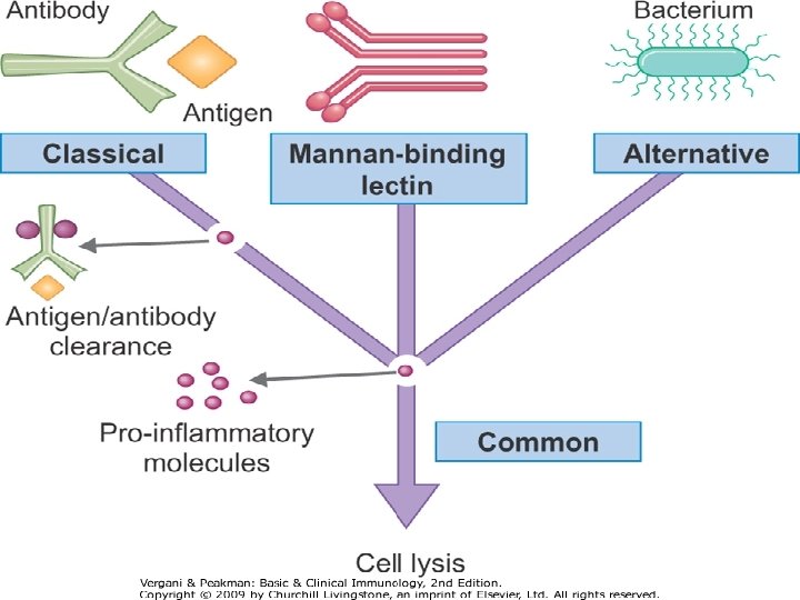
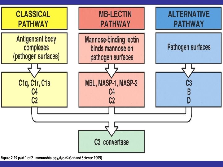
Complement Activation Pathways • The Classical Pathway - Ag- Ab complexes • The Alternative Pathway - Aggregated immunoglobulins and microbial products • The Mannan - Binding Lectin Pathway - Microbial products

The Classical Pathway-1 • Activators: Ag – Ab complexes • Antibodies involved: Ig. G and Ig. M • Activation in an orderly fashion of nine major protein components; C 1 – C 9 • Products of activation are enzymes that catalyze the subsequent step

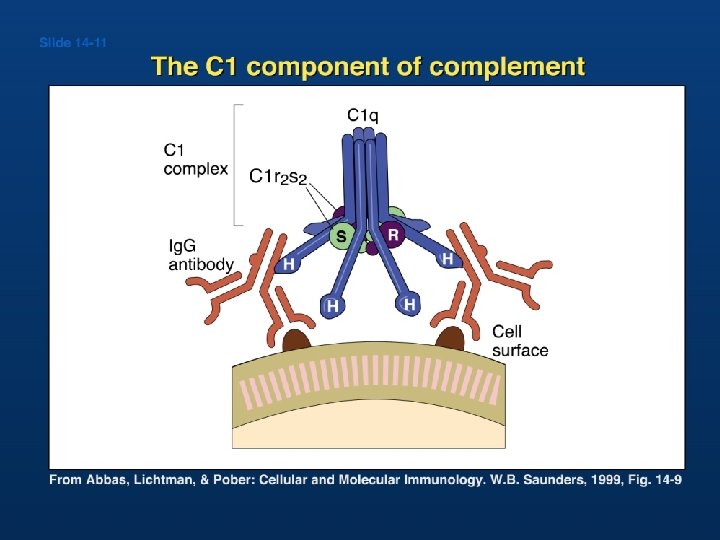
The Classical Pathway-2 • Activation of C 1: § C 1 consists of C 1 q (400. 000 Daltons), C 1 r (95000 Daltons), and C 1 s (85000 Daltons) § Subunits are held together by Calcium ions § C 1 q is a polymer of 6 identical units § C 1 q activation requires binding to a c 1 q- specific receptor on the FC region of at least 2 adjacent molecules of Ig. G or a single molecule of Ig. M, a reaction that requires Calcium ions
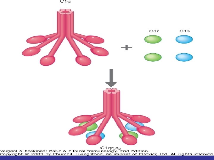

Molecular structure of C 1

Ig. G has only two sites per molecule. In order to achieve effective C 1 q binding and complement activation two Ig. G molecules must bind close to each other

Ig. A and Ig. E cannot activate complement

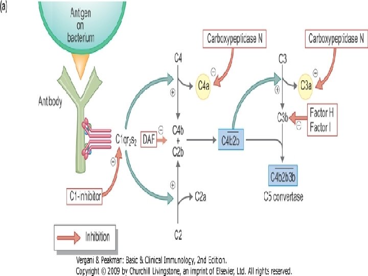
The Classical Pathway-3 • Ig. G 4, Ig. A, and Ig. E do not have complement receptors • Activated C 1 q activates C 1 r which in turn activates C 1 s • Activated C 1 s has esterolytic and proteolytic properties which acts on C 4 splitting it into two fragments; C 4 a and C 4 b • C 4 b complexes with C 1 s forming an active component that acts on C 2 splitting it into C 2 a and C 2 b • C 2 b binds to C 4 b creating a very active complex called the C 3 convertase, where a single molecule can activate hundreds of C 3 molecules

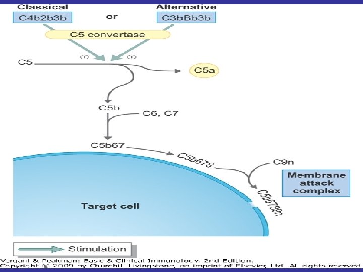
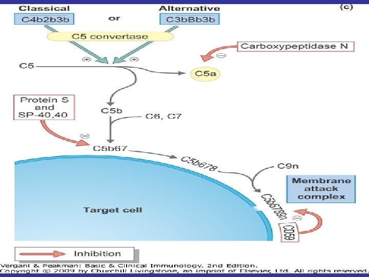
The Classical Pathway-4 • C 3 is split by C 4 b 2 b into C 3 a and C 3 b • C 3 b binds to cells and to C 4 b 2 b to generate C 5 convertase which splits C 5 into C 5 a and C 5 b • C 5 b binds to cells and activates C 6 and C 7 • The complex C 5 b 67 activates C 8 and C 9 forming a giant molecule with a molecular weight of 106 Daltons called the membrane attack complex (MAC)


The Classical Pathway-5 • C 5 b 6789 bound to cells insert themselves into the cell membrane and produce transmembrane channels allowing ions to pass through • The osmotic equilibrium of the cell is disturbed with rapid influx of water into the cell which swells and lyses

C 4 C 1 s C 4 a + C 4 b C 2 C 1 s C 2 a + C 2 b C 4 b and C 2 b combine to form C 4 b 2 b C 3 C 4 b 2 b is a C 3 convertase (there are others) C 3 a + C 3 b binds to C 4 b 2 b to form C 4 b 2 b 3 b which is a C 5 convertase which cleaves C 5 into C 5 a and C 5 b 678 C 5 b 678(9)n
 Pathway-1 • Activators: Bacterial LPS, cell wall of some bacteria, some")
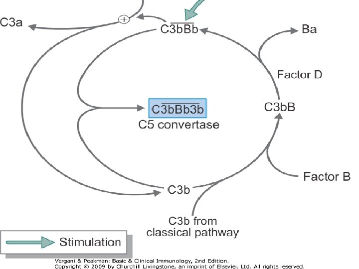
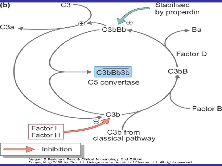
The Alternative (properdin) Pathway-1 • Activators: Bacterial LPS, cell wall of some bacteria, some yeast cells, aggregated Ig. A, and a factor present in cobra venom • Components: C 3 – C 9, factor B, factor D, and Properdin • C 3 b present in trace amounts in serum combines with factor B forming C 3 b. B
 Pathway-2 • C 3 b. B is further activated by serum")
The Alternative (properdin) Pathway-2 • C 3 b. B is further activated by serum factor D which cleaves factor B into Ba and Bb generating C 3 b. Bb • C 3 b. Bb has a C 3 convertase activity splitting C 3 into C 3 a and C 3 b • C 3 b binds to Bb amplifying the loop of activation • Properdin acts to stabilize C 3 b. Bb in the form of C(3 b)n. Bb
-1 • Activators: microorganisms and foreign invaders • Components: C")
The Mannan Binding Lectin (MBL)-1 • Activators: microorganisms and foreign invaders • Components: C 2 – C 9, MASP • MBL recognizes carbohydrate structures through its carbohydrate – recognizing domain (CRD) and then it can interact with an enzyme called MBL – activated serine protease (MASP)
-2 • Activation of MASP leads to enzymatic activation of")
The Mannan Binding Lectin (MBL)-2 • Activation of MASP leads to enzymatic activation of sequential complement components • The MBL/ MASP complex cleaves C 4 into C 4 a and C 4 b and it also cleaves C 2 into C 2 a and C 2 b creating C 3 convertase which cleaves C 3 into C 3 a and C 3 b • The sequence of events then proceeds as for the classical and alternative pathways

Products of C 3 cleavage Adjuvant

Receptors for Complement Proteins

General Functions of Complement

Complement functions related to immune defense • Lysis of cells: This is the original function identified and causes hypotonic cell death by making holes. It is not effective against organisms with rigid cell walls such as fungi and Gram positive bacteria • Opsonization: Antigen coated with C 3 b binds to cells bearing complement receptors and if the cell is a phagocyte the antigen will be phagocytosed.

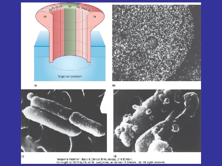
Terminal complement components and the formation of the membrane attack complex

The Membrane Attack Complex C 5 a C 5 70 -100 Å C 8 C 9 C 9 C 9 C 9 C 9 C 5 b C 7 C 6

The contents of the cell leak out through the MAC pore and the cell dies Before complement After complement treatment

• Inflammation: -The anaphylatoxins, C 5 a, C 3 a, and C 4 a of which C 5 a is the most potent bind receptors on mast cells and basophils and cause degranulation with the release of pharmacologically active mediators which induce smooth-muscle contraction and increases in vascular permeability. - Chemoattractants: C 3 a, C 5 a and C 5 b 67 attract and induce monocytes and neutrophils to adhere to vascular endothelial cells, extravasate through the endothelial lining of the capillaries and migrate to the site of complement activation in the tissue.

• Immune clearance: Removes immune complexes from the circulation and deposits them in the liver where they are degraded. • Enhanced immune response: CD 21, part of the co-receptor on the B cell, binds cleaved C 3. Recently it has also been shown that C 3 is required for optimal expansion of T cells during a systemic viral infection. • Virus neutralization: Complement mediates viral neutralization by facilitating viral aggregation and by coating the viral surface.
")
(anaphylotoxins)


Regulation of the Complement Cascade • Short half-time of – C 3 b. Bb – C 5 b • C 1 inhibitor – Inhibits the C 1 s activity • Protein Serum – Binds to C 5 b 67 ® Inhibits Formation of the Membrane Attack Complex

• HRF or CD 59 – Bind to C 8 – Inhibits C 9 binding • Factor H – Binds to C 3 b ®Facilitates binding of Factor I ®cleaves C 3 b to inactive i. C 3 b ®cleaves C 4 b to inactive fragments • Decay Accelerating Factor or CD 56 – Increased dissociation of C 3 convertase (both pathways)
 • Component Deficiencies: Any")
Complement Abnormalities • Hereditary Angioneurotic Edema (C 1 INH Deficiency) • Component Deficiencies: Any component may be affected, most important of which is C 3 deficiency which predisposes to infection by Neisseria meningitidis and Neisseria gonorrhea
, antigen, complement, and sensitized sheep RBCs are")
The Complement Fixation Test • Antibody (lysin), antigen, complement, and sensitized sheep RBCs are required • Complement is fixed to a Ab - Ag-complex • Fixed complement cannot participate in RBC lysis = positive reaction or identification

44 Complement fixation test
- Slides: 44