The Circulatory System What is its Function To


 • Has 2")

 •")
 3.")


 White Blood Cells")

: o Shaped like berets o Contain hemoglobin(an iron containing")
 o Bigger than RBC o Develop in the bone")


 when a blockage")
 • Force of blood + resistance of")


 Arterioles Capillaries")


- Slides: 41
The Circulatory System
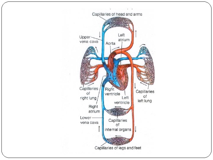
What is it’s Function? • • To transport materials to the cells of the entire body. To eliminate (carry away) waste produced by cells of the body. What goes toward the Nutrients, Oxygen, and cells? Water What is leaving the cells? CO 2 and other wastes
What organs/parts are needed for a circulatory system? Pump • Heart & Pipes 3 Types of Blood Vessels • Arteries • Veins • Capillaries
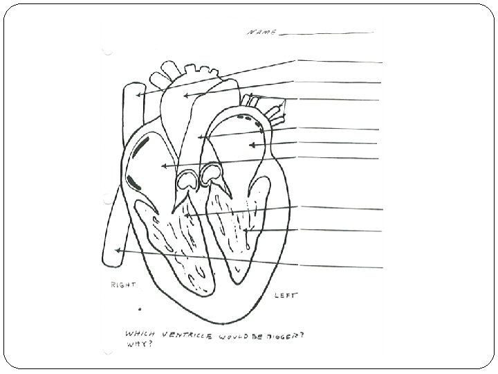
The Human Heart • Has 2 upper chambers called atria (atrium) • Has 2 lower chambers called ventricles • Left side pumps blood to the body • Right side pumps blood to the lungs
The Human Heart 13 http: //www. texasheartinstitute. org/HIC/Anatom y/Anatomy. cfm 1. Aorta 2. Superior Vena Cava 3. Right Atrium 4. Semilunar Valves (pulmonary and aortic) 5. Right Ventricle 6. Inferior Vena Cava 7. Pulmonary Artery 8. Pulmonary Vein 9. Left Atrium 10. Mitral (bicuspid) Valve 11. Left Ventricle
Atria vs. Ventricles • Receives blood from the body and lungs (collecting chamber) • Thin walled • Little muscle • Gets blood from atria • Pumps blood to all parts of the body • Thick walled • Very muscular
Location Oxygenatio Direction of 1. Body blood flow 2. Vena Cava(superior and inferior) 3. Right Atrium 4. Right Ventricle 5. Pulmonary 13 Artery 6. Lungs 7. Pulmonary Vein 8. Left Atrium 9. Left Ventricle http: //www. texasheartinstitute. org/HIC/Anatom 10. Aorta y/Anatomy. cfm 11. Body • O 2 to CO 2 • • • CO 2 O 2 O 2 O 2 to CO 2
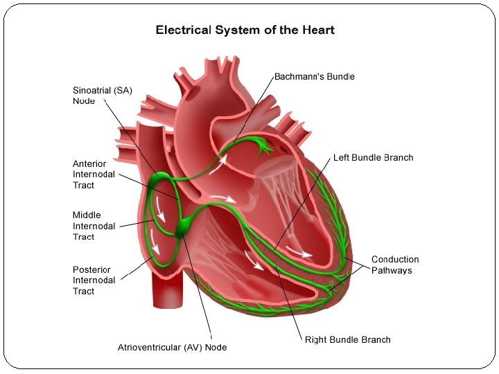
Artificial Pacemakers
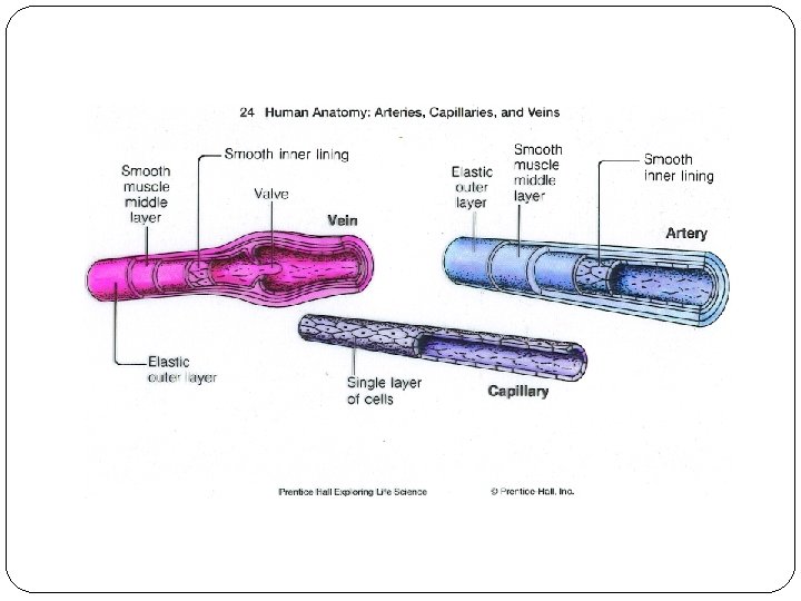
The Blood Vessels 1. Arteries 2. Veins 3. Capillaries
Direction of Flow Speed of Flow Location Arteries Any vessel in which Veins blood flows away from the heart vs. Any vessel in which blood flows to the heart. Fast blood flow Deep within the body’s tissue Method of Heart Movement Slow blood flow On the surface next to muscles Muscles with one way Thickness Thick and elastic to valves. of vessel allow a pulse Thin layer
Capillaries Thin walled vessels which: Drop off: nutrients and oxygen Pick up: CO 2 and wastes Using: the process of osmosis
Questions �Where are capillaries found in the human body? �Put the three blood vessels in order from thickest to thinnest. �Where are the capillaries that pick up digested food?
Blood 4 Components 1. 2. 3. 4. Red Blood Cells (RBC) White Blood Cells (WBC) Platelets Plasma
Blood Plasma: • Yellowish Liquid • 92% Water • Contains dissolved nutrients and wastes
Blood Red Blood Cells (RBC): o Shaped like berets o Contain hemoglobin(an iron containing protein) to which oxygen binds o Made in bone marrow o 120 day life span o Broken down in your liver and spleen when old
Blood White Blood Cells (WBC) o Bigger than RBC o Develop in the bone marrow o Defend against bacteria and viruses. o A part of your immune system 4
Blood Platelets o Bits of cells from bone marrow o Last about 10 days o They prevent bleeding by collecting around the cut, release the enzyme fibrin which weaves fibers across the cut, trapping RBC & plasma. Plasma sets and hardens forming a clot called a scab. A scab is a surface blood clot. 3
Blood Lymph o As blood moves through capillaries, plasma leaks out and surrounds the body’s cells. It collects in a system of vessels and is now called lymph. The lymph vessels transport lymph back into the blood stream. Lymph nodes remove bacteria from the lymph. 1
Blood Groups You can not transfer all blood to every person because some types have an anti-chemical that causes the cells to clump together. This could cause death.
Blood Groups There are 4 different blood groups — Has an A protein attached to the outer edge of the RBC — Has a B protein attached to the Type B outer edge of the RBC Type AB — Has an A & B protein attached to the outer edge of the RBC Type O — Has no A or B protein attached. Type A
Blood Groups Blood Group A Proteins on RBC A Can Accept Clumping Chemicals Transfusio in Plasma ns from Anti B A, O Group The. Bprotein on Clumping the RBCchemicals is A are. B, like B Anti O like a nation’smissiles flag. It that attack any A, B, AB to other A, red B blood Noneor RBC with a signals foreign objects AB, O cells that we are different on the flag. Anti(like A, “B” O side. proteins/flags) None O same Anti B
Blood Groups Rh Factor J. M. Garg Rh = rhesus monkey • Surface of human RBC has a group of 18 proteins • If a person has any one of these proteins they are Rh+ • If a person has none they are Rh • Rh- can donate to Rh+, but Rh+ cannot donate to Rh-
Blood Groups Blood Group A Proteins on RBC A B B AB O Can Accept Clumping Chemicals Transfusio in Plasma ns from Anti B A, O Group Anti A B, O A, B None A, B, AB, O None Anti A, Anti B O
Cardiovascular Disease Atherosclerosis • Thickening of the inner lining of the arteries • Caused when cholesterol, fatlike substances, collect on the inner lining of the artery • The muscle cells, of the heart, that do not get enough O 2, die.
Cardiovascular Disease Atherosclerosis • Leads to a heart attack, (cardiac arrest) when a blockage of blood flow occurs in an artery supplying blood to the heart muscle • Leads to a stroke, when the blockage occurs in an artery Copyright © 2011 Nephron supplying blood to the brain
Cardiovascular Disease Hypertension – (high blood pressure) • Force of blood + resistance of blood vessel walls. • Increases the stress on the heart muscle. • Called “silent killer” – no symptoms
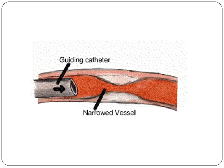
Medical Fixes Angioplasty
Medical Fixes Bypass Surgery
Imagine your are a blood cell passing into the lungs. List all the structures in order that you pass through before you enter the lungs for a second time.
Nasal Passage Trachea Bronchioles Alveoli
Capillaries Pulmonary Vein Left Atrium Bicuspid Valve Left Ventricle
Aortic Valve Aorta Arteries (left bicep) Arterioles Capillaries
Through DIFFUSION, we passed through the capillaries into one of Mr. Levine’s brain cells. Oxyge n Glucose Respirati on ATP
Respirati on CO 2 H 2 O You are now a part of CO 2 molecule. What structures do you encounter on your way out of Mr. Levine’s body?
Capillaries Venules Veins Superior Vena Cava Right Atrium
Tricuspid Valve Right Ventricle Pulmonary Valve Pulmonary Artery Lungs (and your exhaled through the capillaries)