The Circulatory System By Scientist Cindy The cardiovascular

The Circulatory System By Scientist Cindy

The cardiovascular system functions • to circulate blood through the body. • The blood carries vital nutrients, oxygen and hormones to the cells, and also picks up unwanted toxins, waste and carbon dioxide from the cells.

Components of the Cardiovascular Syste m • The 3 Components of the Cardiovascular System are. . . 1) The Heart - The function of the heart is to pump the blood around the body. It does this by creating a pressure gradient that forces the movement of the blood. This Photo by Unknown Author is licensed under CC BY-SA

Components of the Cardiovascular Syste m • The 3 Components of the Cardiovascular System are. . . 2) The Blood - The function of the blood is to deliver oxygen and nutrients to the cells of the body and to take away the unwanted waste products and carbon dioxide from the cells. This Photo by Unknown Author is licensed under CC BY

Components of the Cardiovascular System • The 3 Components of the Cardiovascular System are. . . 3) The Blood Vessels - The function of the blood vessels is to provide the network of passage ways that the blood can travel in. In short, the blood vessels allow the blood to travel through the body.

The cardiovascular system circulates blood around two loops. • THE PULMONARY CIRCUIT Functions to bring blood from the heart to the lungs and back to the heart • THE SYSTEMIC CIRCUIT - Functions to bring blood from the heart to all the parts of the body (except the lungs) and then back to the heart.

The cardiovascular system circulates blood around two loops. • In pulmonary circulation, deoxygenated blood travels from the heart to the lungs to gain oxygen and get rid of carbon dioxide before returning to the heart. • In contrast, in systemic circulation, oxygenated blood is pumped from the heart to the body and deoxygenated blood is returned back to the heart. .

THE SYSTEMIC CIRCUIT • Systemic Circulation is the movement of blood from the heart through ALL OF THE PARTS OF THE BODY except THE LUNGS. • As the blood travels around the body, the blood delivers oxygen and nutrients to the cells of the body and picks up the unwanted waste products and carbon dioxide from the cells, while bringing deoxygenated blood back to the heart.

THE SYSTEMIC CIRCUIT • The systemic circuit begins when the oxygenated blood is pumped out of the left ventricle to the aorta (the largest artery in the body). • The aorta branches into major arteries that travel to the upper and lower body.

Superi or Ven a Cava Inferio r Vena Cava THE SYSTEMIC CIRCUIT • Once the blood has delivered the nutrients and oxygen to the and picked up the carbon dioxide and waste products from the cells, this deoxygenated blood returns to the heart through the superior and inferior vena cava.

THE SYSTEMIC CIRCUIT • The systemic circuit supplies oxygenated blood to the organ system. • Oxygenated blood from the lungs is returned to the left atrium, then the ventricle contracts and pumps blood into the aorta. • Systemic arteries split from the aorta and direct blood into the capillaries. • Cells consume the oxygen and nutrients and add carbon dioxide, wastes, enzymes and hormones. • The veins drain the deoxygenated blood from the capillaries and return the blood to the right atrium.

the pulmonary circuit • In the pulmonary circuit, blood is pumped to the lungs from the right ventricle of the heart. • It is carried to the lungs via pulmonary arteries. At lungs, oxygen in the alveolae diffuses to the capillaries surrounding the alveolae and carbon dioxide inside the blood diffuses to the alveolae. • As a result, blood is oxygenated which is then carried to the heart's left half -to the left atrium via pulmonary veins. • Oxygen rich blood is prepared for the whole organs and tissues of the body. This is important because mitochondria inside the cells should use oxygen to produce energy from the organic compounds.

The Vessels of the Circulatory System • The 2 Main Types of Blood Vessels are • 1) ARTERIES • 2) VEINS

ARTERIES • Arteries are the blood vessels that are carrying blood AWAY FROM the heart. • The arteries have high pressure and thick arterial walls.

VEINS • Veins are the blood vessels that are carrying blood TOWARDS the heart. • The veins have thin walls and low pressure since they are further away from the heart. • For this reason, the veins have one-way valves that prevent the blood from flowing backward.

The Vessels of the Circulatory System • Although most veins are shown in BLUE and most arteries are shown in RED, the color IS NOT an indication of whether or not it is an artery or a vein!

The Vessels of the Circulatory System • Instead, the coloring means the following: • The RED coloring indicates that the blood carried in that vessel is OXYGENATED. • The BLUE coloring indicates that the blood carried in that vessel is DEOXYGENATED.

the pulmonary circuit • HOWEVER, in the pulmonary circuit of the cardiovascular system, this IS NOT the case. In the pulmonary circuit of the cardiovascular system, the pulmonary arteries contain deoxygenated blood that travels from the heart to the lungs. • When this blood passes by the lungs (the alveoli of the lungs), it gains oxygen molecules and releases carbon dioxide molecules. • At this point, the blood becomes oxygenated and appears more reddish in color. • This oxygenated blood then travels from the lungs back to the heart through the PULMONARY VEINS.

The Heart • The heart is a hollow, muscular organ about the size of a fist. It is responsible for pumping blood through the blood vessels by repeated, rhythmic contractions. • The heart is composed of cardiac muscle, an involuntary muscle tissue that is found only within this organ. • The term "cardiac" (as in cardiology) means "related to the heart” and comes from the Greek word kardia, for "heart. "

It has a four-chambered, double pump and is located in the thoracic cavity between the lungs. The cardiac muscle is self-exciting, meaning it has its own conduction system. The Heart This is in contrast with skeletal muscle, which requires either conscious or reflex nervous stimuli. The heart's rhythmic contractions occur spontaneously, although the frequency or heart rate can be changed by nervous or hormonal influence such as exercise or the perception of danger.

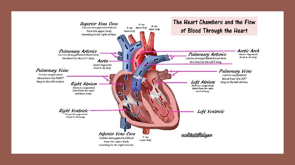
• The heart has four chambers: • • • The Right Atrium The Right Ventricle The Left Atrium The Left Ventricle The two atria lie superiorly to the two ventricles. • The atria are smaller with thin walls, while the ventricles are larger with thick muscular walls. Heart Chambers

There are two atria; the right atrium and the left atrium. Chambers The right atrium receives deoxygenated (oxygen-poor) blood from the body. The left atrium receives oxygenated (oxygenrich) blood coming from the blood vessels surrounding the lungs. which has been oxygenated and is ready to be sent to the body.

• The word "atrium" comes from the Latin word for "entrance". • The atria (plural for atrium) of the heart function to RECEIVE blood. Atria • The right atrium of the heart is the chamber that receives deoxygenated blood from the body. • The left atrium of the heart is the chamber that receives oxygenated blood from the lungs.

Ventricles • The word "ventricle" comes from the Latin word for "belly". • This origin probably comes from the fact that the ventricles of the heart are the larger of the 4 chambers. • They are muscular and perform the rhythmic contraction and relaxation of the cardiac muscle that creates the "pumping" action of the heart. The ventricles both SEND blood out of the heart.

• The word "ventricle" comes from the Latin word for "belly". Ventricles • This origin probably comes from the fact that the ventricles of the heart are the larger, more rounded chambers of the heart. • The ventricles receive blood from the atria and the send that blood out of the heart. The ventricles create the "pumping" action of the heart by the rhythmic contraction and relaxation of the cardiac muscle (myocardium).

• The right ventricle receives the deoxygenated blood from the right atrium and sends that blood out to the lungs to pick up oxygen. The blood picks up oxygen and return to the heart at the left atrium. • The left ventricle receives the oxygenated blood from the left atrium and sends that blood out to the body. Ventricles

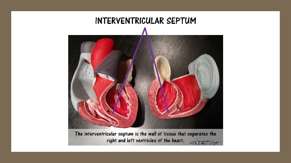
Ventricles • The ventricle is a heart chamber which collects blood from an atrium and pumps it out of the heart. • There are two ventricles: the right ventricle pumps blood into the pulmonary circulation for the lungs, and the left ventricle pumps blood into the systemic circulation for the rest of the body. • Ventricles have thicker walls than the atria, and thus can create the higher blood pressure. • Comparing the left and right ventricle, the left ventricle has thicker walls because it needs to pump blood to the whole body.

• The left atrium receives oxygenated blood from the left and right pulmonary veins which carries blood coming from the blood vessels surrounding the lungs.

The Flow of Blood Through the Chambers of the Heart • The right atrium receives the deoxygenated blood that has traveled through the body. This blood has a low level of oxygen and a high level of carbon dioxide. This blood also has low pressure since it has traveled a good distance since it got its last "push" from the heart. • This deoxygenated blood from the lower and upper body travels to the right atrium, and then travels to the right ventricle. When the right ventricle contracts, it pushes the deoxygenated blood out of the heart and sends it to the lungs. (This is the pulmonary circuit).

The Flow of Blood Through the Chambers of the Heart • As this blood passes by the lungs, it picks up oxygen and gets rid of carbon dioxide. • The oxygenated blood from the lungs then travels back to the heart to get another "push" before it goes to the body (this is the systemic circuit). • The left atrium receives the oxygenated blood coming from the lungs. This blood enters the left atrium and then travels to the left ventricle. • The left ventricle contracts to push the oxygenated blood out of the heart and to the body in order to deliver oxygen and nutrients to the cells and to pick up unwanted substances like waste and carbon dioxide.


The Flow of Blood Through the Chambers of the Heart • The blood vessels that transport blood to and from all the body make up the SYSTEMIC CIRCUIT. • So the 2 chambers of the heart that receive blood are the ATRIA (atria= plural; atrium= singular) and the two chambers of the heart that send blood are the VENTRICLES. • The ventricles contract and relax rhythmically to create the pumping action of the heart.

• The right atrium receives deoxygenated blood from the upper portions of the body (the head, neck and arms) through the superior vena cava. Superior Vena Cava

• The right atrium receives deoxygenated blood from the lower portions of the body (the torso and legs) through the inferior vena cava. Inferior Vena Cava

The Valves of the Heart

The Valves of the Heart

The Flow of Blood Through the Chambers of the Heart

The Valves of the Heart The function of the heart valves is to assure that blood flows in only one direction through the heart. When there is an increase in pressure of the blood, that pressure acts to push the valve open. • The valves only open in one direction, which prevents back flow and guides the blood to flow in the proper direction. •

The four valves of the heart are as follows: • The Tricuspid Valve is an atrioventricular (AV) valve that allows for the one -way movement of blood from the right atrium to the right ventricle. • The Pulmonary Semilunar Valve is a semilunar (SV) valve that allows for the one-way movement of blood from the right ventricle to the pulmonary arteries (which travel to the lungs). • The Mitral (Bicuspid) Valve is an atrioventricular (AV) valve that allows for the one-way movement of blood from the left atrium to the left ventricle. • The Aortic Semilunar Valve is a semilunar (SV) valve that allows for the one -way movement of blood from the left ventricle to the aorta (which travel to the body).

The four main valves of the heart are as follows: • The Tricuspid Valve is an atrioventricular (AV) valve that allows for the one-way movement of blood from the right atrium to the right ventricle.
")
The four main valves of the heart are as follows: • The Mitral (Bicuspid) Valve is an atrioventricular (AV) valve that allows for the one-way movement of blood from the left atrium to the left ventricle.

The four main valves of the heart are as follows: • The Pulmonary Semilunar Valve is a semilunar (SV) valve that allows for the one -way movement of blood from the right ventricle to the pulmonary arteries (which travel to the lungs).

The four main valves of the heart are as follows: • The Aortic Semilunar Valve is a semilunar (SV) valve that allows for the one-way movement of blood from the left ventricle to the aorta (which travel to the body).

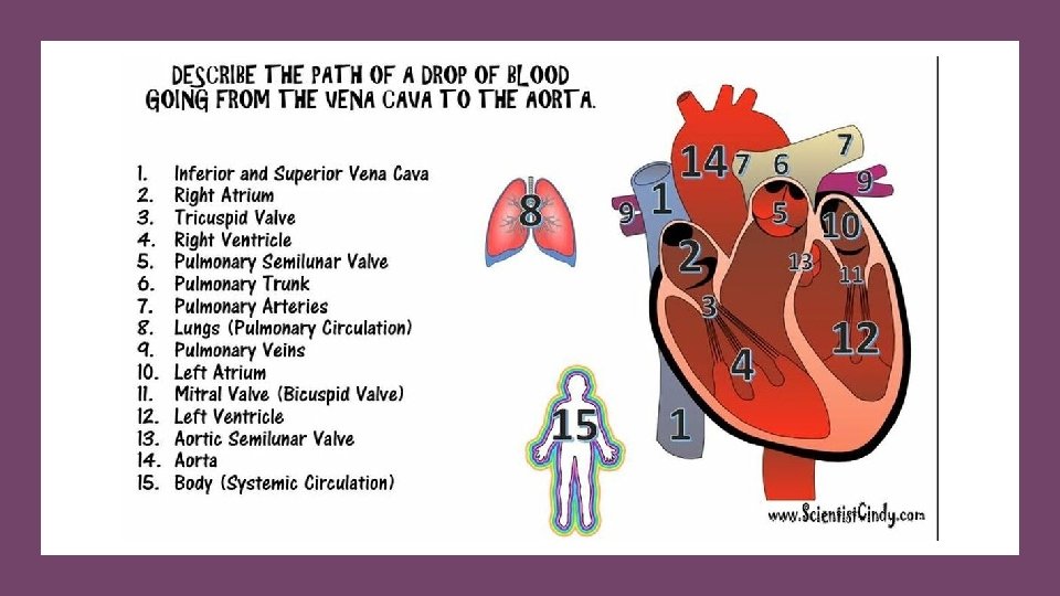
Deoxygenated blood comes from the body and goes to the right atrium. The blood then travels through the TRICUSPID VALVE to get to the right ventricle. Once the right ventricle contracts, this blood is then pumped through the PULMONARY SEMILU NAR VALVE to the pulmonary arteries which will carry the blood to the lungs. Oxygenated blood comes from the lungs and enters the left atrium. The blood then travels through the MITRAL (BICUSPID) VALVE to the left ventricle. When the left ventricle contracts, the blood is pumped through the AORTIC SEMILUNAR VALVE to the aorta where it will travel to the body.

VEINS = Veins are the blood vessels that are carrying blood TOWARDS the heart. The veins have low pressure since they are further away from the heart. Veins have one-way valves that prevent the blood from flowing backward.

capillaries • Inside the capillaries, exchange of oxygen and carbon dioxide takes place. • Red blood cells inside the capillary releases their oxygen which passes through the wall and into the surrounding cells. • No cells of the body can be more than just a few cells away from tissue. • The tissue then releases waste, such as carbon dioxide, which then passes through the wall and into the red blood cells.

• Arteries carry blood to smaller blood vessels called arterioles. • From the arterioles, the blood then enters one or more capillaries. This Photo by Unknown Author is licensed under CC BY-SA

capillaries • The capillaries are the sites of gas exchange and nutrient/waste exchange. • The walls of capillaries are only one cell-layer thick. The walls of the capillaries must be extremely thin in order to allow for diffusion of gasses and other substances to occur. • The capillaries are so thin that the blood cells can only pass in single file. This Photo by Unknown Author is licensed under CC BY-SA

Arterioles • An arteriole is a small artery that extends and leads to capillaries. • Arterioles have thick smooth muscular walls. • These smooth muscles are able to contract (causing vessel constriction) and relax (causing vessel dilation). • This contracting and relaxing affects blood pressure; the higher number of vessels dilated, the lower blood pressure will be. • Arterioles are just visible to the naked eye.

Capillaries • Capillaries are the smallest of a body’s vessels; they connect arteries and veins, and most closely interact with tissues. • They are very prevalent in the body; total surface area is about 6, 300 square meters. Because of this, no cell is very far from a capillary, no more than 50 micrometers away.

Capillaries • The walls of capillaries are composed of a single layer of cells, the endothelium, which is the inner lining of all the vessels. • This layer is so thin that molecules such as oxygen, water and lipids can pass through them by diffusion and enter the tissues. • Waste products such as carbon dioxide and urea can diffuse back into the blood to be carried away for removal from the body.

Capillaries • The "capillary bed" is the network of capillaries present throughout the body. • These beds are able to be “opened” and “closed” at any given time, according to need. • This process is called autoregulation and capillary beds usually carry no more than 25% of the amount of blood it could hold at any time. • The more metabolically active the cells, the more capillaries it will require to supply nutrients.
 The Aorta 2)")
The Arteries • The main arteries of the heart include 1) The Aorta 2) The Pulmonary Trunk 3) The Pulmonary Arteries 4) The Coronary Arteries

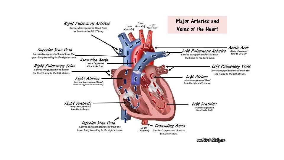
The Aorta • The largest artery in the body is the AORTA. • It is the largest and strongest artery in the body and it marks the beginning of the systemic circuit (HEART --> BODY --> HEART). • Oxygenated blood gets pumped out of the left ventricle through the aortic semilunar valve, into the aorta. • The aorta is the largest of the arteries in the systemic circuit

The Regions of the Aorta • The aorta is the largest artery in the body. The function of the aorta is to send oxygenated blood to the body. • That means all of the blood in the systemic circuit of the circulatory system has to go through the aorta.

The aorta has different regions • The first segment of the aorta ascends upward and is appropriately called the ascending aorta.

The Regions of the Aorta • The next segment makes a U -turn and forms and arch. This portion is called the aortic arch. • The aortic arch has 3 vessels that project off of it superior. These 3 vessels collectively, function to bring oxygenated blood to the upper portions of the body.

The Regions of the Aorta • After the aortic arch, the aorta turns inferiorly and descends as the descending aorta. • The portion of the descending aorta that gives through the thoracic cavity ids called the thoracic aorta. • Once thoracic aorta travels through the diaphragm, it leave thoracic cavity and enters into the abdominal cavity. • This portion of the descending aorta is called the abdominal aorta.

• The aortic arch contains 3 branches • the brachiocephalic trunk • the left common carotid artery • the left subclavian artery The Branches of the Aortic Arch

The Branches of the Aortic Arch • The 3 branches of the aortic arch function, collectively, to carry oxygenated blood to the upper regions of the body. • The first branch is the brachiocephalic trunk. • The brachiocephalic trunk bifurcates (splits into 2) to form the right subclavian artery and the right common carotid artery.

The Branches of the Aortic Arch • The right subclavian artery supplies blood to the right arm and the right common carotid artery will continue to travel superiorly and supply blood to the right side of the head, neck and brain.

The Branches of the Aortic Arch • The second branch is the left common carotid artery and it supplies blood to the left side of the head, neck and brain. • The third branch is the left subclavian artery and it supplies blood to the left arm. • The descending aorta supplies blood to the lower portions of the body.

• Another large artery you see in the heart is colored BLUE because it is carrying deoxygenated blood from the right ventricle to the lungs. • This artery is called the pulmonary trunk. PULMONARY TRUNK AND PULMONARY ARTERIES • The pulmonary trunk branches off to form the right and left pulmonary arteries. The right pulmonary artery carries the blood to the right lung and the left pulmonary artery carries blood to the left lung.

The Brachiocephalic Veins • The right and left brachiocephalic veins come together to form the superior vena cava. • The word "brachio-" means "upper arm" and the word "cephalic" means "head". • The right brachiocephalic vein brings deoxygenated blood from the right side of the head and the right arm, to the superior vena cava. • The left brachiocephalic vein brings deoxygenated blood from the left side of the head and neck and the left arm, to the superior vena cava

• Oxygenated blood coming from the lungs returns to the heart through the right and left pulmonary veins. • The veins are colored RED, because these veins carry oxygenated blood. • The right pulmonary veins bring oxygenated blood from the right lung to the left atrium of the heart. The Pulmonary Veins • The left pulmonary veins bring oxygenated blood from the left lung to the left atrium of the heart.

CORONARY ARTERIES • Although blood fills the chambers of the heart, the muscle tissue of the heart, or myocardium, is so thick that it requires coronary blood vessels to deliver blood deep into the myocardium. • The vessels that supply blood high in oxygen to the myocardium are known as coronary arteries. • The vessels that remove the deoxygenated blood from the heart muscle are known as cardiac veins.

CORONARY ARTERIES • The coronary arteries that run on the surface of the heart are called epicardial coronary arteries • These relatively narrow vessels are commonly affected by atherosclerosis and can become blocked, causing angina (heart pain) or a myocardial infarction (heart attack). • The coronary arteries are classified as "end circulation", since they represent the only source of blood supply to the myocardium.

coronary arteries: • There are two main coronary arteries: • Right coronary artery • Left coronary artery Both of these arteries originate from the beginning (root) of the aorta, immediately above the aortic valve.

coronary arteries: • The heart itself needs a supply of blood. • There are two arteries that begin at the base of the aorta called the right and left coronary arteries. • The right coronary artery supplies oxygenated blood to the cardiac muscles on the right side of the heart and the left coronary artery supplies oxygenated blood to the cardiac muscles on the left side of the heart.

• • Systemic arteries split from the aorta and direct blood into the capillaries. Cells consume the oxygen and nutrients and add carbon dioxide, wastes, enzymes and hormones. The veins drain the deoxygenated blood from the capillaries and return the blood to the right atrium. THE VEINS

• The largest veins of the body are the superior and inferior vena cava. • The superior vena cava brings deoxygenated blood from the upper body to the right atrium. • The inferior vena cava brings deoxygenated blood from the lower body to the right atrium. THE VEINS

VEINS • Veins carry blood to the heart. • The veins lack the smooth muscle that we see in arteries. • Since veins are vessels that are headed back to the heart, it has been a long time (and distance) from its last “push" from the heart. • For this reason, veins have low blood pressure compared to arteries. • Larger veins have one-way valves called venous valves to prevent the backflow of blood.

• Oxygenated blood coming from the lungs returns to the heart through the right and left pulmonary veins. • The right pulmonary veins bring oxygenated blood from the right lung to the left atrium of the heart. pulmonary veins • The left pulmonary veins bring oxygenated blood from the left lung to the left atrium of the heart.

Venules • A venule is a small vein that allows deoxygenated blood to return from the capillary beds to the larger blood veins, except in the pulmonary circuit were the blood is oxygenated. • Venules have three layers; they have the same makeup as arteries with less smooth muscle, making them thinner.
 is")
TISSUE LAYERS OF THE HEART • The pericardium (lining of the pericardial cavity) is the thick, membranous sac that surrounds the heart. It protects and lubricates the heart. • There are two layers to the pericardium: the fibrous pericardium and the serous pericardium. • The serous pericardium is divided into two layers; in between these two layers there is a space called the pericardial cavity.

The walls of the heart have 3 layers. • The Epicardium • The Visceral Layer of the Serous Pericardium Makes Up the Epicardium • The Myocardium • Thick Layer of Cardiac Muscle Tissue • The Endocardium • Inner lining

The walls of the heart have 3 layers. • Epicardium The outer most layer next to the myocardium is known as the Epicardium. • This is the outer layer after endocardium and myocardium that consists of a thin layer of connective tissue and fat.

The walls of the heart have 3 layers. • Endocardium The endocardium is the innermost lining of the heart which consists of the endothelial cells forming a smooth membrane in places, and a pocked and tribeculated surface in others (mainly the ventricles, or lower pumping chambers).

The walls of the heart have 3 layers. • Myocardium The myocardium is the muscular tissue of the heart. The myocardium is composed of specialized cardiac muscle cells with an ability not possessed by muscle tissue elsewhere in the body. Cardiac muscle, like other muscles, can contract, but it can also conduct electricity, like nerves. The blood to the myocardium is supplied by the coronary arteries.

The walls of the heart have 3 layers. • If these arteries are occluded by atherosclerosis and/or thrombosis, this can lead to angina pectoris or myocardial infarction due to ischemia (lack of oxygen). Failure of the heart to contract properly (for various reasons) is termed heart failure, generally leading to fluid retention, edema, pulmonary edema, renal insufficiency, hepatomegaly, a shortened life expectancy and decreased quality of life.
 • The proper name for Red Blood Cells is")
THE RED BLOOD CELLS (ERYTHROCYTES) • The proper name for Red Blood Cells is Erythrocytes. Red blood cells are short-lived and do not have a nucleus. • The main function of the erythrocytes is to • transport oxygen to the cells of the body • take away carbon dioxide from the cells of the body
 • The red blood cells are rather easy to")
THE RED BLOOD CELLS (ERYTHROCYTES) • The red blood cells are rather easy to spot on slides, because they do not have a nucleus! Red blood cells (like all blood cells) form from hematopoietic stem cells in the bone marrow. • As they mature, they lose their nucleus, their DNA and many of their organelles.
")
THE WHITE BLOOD CELLS (LEUKOCYTES)
 • White blood cells help to protect your body")
THE WHITE BLOOD CELLS (LEUKOCYTES) • White blood cells help to protect your body against infection and fight disease. • Unlike the red blood cells, white blood cells DO have a nucleus.

Platelets • The platelets appear as small darkly stained fragments on the slide. • These are blood clotting factors.
 • Carry oxygen to the")
Composition of the Blood • Erythrocytes (Red Blood Cells) • Carry oxygen to the cells • Carry carbon dioxide away from the cells • Lack nuclei and mitochondria • Short life span - 120 -day life span • Contain hemoglobin and transferrin
 • Have nuclei and mitochondria")
Composition of the Blood • Leukocytes (White Blood Cells) • Have nuclei and mitochondria • 2 Categories of Leukocytes • Granular leukocytes: neutrophils, eosinophils, and basophils • Aggranular leukocytes: monocytes and lymphocytes

Hematopoiesis Process of blood cell formation: • Leukopoiesis: white blood cells • Red bone marrow and lymphoid tissues • Cytokine regulation Formed Elements in the Blood - Leukopoiesis

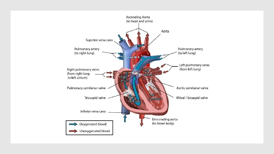
• Right atrium: receives deoxygenated blood from the body • Left atrium: receives oxygenated blood from the lungs • Right ventricle: pumps deoxygenated blood to the lungs • Left ventricle: pumps oxygenated blood to the body Structure of the Heart

Pulmonary and Systemic Circulations • Pulmonary: between heart and lungs • Blood pumps to lungs via pulmonary arteries. • Blood returns to heart via pulmonary veins.
- Slides: 95