
THE CHANGING PATIENT PATHWAY IN BREAST CANCER FROM

THE CHANGING PATIENT PATHWAY IN BREAST CANCER: FROM GP TO CURE MR TERENCE J. BOYLE Consultant Breast Surgeon BEACON HOSPITAL Clinical Lead - Beacon Breast Centre & ST JAMES’S HOSPITAL Clinical Lead - Breast Care Department
OUTLINE • Presentation/initial work-up – Triple Assessment • Subsequent investigation – Staging: Breast, axilla, systemic • Characterising the disease – Tumour GRADE, Disease STAGE – Biomarker Profile • Treatment strategies – Surgery, systemic therapies, radiotherapy • Follow-up/ongoing care issues – Psychological support (NB: Specialist Nurses) – Physiotherapy, chronic pain, ‘reconstruction’
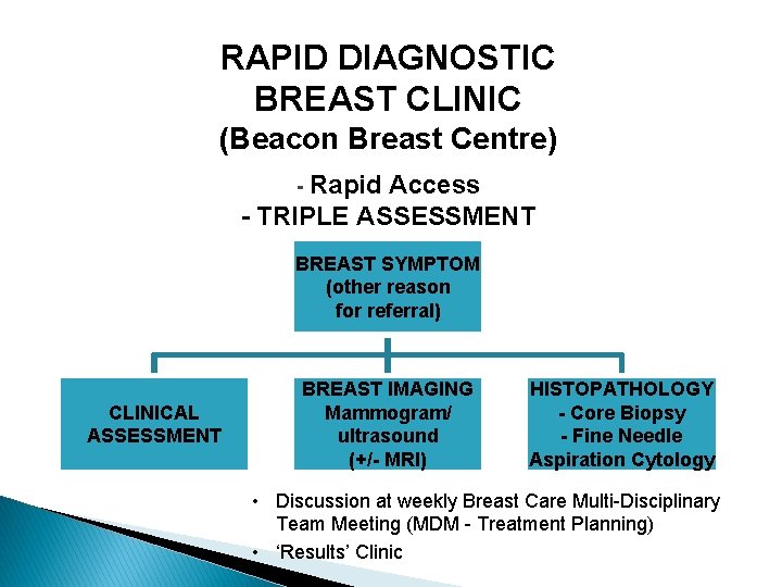
RAPID DIAGNOSTIC BREAST CLINIC (Beacon Breast Centre) - Rapid Access - TRIPLE ASSESSMENT BREAST SYMPTOM (other reason for referral) CLINICAL ASSESSMENT BREAST IMAGING Mammogram/ ultrasound (+/- MRI) HISTOPATHOLOGY - Core Biopsy - Fine Needle Aspiration Cytology • Discussion at weekly Breast Care Multi-Disciplinary Team Meeting (MDM - Treatment Planning) • ‘Results’ Clinic
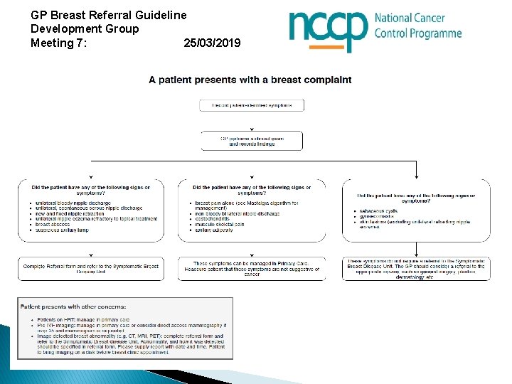
GP Breast Referral Guideline Development Group Meeting 7: 25/03/2019
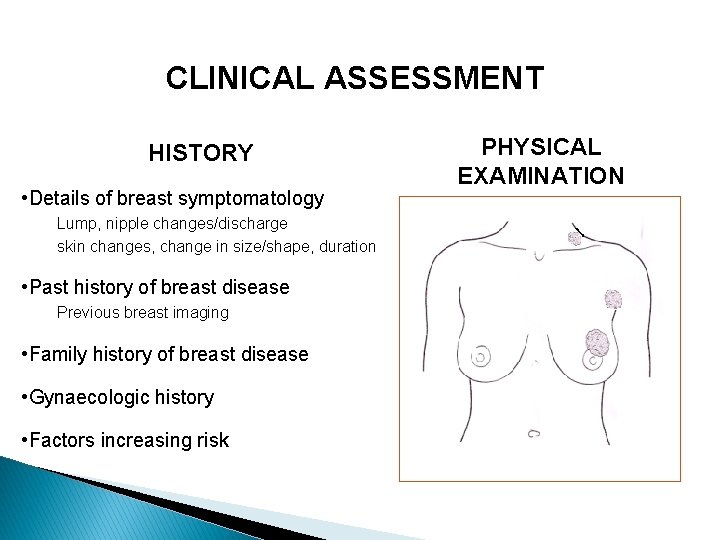
CLINICAL ASSESSMENT HISTORY • Details of breast symptomatology Lump, nipple changes/discharge skin changes, change in size/shape, duration • Past history of breast disease Previous breast imaging • Family history of breast disease • Gynaecologic history • Factors increasing risk PHYSICAL EXAMINATION
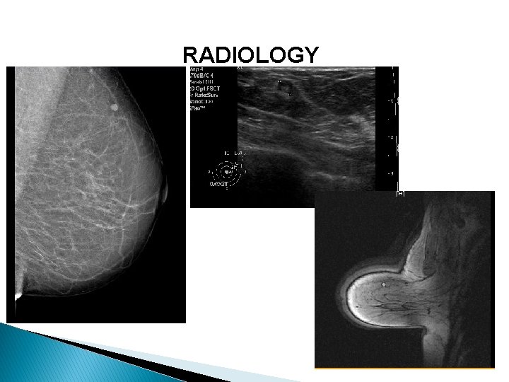
RADIOLOGY
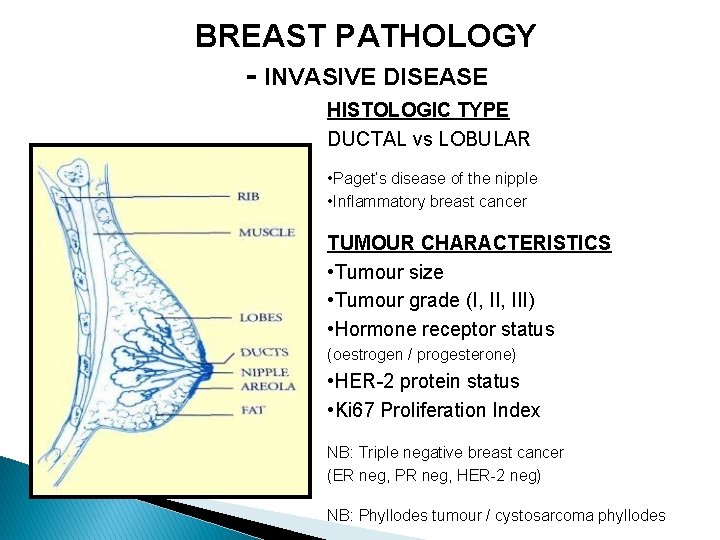
BREAST PATHOLOGY - INVASIVE DISEASE HISTOLOGIC TYPE DUCTAL vs LOBULAR • Paget’s disease of the nipple • Inflammatory breast cancer TUMOUR CHARACTERISTICS • Tumour size • Tumour grade (I, III) • Hormone receptor status (oestrogen / progesterone) • HER-2 protein status • Ki 67 Proliferation Index NB: Triple negative breast cancer (ER neg, PR neg, HER-2 neg) NB: Phyllodes tumour / cystosarcoma phyllodes
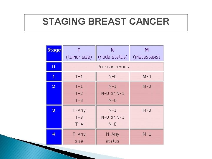
STAGING BREAST CANCER
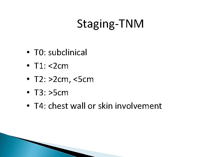
Staging-TNM • • • T 0: subclinical T 1: <2 cm T 2: >2 cm, <5 cm T 3: >5 cm T 4: chest wall or skin involvement
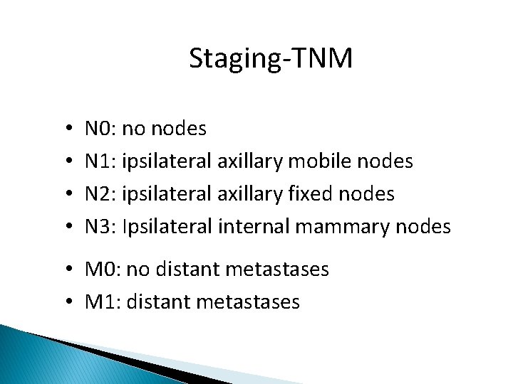
Staging-TNM • • N 0: no nodes N 1: ipsilateral axillary mobile nodes N 2: ipsilateral axillary fixed nodes N 3: Ipsilateral internal mammary nodes • M 0: no distant metastases • M 1: distant metastases

MANAGEMENT OF BREAST CANCER DIAGNOSIS (Triple Assessment) Characterisation of Breast Disease Axillary assessment Systemic staging investigations Treatment Planning: Surgery, Systemic therapy, Radiotherapy
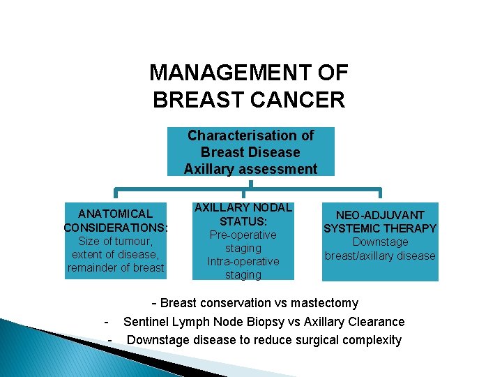
MANAGEMENT OF BREAST CANCER Characterisation of Breast Disease Axillary assessment ANATOMICAL CONSIDERATIONS: Size of tumour, extent of disease, remainder of breast AXILLARY NODAL STATUS: Pre-operative staging Intra-operative staging NEO-ADJUVANT SYSTEMIC THERAPY Downstage breast/axillary disease - Breast conservation vs mastectomy - Sentinel Lymph Node Biopsy vs Axillary Clearance - Downstage disease to reduce surgical complexity
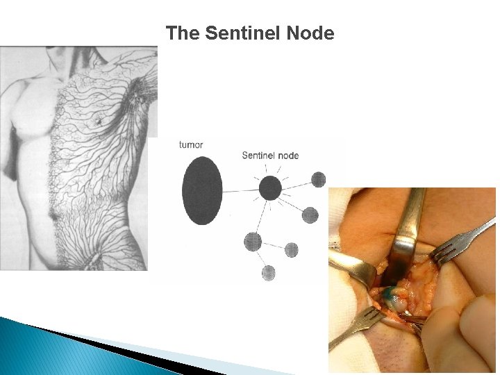
The Sentinel Node
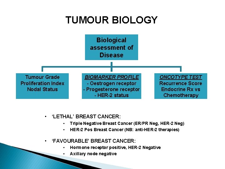
TUMOUR BIOLOGY Biological assessment of Disease Tumour Grade Proliferation Index Nodal Status • ONCOTYPE TEST Recurrence Score Endocrine Rx vs Chemotherapy ‘LETHAL’ BREAST CANCER: • • • BIOMARKER PROFILE - Oestrogen receptor - Progesterone receptor - HER-2 status Triple Negative Breast Cancer (ER/PR Neg, HER-2 Neg) HER-2 Pos Breast Cancer (NB: anti-HER-2 therapies) ‘FAVOURABLE’ BREAST CANCER: • • Hormone receptor positive, HER-2 Negative Axillary node negative
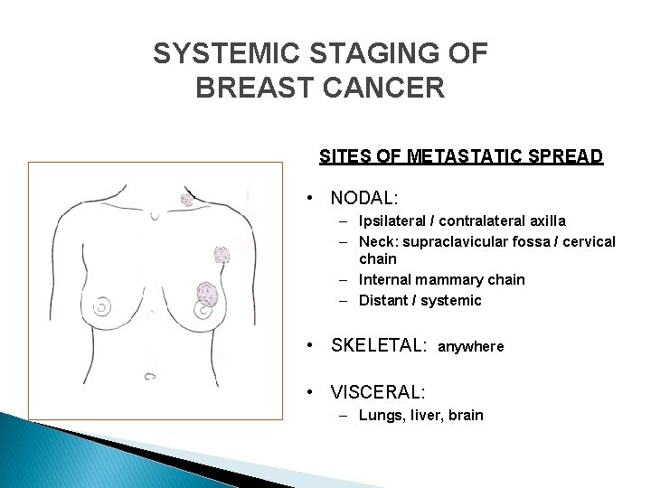
SYSTEMIC STAGING OF BREAST CANCER SITES OF METASTATIC SPREAD • NODAL: – Ipsilateral / contralateral axilla – Neck: supraclavicular fossa / cervical chain – Internal mammary chain – Distant / systemic • SKELETAL: anywhere • VISCERAL: – Lungs, liver, brain
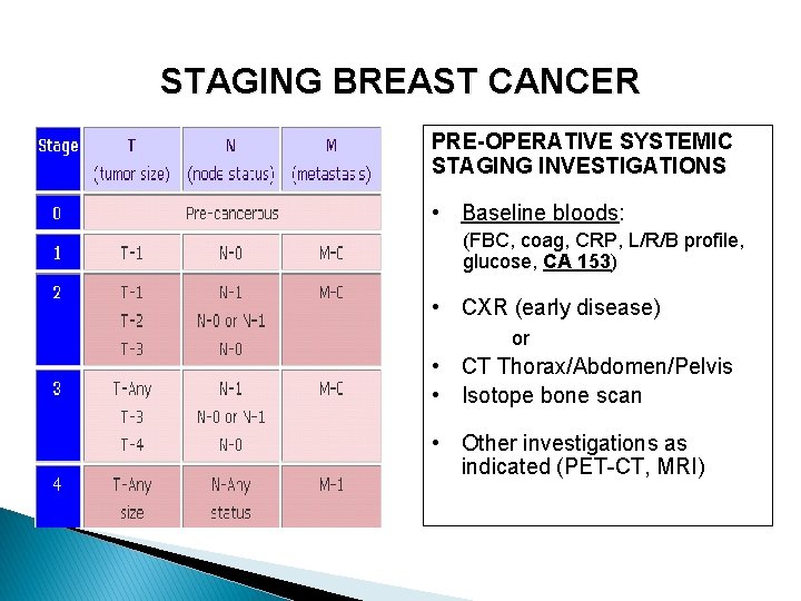
STAGING BREAST CANCER PRE-OPERATIVE SYSTEMIC STAGING INVESTIGATIONS • Baseline bloods: (FBC, coag, CRP, L/R/B profile, glucose, CA 153) • CXR (early disease) or • CT Thorax/Abdomen/Pelvis • Isotope bone scan • Other investigations as indicated (PET-CT, MRI)
TREATMENT STRATEGIES FOR BREAST CANCER 1. SURGICAL THERAPY • 2. Primary vs Delayed CHEMOTHERAPY • • • 3. Adjuvant (post-operative) Neo-adjuvant (primary / pre-operative) Palliative HORMONAL THERAPY • 4. Disease must be ER / PR positive IMMUNOTHERAPY • Disease must overexpress HER-2 5. RADIOTHERAPY 6. NOVEL THERAPIES (Anti-angiogenics etc)
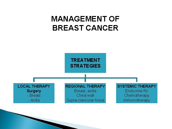
MANAGEMENT OF BREAST CANCER TREATMENT STRATEGIES LOCAL THERAPY Surgery - Breast - Axilla REGIONAL THERAPY Breast, axilla Chest wall Supra-clavicular fossa SYSTEMIC THERAPY Endocrine Rx Chemotherapy Immunotherapy
Molecular profiling to determine if patient will benefit from Chemotherapy? § § § Gene assay performed on paraffin-embedded archival breast cancer tissue Predicts risk of recurrence at 10 yrs Data generated from retrospective analysis of archival tissue Gene set has been validated across multiple validation sets using prospectively defined end points (NSABP B 14 & Kaiser Permanente) Final gene set: relapse-free interval correlations across three independent studies: testing 250 genes in 447 patients
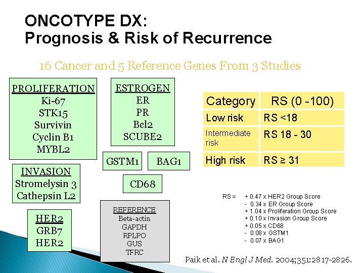
ONCOTYPE DX: Prognosis & Risk of Recurrence 16 Cancer and 5 Reference Genes From 3 Studies PROLIFERATION Ki-67 STK 15 Survivin Cyclin B 1 MYBL 2 INVASION Stromelysin 3 Cathepsin L 2 HER 2 GRB 7 HER 2 ESTROGEN ER PR Bcl 2 SCUBE 2 GSTM 1 BAG 1 Category RS (0 -100) Low risk RS <18 Intermediate risk RS 18 - 30 High risk RS ≥ 31 CD 68 RS = REFERENCE Beta-actin GAPDH RPLPO GUS TFRC + 0. 47 x HER 2 Group Score - 0. 34 x ER Group Score + 1. 04 x Proliferation Group Score + 0. 10 x Invasion Group Score + 0. 05 x CD 68 - 0. 08 x GSTM 1 - 0. 07 x BAG 1 Paik et al. N Engl J Med. 2004; 351: 2817 -2826.
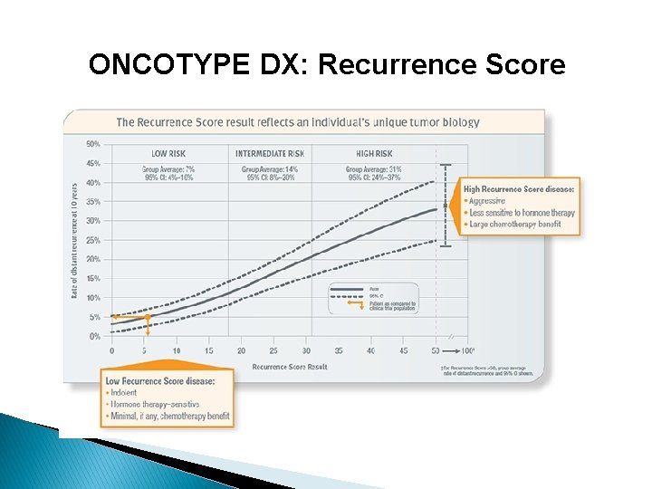
ONCOTYPE DX: Recurrence Score
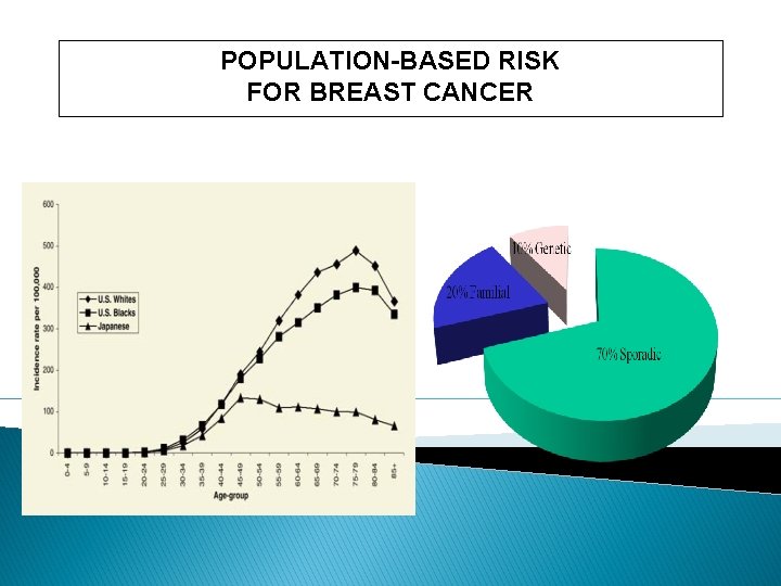
POPULATION-BASED RISK FOR BREAST CANCER
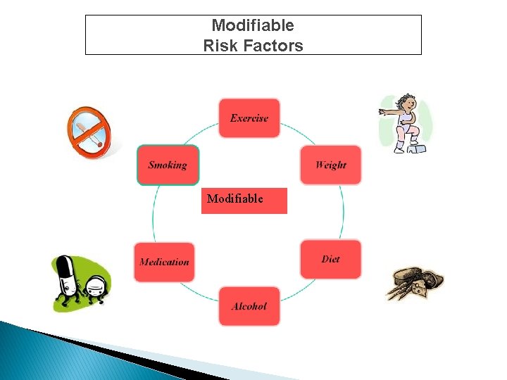
Modifiable Risk Factors Modifiable
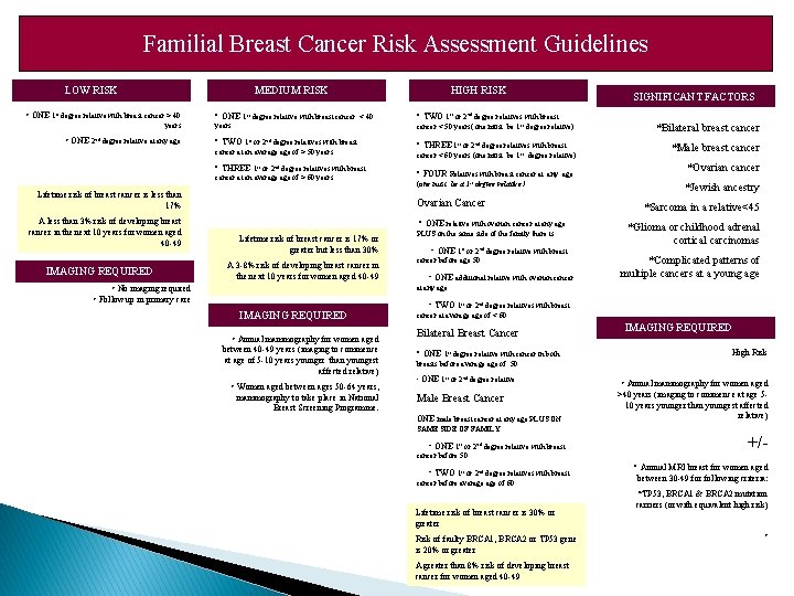
Familial Breast Cancer Risk Assessment Guidelines LOW RISK MEDIUM RISK • ONE 1 st degree relative with breast cancer > 40 years • ONE 2 nd degree relative at any age • ONE 1 st degree relative with breast cancer HIGH RISK < 40 < cancer < 50 years( one must be 1 st degree relative) • TWO 1 st or 2 nd degree relatives with breast • THREE 1 st or 2 nd degree relatives with breast cancer at an average of > 60 years Lifetime risk of breast cancer is less than 17% IMAGING REQUIRED • TWO 1 st or 2 nd degree relatives with breast 40 years cancer at an average of > 50 years A less than 3% risk of developing breast cancer in the next 10 years for women aged 40 -49 SIGNIFICANT FACTORS 1 st 2 nd or degree relatives with breast cancer < 60 years (one must be 1 st degree relative) • FOUR Relatives with breast cancer at (one must be a 1 st any age degree relative ) Ovarian Cancer Lifetime risk of breast cancer is 17% or greater but less than 30% A 3 -8% risk of developing breast cancer in the next 10 years for women aged 40 -49 • ONE relative with ovarian cancer at any age PLUS on the same side of the family there is: • ONE 1 st or 2 nd degree relative with breast cancer before age 50 • ONE additional relative with ovarian cancer *Bilateral breast cancer *Male breast cancer *Ovarian cancer *Jewish ancestry *Sarcoma in a relative<45 *Glioma or childhood adrenal cortical carcinomas *Complicated patterns of multiple cancers at a young age at any age • No imaging required • Follow up in primary care IMAGING REQUIRED • Annual mammography for women aged between 40 -49 years (imaging to commence at age of 5 -10 years younger than youngest affected relative) • Women aged between ages 50 -64 years, mammography to take place in National Breast Screening Programme. • TWO 1 st or 2 nd degree relatives with breast cancer at average of < 60 Bilateral Breast Cancer • ONE 1 st degree relative with cancer in both IMAGING REQUIRED High Risk breasts before average of 50 • ONE 1 st or 2 nd degree relative Male Breast Cancer ONE male breast cancer at any age PLUS ON SAME SIDE OF FAMILY: • ONE 1 st or 2 nd degree relative with breast cancer before 50 • TWO 1 st or 2 nd degree relatives with breast cancer before average of 60 Lifetime risk of breast cancer is 30% or greater Risk of faulty BRCA 1, BRCA 2 or TP 53 gene is 20% or greater A greater than 8% risk of developing breast cancer for women aged 40 -49 • Annual mammography for women aged >40 years (imaging to commence at age 510 years younger than youngest affected relative) +/ • Annual MRI breast for women aged between 30 -49 for following criteria: *TP 53, BRCA 1 & BRCA 2 mutation carriers (or with equivalent high risk) •
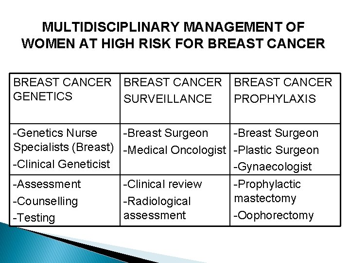
MULTIDISCIPLINARY MANAGEMENT OF WOMEN AT HIGH RISK FOR BREAST CANCER GENETICS SURVEILLANCE PROPHYLAXIS -Genetics Nurse -Breast Surgeon Specialists (Breast) -Medical Oncologist -Plastic Surgeon -Clinical Geneticist -Gynaecologist -Assessment -Counselling -Testing -Clinical review -Radiological assessment -Prophylactic mastectomy -Oophorectomy

Thank you!
- Slides: 26