THE CENTRAL NERVOUS SYSTEM Emily BeaumontMc Allen Aims

THE CENTRAL NERVOUS SYSTEM Emily Beaumont-Mc. Allen

Aims and Objectives Aim: To introduce the mammalian central nervous system. Ob 1: To identify the features and structures of the central nervous system and nervous tissue Ob 2: To identify the divisions of the brain Ob 3: To be able to explain the functions of the central nervous system Ob 4: To be able to compare the mammalian central nervous system and a reptilian nervous system structure and functions.

Nervous System Central Nervous System Brain Peripheral Nervous System Autonomic Spinal Cord Sympathetic Somatic Parasympathetic

Central Nervous System Consists of the brain and spinal cord Made of masses of nerve tissue, protected by membranes & the cranium or skull

The Nervous System ■ The role of the nervous system is to: – Regulate body functions using impulses – Interpret sensory information – Co-ordinate the actions of the body – Store information in the form of memories – Process learning

The Mammalian Brain ■ What is the role of the brain? ■ The information transmitted by neurones is relayed to the brain ■ The brain then processes this information and uses it to regulate the body’s processes ■ What parts of the brain are you already aware of? ■ The brain in separated into several parts, see the next slide for an illustration.
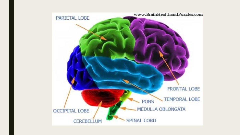

TASK: Match the structure to the function Part Function Cerebrum Controls and processes movement and memory provides ‘personality’ Hypothalamus Controls autonomic activity and the pituitary gland Pituitary Gland Releases hormones to control other glands in the body Cerebral Hemispheres Thinking and language production and interpretation Medulla Oblongata Connects the brain and the spinal cord Brain Stem Controls sleep, breathing and heart rate Cerebellum Controls hand-eye co-ordination, balance and muscle tone Thalamus Receives and processes sensory information

General structure of a Neuron

Nervous Tissue The Nervous systems are made up on nervous tissue. This contains Neurons (Nerve cells) These are three types. Motor neurons connects a muscle (target organ) with an interneuron. Sensory neurons connects a sensory organ with an interneuron. Interneurons connect motor and sensory neurons.

The Synapse When nerve cells have to pass a message to another nerve cell the message has to cross

Structures of a Neuron Using the diagram of the Neuron, briefly describe what you think the functions would be of the structures of the neuron. Structure Dendrites Axon Myelin Sheath (made up of Schwann cells) Cell body and nucleus Synapse (gap between the axon and another neuron dendrite) Nodes of Ranvier (tiny gaps between the myelin sheath) Function

Nodes of Ranvier Speed of nerve impulses Nerve impulses can travel very quickly – at 100 metres per second or even faster. However, different impulses travel at different speeds and are processed in different ways

Fast pain moves quickly from the receptor to the brain. This prompts an immediate response to reduce pain or the risk of injury – for example, taking a hand off something hot. Nerves that move a pain message fast have a large axon diameter. This gives less resistance to the movement of the message. Just as water travels faster in a bigger pipe, the myelin sheath also helps to move the message fast because of the saltatory conduction it creates. Slow pain travels from the receptor to the hypothalamus which can release endorphins to reduce the pain. We tend to experience slow pain as a chronic dull, aching pain. Nerves that transport slow pain have smaller axon diameter and don’t have a myelinated sheath.

More Cell stuff Central Nervous System neurons are insulated by myelin shealths, these are produced by OLIGODENDROCYTE cells. These are a type of GLIAL cell. Astrocytes – These form the blood-brain barrier by holding together neurons and blood vessels Oligodendrocytes These form the myelin sheath around axons of the CNS. Microglia – Phagocyte cells that migrate through the CNS removing foreign matter and degenerated brain tissue Ependymal cells – Epithelial cells that line the brain and central canal of the spinal cord, form cerebrospinal fluid and aid in its circulation
- Slides: 15