
The Center for Health in the Human Ecosystem
- Slides: 19
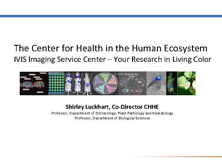
The Center for Health in the Human Ecosystem IVIS Imaging Service Center – Your Research in Living Color Shcherbakova, DM. . Nat. Met. 2013 Shirley Luckhart, Co-Director CHHE Professor, Department of Entomology, Plant Pathology and Nematology Professor, Department of Biological Sciences
IVIS Imaging Service Center contacts – Shirley, Brandi Torrevillas, Kiah Stewart Brandi Torrevillas, Manager Luckhart lab Kiah Stewart, Manager Lab Animal Research Facility
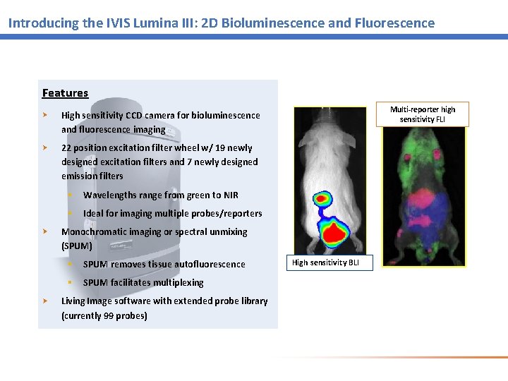
Introducing the IVIS Lumina III: 2 D Bioluminescence and Fluorescence Features Multi-reporter high sensitivity FLI High sensitivity CCD camera for bioluminescence and fluorescence imaging 22 position excitation filter wheel w/ 19 newly designed excitation filters and 7 newly designed emission filters § Wavelengths range from green to NIR § Ideal for imaging multiple probes/reporters Monochromatic imaging or spectral unmixing (SPUM) § SPUM removes tissue autofluorescence § SPUM facilitates multiplexing Living Image software with extended probe library (currently 99 probes) High sensitivity BLI
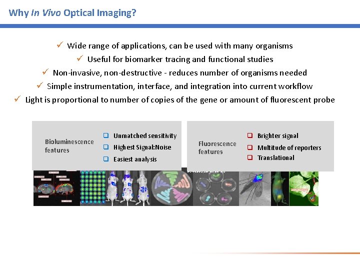
Why In Vivo Optical Imaging? ü Wide range of applications, can be used with many organisms ü Useful for biomarker tracing and functional studies ü Non-invasive, non-destructive - reduces number of organisms needed ü Simple instrumentation, interface, and integration into current workflow ü Light is proportional to number of copies of the gene or amount of fluorescent probe Bioluminescence features q Unmatched sensitivity q Highest Signal: Noise q Easiest analysis Fluorescence features Shcherbakova, DM. . Nat. Met. 2013 q Brighter signal q Multitude of reporters q Translational
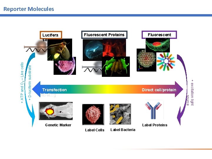
Reporter Molecules + D-luciferin substrate Fluorescent Proteins Fluorescent dyes Transfection Direct cell/protein labeling Label Proteins Genetic Marker Label Cells Label Bacteria + excitation light source + ATP and O 2 – Live cells Lucifera se
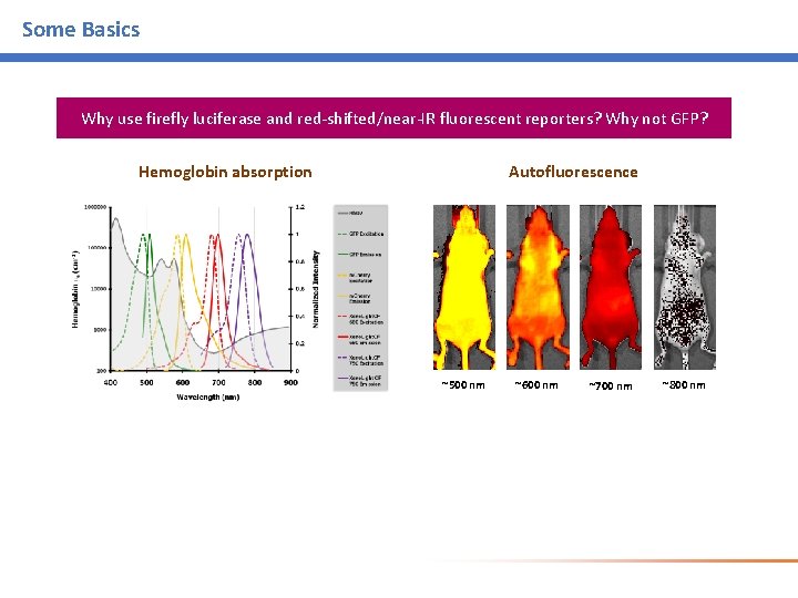
Some Basics Why use firefly luciferase and red-shifted/near-IR fluorescent reporters? Why not GFP? Autofluorescence Hemoglobin absorption ~500 nm ~600 nm ~700 nm ~800 nm
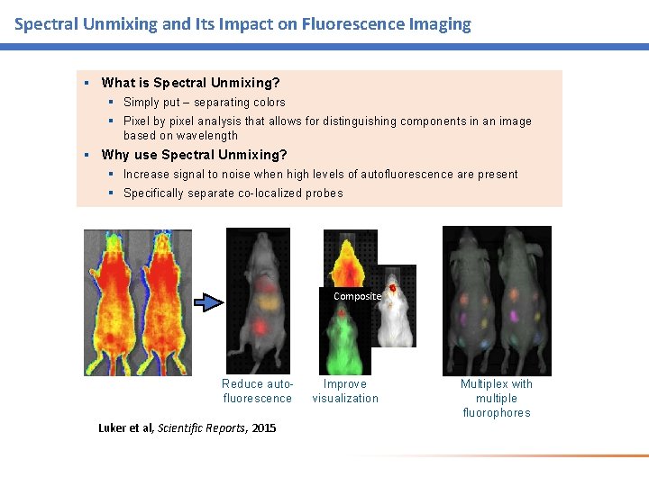
Spectral Unmixing and Its Impact on Fluorescence Imaging § What is Spectral Unmixing? § Simply put – separating colors § Pixel by pixel analysis that allows for distinguishing components in an image based on wavelength § Why use Spectral Unmixing? § Increase signal to noise when high levels of autofluorescence are present § Specifically separate co-localized probes Composite Reduce autofluorescence Luker et al, Scientific Reports, 2015 Improve visualization Multiplex with multiple fluorophores
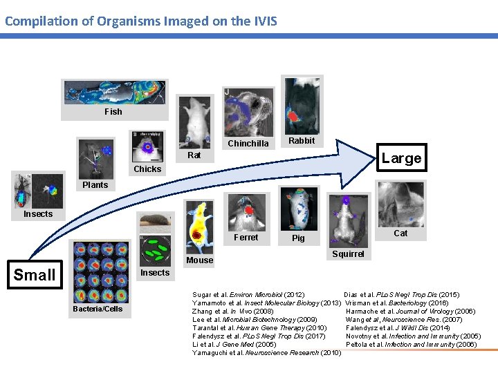
Compilation of Organisms Imaged on the IVIS Fish Chinchilla Rabbit Large Rat Chicks Plants Insects Ferret Mouse Small Cat Pig Squirrel Insects Bacteria/Cells Sugar et al. Environ Microbiol (2012) Dias et al. PLo. S Negl Trop Dis (2015) Yamamoto et al. Insect Molecular Biology (2013) Vrisman et al. Bacteriology (2016) Zhang et al. In Vivo (2008) Harmache et al. Journal of Virology (2006) Lee et al. Microbial Biotechnology (2009) Wang et al, Neuroscience Res. (2007) Tarantal et al. Human Gene Therapy (2010) Falendysz et al. J Wildl Dis (2014) Falendysz et al. PLo. S Negl Trop Dis (2017) Novotny et al. Infection and Immunity (2005) Li et al. J Gene Med (2005) Peltola et al. Infection and Immunity (2006) Yamaguchi et al. Neuroscience Research (2010)
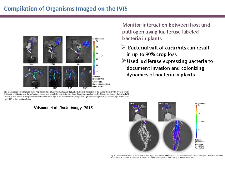
Compilation of Organisms Imaged on the IVIS Monitor interaction between host and pathogen using luciferase labeled bacteria in plants Ø Bacterial wilt of cucurbits can result in up to 80% crop loss ØUsed luciferase expressing bacteria to document invasion and colonizing dynamics of bacteria in plants Vrisman et al. Bacteriology. 2016.
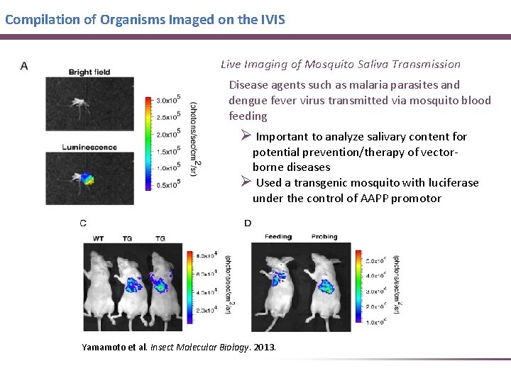
Compilation of Organisms Imaged on the IVIS Live Imaging of Mosquito Saliva Transmission Disease agents such as malaria parasites and dengue fever virus transmitted via mosquito blood feeding Ø Important to analyze salivary content for potential prevention/therapy of vectorborne diseases Ø Used a transgenic mosquito with luciferase under the control of AAPP promotor Yamamoto et al. Insect Molecular Biology. 2013.
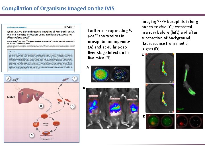
Compilation of Organisms Imaged on the IVIS Luciferase-expressing P. yoelii sporozoites in mosquito homogenate (A) and at 48 hr postliver stage infection in live mice (B) Imaging YFP+ basophils in long bones ex vivo (C); extracted marrow before (left) and after subtraction of background fluorescence from media (right) (D) C A B D
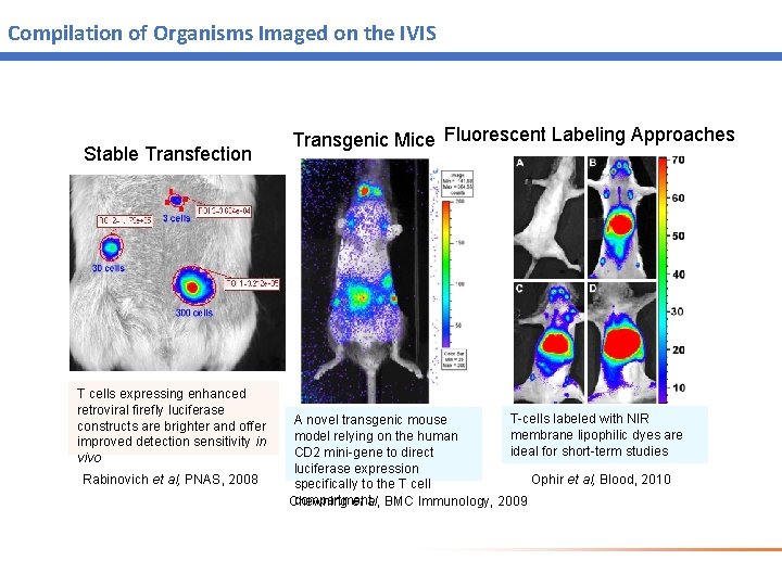
Compilation of Organisms Imaged on the IVIS Stable Transfection T cells expressing enhanced retroviral firefly luciferase constructs are brighter and offer improved detection sensitivity in vivo Rabinovich et al, PNAS, 2008 Transgenic Mice Fluorescent Labeling Approaches T-cells labeled with NIR A novel transgenic mouse membrane lipophilic dyes are model relying on the human ideal for short-term studies CD 2 mini-gene to direct luciferase expression Ophir et al, Blood, 2010 specifically to the T cell compartmental, BMC Immunology, 2009 Chewning et
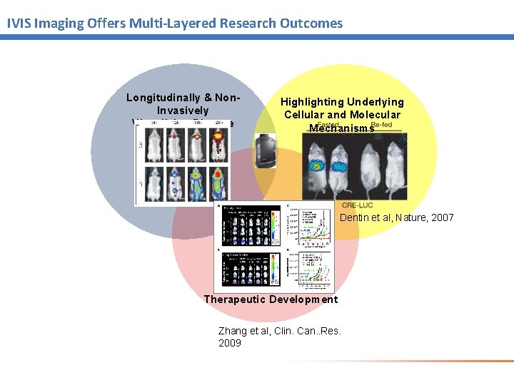
IVIS Imaging Offers Multi-Layered Research Outcomes Longitudinally & Non. Invasively Visualizing Disease Highlighting Underlying Cellular and Molecular Mechanisms Dentin et al, Nature, 2007 Therapeutic Development Zhang et al, Clin. Can. . Res. 2009
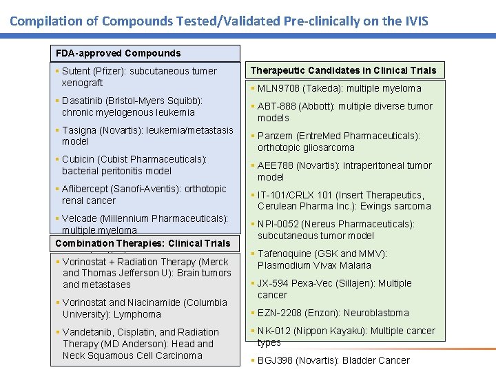
Compilation of Compounds Tested/Validated Pre-clinically on the IVIS FDA-approved Compounds § Sutent (Pfizer): subcutaneous tumer xenograft § Dasatinib (Bristol-Myers Squibb): chronic myelogenous leukemia § Tasigna (Novartis): leukemia/metastasis model § Cubicin (Cubist Pharmaceuticals): bacterial peritonitis model § Aflibercept (Sanofi-Aventis): orthotopic renal cancer § Velcade (Millennium Pharmaceuticals): multiple myeloma Combination Therapies: Clinical Trials § Prolia (Amgen): bone metastases § Vorinostat + Radiation Therapy (Merck and Thomas Jefferson U): Brain tumors and metastases § Vorinostat and Niacinamide (Columbia University): Lymphoma § Vandetanib, Cisplatin, and Radiation Therapy (MD Anderson): Head and Neck Squamous Cell Carcinoma Therapeutic Candidates in Clinical Trials § MLN 9708 (Takeda): multiple myeloma § ABT-888 (Abbott): multiple diverse tumor models § Panzem (Entre. Med Pharmaceuticals): orthotopic gliosarcoma § AEE 788 (Novartis): intraperitoneal tumor model § IT-101/CRLX 101 (Insert Therapeutics, Cerulean Pharma Inc. ): Ewings sarcoma § NPI-0052 (Nereus Pharmaceuticals): subcutaneous tumor model § Tafenoquine (GSK and MMV): Plasmodium Vivax Malaria § JX-594 Pexa-Vec (Sillajen): Multiple cancer § EZN-2208 (Enzon): Neuroblastoma § NK-012 (Nippon Kayaku): Multiple cancer types § BGJ 398 (Novartis): Bladder Cancer
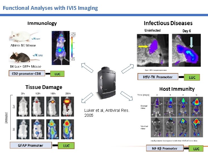
Functional Analyses with IVIS Imaging Luker et al, Antiviral Res. 2005
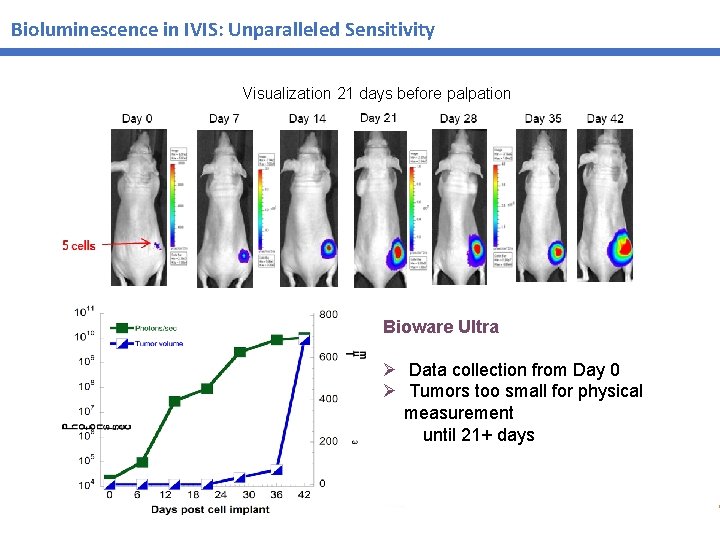
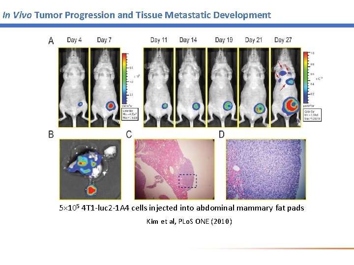
Bioluminescence in IVIS: Unparalleled Sensitivity Visualization 21 days before palpation Bioware Ultra Ø Data collection from Day 0 Ø Tumors too small for physical measurement until 21+ days
In Vivo Tumor Progression and Tissue Metastatic Development 5× 10⁵ 4 T 1 -luc 2 -1 A 4 cells injected into abdominal mammary fat pads Kim et al, PLo. S ONE (2010)
Technology Access Grants – apply now! Applications are now open for CHHE IVIS Imaging Service Center Technology Access Grants (TAGs). The CHHE will fund up to five (5) TAGs for UI investigators interested in using this facility for research. Up to $1, 200 per project will be awarded for hours of training provided by the service center, for use of the IVIS Lumina III and for data analysis completed on service center computers. UI investigators, postdoctoral fellows, graduate students and other research group members are eligible to apply. Applications will be reviewed starting April 15, 2019, and continue until five TAGs are awarded. We anticipate the projects will be implemented during the period from May 15, 2019 through July 31, 2020.
Thank you!