THE CELL NUCLEUS The nucleus pl nuclei latin
 is")


")


")


 and other short (p)")


")


")


- Slides: 68
THE CELL NUCLEUS The nucleus (pl. nuclei; latin nucleus or nuculeus, meaning kernel) is a membrane-enclosed organelle found in eukaryotic cell. � It contains most of the cell's genetic material. � Genetic material is organized as multiple long linear DNA molecules in complex with proteins, histones to form chromosomes. � The genes within these chromosomes are the cell's nuclear genome. � The function of the nucleus is to maintain the integrity of these genes and to control the activities of the cell � Nucleus regulates gene expression therefore, it is a control center of the cell. � The genetic material is surrounded by the nuclear membrane known as nuclear envelop �
HISTORY AND DISCOVERY � � � � The first organelle discovered in the cell was nucleus. Antonie van Leeuwenhoek (1632 – 1723). "Lumen", the nucleus, in RBCs of salmon Franz Bauer (1804) described nucleus in more detail. Robert Brown (Scottish) (1831) described it in a talk at meeting of the Linnean Society of London. He was studying orchids and observed an opaque area, which he called the areola or nucleus, in the cells of the flower's outer layer. Matthias Schleiden (1838), suggested that nucleus plays a role in generating cells, and called it "Cytoblast" (cell builder). Franz Meven was a strong opponent of this view, having already described cells multiplying by division and believing that many cells would have no nuclei. Robert Remak (1852) and Rudolf Virchow (1855) decisively propagated the new paradigm that cells are generated solely by cells. The function of the nucleus remained unclear by then. Oscar Hertwig (1877 - 1878) published several studies on the fertilization of sea urchin eggs, showing that the nucleus of the sperm enters the oocyte and fuses with its nucleus. This was the first time it was suggested that an individual develops from a (single) nucleated cell.
FUNCTIONS OF THE NUCLEUS � The function of the nucleus as carrier of genetic information became clear after mitosis was discovered and the Mendelian rules were rediscovered at the beginning of the 20 th century � The chromosome theory of heredity was therefore developed � Site of transcription of DNA into m. RNA, r. RNA and t. RNA � Site of protein-r. RNA assembly into ribosomal subunits
STRUCTURE OF NUCLEUS � The nucleus is the largest cellular organelle in eukaryotic cell. � The average diameter of the nucleus is approximately 6 μm, which occupies about 10% of the total cell volume. � The viscous liquid within it is called nucleoplasm. � It appears as a dense, roughly spherical organelle
LIGHT MICROGRAPHS OF CELL NUCLEUS
NUCLEUS IN PROPHASE OF MITOSIS
NUCLEUS OF NERVE CELL
� Nucleus is surrounded by double unit membrane called nuclear envelope � Nuclear envelope is perforated by nuclear pores. � Pores have a channel made of transport proteins for transfer of molecules like proteins and RNA inside actively and small molecules like ions out side � Sub-nuclear bodies exist, made up of unique proteins and RNA molecules the nucleolus � Nucleolus is a site of production of ribosome that are exported to the cytoplasm where they act as sites of m. RNA translation into proteins
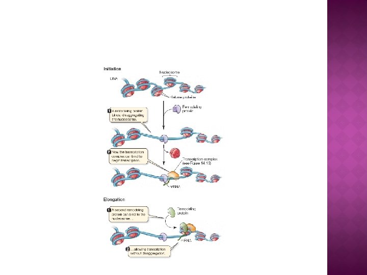
� � � � DNA- protein complex is known chromatin which can be seen in side the nucleus as dense granular material Chromatin are of two types Euchromatin that is less compact DNA form, and contains genes that are frequently expressed by the cell The other type heterochromatin the more compact form, and contains DNA that is infrequently transcribed Heterochromatin is further categorized into facultative heterochromatin consisting of genes that are organized only in certain cell types or at certain stages of development Constitutive heterochromatin consists of chromosome structural components such as telomers and centromeres During interphase the chromatin organizes itself into discrete individual patches called chromosomes Active genes are towards the boundary of chromosome
GRAPHIC STRUCTURE OF NUCLEUS
TEM OF NUCLEUS- RER CONTINUUM
SEM OF NUCLEUS-RER CONTINUUM
TEM IMAGE OF NUCLEUS
ELECTRON MICROGRAPHS OF CHROMATIN (DNA PROTEIN COMPLEX)
NUCLEAR ENVELOPE � The nuclear envelope, consists of two cellular membranes, an inner and an outer membrane, arranged parallel to one another and separated by 10 -50 nm perinuclear space � Serves as a barrier to prevent macromolecules from diffusing freely between the nucleoplasm and the cytoplasm � The outer nuclear membrane is continuous with the membrane of the rough endoplasmic reticulum (RER), and is also studded with ribosomes � The space between the membranes is called the perinuclear space and is continuous with the lumen of RER
NUCLEUS-RER CONTINUITY
NUCLEAR MEMBRANE
NUCLEUS OF MERISTEMATIC CELL
NUCLEAR PORE Nuclear pores provide aqueous channels through the envelope, are composed of multiple proteins known as nucleoporins. � The pores are 25 million Da in MW and consist of around 50 (Yeast) to several hundred proteins (vertebrates). � The pores are 100 nm ф; the gap through which molecules freely diffuse is 9 nm wide. This size selectively allows the passage of small water-soluble molecules while preventing larger molecules, such as nucleic acids and larger proteins � These large molecules must be actively transported into the nucleus instead. � The nucleus of a typical mammalian cell have about 3000 to 4000 pores �
SEM OF NUCLEAR PORE
SEM MICROGRAPH OF NUCLEAR MEMBRANE
SEM MICROGRAPH OF NUCLEAR PORES
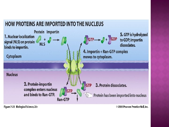
� � � Pore contains an eightfold-symmetric ring-shaped structure at a position where the inner and outer membranes fuse. Attached to the ring is a structure called the nuclear basket that extends into the nucleoplasm, and a series of filamentous extensions that reach into the cytoplasm. Both structures serve to mediate binding to nuclear transport proteins Most proteins, ribosomal subunits, and some DNAs are transported through the pore complexes in a process mediated by a family of transport factors known as karyoporines Those karyoporines that mediate movement into the nucleus are also called importins, whereas those that mediate movement out of the nucleus are called exportins
NUCLEAR BASKET
NUCLEAR ENVELOP
NUCLEAR TRAFFICKING
NUCLEAR TRAFFICKING
� Steroid hormones and other small lipidsoluble molecules involved in intercellular signalling, can diffuse through the cell membrane and into the cytoplasm, where they bind with nuclear receptor proteins that are transported into the nucleus. � There they serve as transcriptional factors when bound to their receptors or function as histone deacetylases that repress gene expression
LAMINA �A network of protein filaments provide the nucleus with mechanical support are known as nuclear lamina forms an organized meshwork on the internal face of the envelope, while less organized support is provided on the cytosolic face of the envelope. � Both systems provide structural support for the nuclear envelope and anchoring sites for chromosomes and nuclear pores � The nuclear lamina is composed of lamin proteins which are synthesized in the cytoplasm and later transported into the nucleus � Nuclear matrix (frame work of protein fibers) and lamina may help synthesize the genetic material efficiently
FIBROUS PROTEIN OF LAMINA
NUCLEAR LAMINA
NUCLEOLUS � In non dividing nucleus a darkly stained structure is nucleolus (pl; nucleoli) � There may be one or more in single nucleus � Consists of granules and fibers of chromatin � Site of r. RNA synthesis and assembling the imported proteins with r. RNA to make ribosomal large and small subunits � These subunits then exit the nucleus into the cytoplasm and assemble into ribosome the site of protein synthesis
FLUORESCENT IMAGE OF NUCLEOLI
CONFOCAL IMAGE OF NUCLEOLI
TEM IMAGE OF NUCLEOLI
TEM IMAGES OF NUCLEUS AND NUCLEOLI
� When observed with TEM, the nucleolus can be seen to consist of three distinguishable regions � The innermost fibrillar centers (FCs), surrounded by the dense fibrillar component (DFC), which in turn is bordered by the granular component (GC) � Transcription of the r. DNA occurs either in the FC or at the FC-DFC boundary, and, therefore, when r. DNA transcription in the cell is increased, more FCs are detected. � Most of the cleavage and modification of r. RNAs occurs in the DFC, while the latter steps involving protein assembly onto the ribosomal subunits occur in the granular component
CHROMOSOMES � DNA is organized into discrete units during cell division called chromosomes that carry the genetic information � Chromosome consists of one long molecule of DNA associated with many proteins � Proteins allow the DNA to coil around to reduce its length to fit into the nucleus � In dividing cells chromatin condensed to form chromosomes � Their # is species specific: Human (46), fruit fly (8) diploid number and 23 and 4 haploid number respectively
� Chromosome consists of two chromatids having one long (q) and other short (p) arm � Two arms are joined at a region known as centromere � The ends of chromosome are known telomeres
CHROMOSOME
CHROMOSOME
ELECTRON MICROGRAPHS OF CHROMATIN (DNA PROTEIN COMPLEX)
FISH IMAGE OF CHROMOSOMES
CHROMOSOME COMPARISON HUMAN CHIMPANZEE
HUMAN CHROMOSOMES
HUMAN CHROMOSOMES
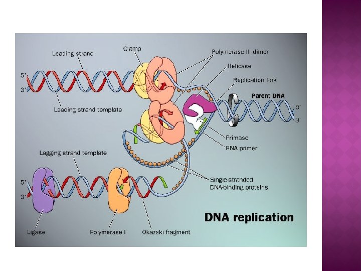
FUNCTIONS OF NUCLEUS � Site of DNA replication � Site of m. RNA synthesis � Site of t. RNA synthesis � Site of r. RNA synthesis � Site of assembly of ribosomal subunits � Nucleus directs protein synthesis by synthesizing m. RNA per instructions of DNA � m. RNA is transported to cytoplasm via nuclear pores � In the cytoplasm m. RNA attaches with the ribosomes and translate m. RNA’s message into primary structure of specific proteins via translation
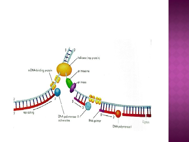
DNA REPLICATION
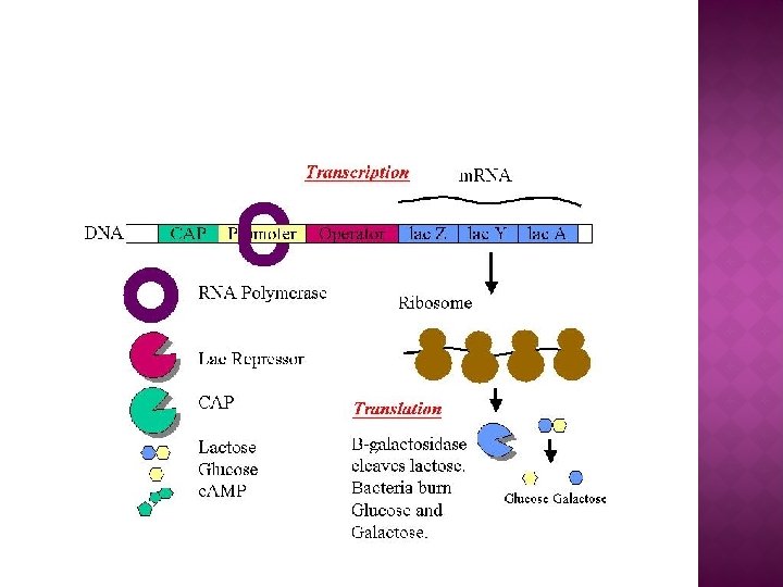
TRANSCRIPTION (MASSENGER RNA SYNTHESIS)
TRANSCRIPTION
TRANSCRIPTION AND TRANSLATION
CELL DEATH � � � During its lifetime, a nucleus may be broken down, either in the process of cell division or as apoptosis (process of programmed cell death) During these events the nuclear envelope and lamina — can be systematically degraded In most cells, the disassembly of the nuclear envelope marks the end of the prophase of mitosis However, this disassembly of the nucleus is not a universal feature of mitosis and does not occur in unicellular eukaryotes (yeasts) undergo so-called closed mitosis in which the nuclear envelope remains intact. In close mitosis, the daughter chromosomes migrate to opposite poles of the nucleus, which then divides in two. The cells of higher eukaryotes undergo open mitosis in which nuclear envelope breaks down. The daughter chromosomes then migrate to opposite poles of the mitotic spindle, and new nuclei reassemble around them.
TRANSFORMATION OF FOREIGN DNA INTO GENOMIC DNA
TRANSCRIPTION AND TRANSLATION
DNA REPLICATION
DNA REPLICATION
TRANSCRIPTION
POST TRANSCRIPTIONAL CHANGES
TRANSCRIPTION AND TRANSLATION
NUCLEUS