The cell Histology is a medical basic science

The cell

Histology: is a medical basic science dealing with the microscopic anatomy of cells, tissues and organs, in order to correlate their structure with their function. Study of histology is dependent upon several tools: microscopes to examine tissues Microtechniques which means preparation of tissues to make them suitable for microscopic examination. • The cell is the smallest structural and functional unit capable of independent existence in all the living organisms. • The human body is composed of more than 200 different types of cells however all cells have a common basic structure. • Generally, the cell is formed of 2 major components: vnucleus vcytoplasm. The cytoplasm is a dense fluid matrix= the cytosol contains the cell organelles , the cell inclusions and cytoskeleton. • Each cell is bounded by the cell membrane, separating the cytoplasm from its extracellular environment.
 • It is a vital intact cell membrane")
(Cell membrane = PLASMA MEMBRANE= PLASMALEMMA) • It is a vital intact cell membrane is essential to cell survival , dynamic , stable , semipermable membrane that surrounds the cell. v. LM picture : Not seen invisible by light microscope because it is too thin (8. 5 nm) condensation of the stain on the outer surface of the cell membrane marks its site in histological sections. v. EM picture • At low magnification the plasma membrane appears as thin dense line • With higher magnification, this line appears as a trilaminar structure, with an outer (= extracellular leaflet) and an inner (= cytoplasmic leaflet) electron dense lines and a middle electron lucent zone in between. 3

Molecular structure of the cell membrane ØThese biological membranes consist mainly of bimolecular layer of phospholipids in which specialized proteins are suspended in association with surface carbohydrates. Ø Membrane phospholipids and the associated proteins are usually present in a 1: 1 proportion by weight. 1 - Lipid molecules of the cell membrane: • Phospholipids: each phospholipid molecule consists of: • One polar hydrophilic head that faces the aqueous media on either side of the membrane (the tissue fluid at the outer surface of the membrane or the cytosol at the inner surface of the membrane). • Two long non-polar hydrophobic fatty acid tails project towards the center of the membrane facing each other. They form weak non-covalent bonds with each other, holding the bilayer together. • - The phospholipid molecules are responsible for the semi-permeability of the cell membrane. They prevent passage of water soluble substances and ions; however they allow passage of small non-polar and fat soluble substances. • • • Cholesterol: They are incorporated within the lipid bilayer. Membrane cholesterol molecules are responsible for : Stability of the membrane components Regulation of membrane fluidity in body temperature. 4

The trilaminar structure of the cell membrane seen in EM after fixation with osmium tetroxide is caused by the deposition of osmium on the hydrophilic heads of the phospholipid molecules resulting in two electron dense lines, while the hydrophobic tails of the lipid molecules remain unstained resulting in the intervening pale zone.

Protein molecules of the cell membrane: • they are present in two forms: v. Integral membrane proteins: • They are embedded within the lipid bilayer. Most of these proteins traverse the whole thickness of the membrane and are called transmembrane proteins. • There are six functional forms of integral membrane proteins : 1. Pumps serve to transport ions (Na+, K+) actively across the membrane. 2. Channels serve to transport certain substances passively across the membrane. 3. Receptors allow binding and recognition of specific substances (ligands) e. g. hormone on the extracellular surface of the membrane. 4. Enzymes membrane proteins possess various enzymatic activities for example: ATP synthase of the inner mitochondrial membrane and some types of digestive enzymes in the small intestine. 5. Linkers serve to anchor the intracellular cytoskeleton to the extracellular matrix. 6. Structural proteins form junctions between neighboring cells. v. Peripheral membrane proteins: • They are not embedded into lipid bilayer but they are loosely-associated with membrane surface. • They are usually located on the cytoplasmic surface of the membrane. • They form a link between the cell membrane & the cytoplasmic components.

Carbohydrate molecules= cell coat or glycocalyx. • The carbohydrate residues are present as glycoproteins and glycolipids of the cell membrane. • They are always oriented towards the outside of the membrane with their branching saccharides extend some distance beyond the cell surface The cell coat is responsible for: The glycocalyx of the plasma membrane vary from species to species and even from one cell type to another. The diversity of the molecules and their location on the cell's surface enable membrane carbohydrates to function as markers that distinguish one cell from another ØThe cell coat is represented by the "fuzzy" material on the outer surface of the membrane. ØCellular recognition e. g. the glycocalyx on the surface of red blood cells (RBCs) determines the four human blood groups. ØCell-to-cell adhesion. ØFormation of receptor sites for ligands by the glycoproteins of the cell membrane. 7

Functions of cell membrane v. Acting as an interface between the cytoplasm and the external environment. v. Maintaining the structural integrity of the cell v. Controlling movements of substances in and out of the cell vtransport systems for specific molecules v. Regulating cell-cell interactions v. Recognizing antigens, foreign cells and altered cells • Fluid mosaic model of the cell membrane The membrane is composed of a sea of lipids (fluid) in which proteins (mosiac) are moving and floating like icebergs.

Receptor – mediated
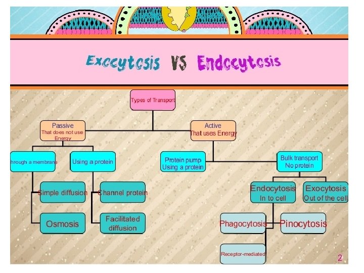

VESICULAR TRANSPORT OF macromolecules ACROSS THE CELL MEMBRANE • Mass transfer of materials through the cell membrane occurs by changes in the plasma membrane at localized sites. It involves 2 processes; the endocytosis and the exocytosis. 1. ENDOCYTOSIS • Endocytosis is the uptake of material, a process by which a cell ingests macromolecules, particulate matter and other substances from the extracellular space. • It is an active process that involves invagination of the cell membrane to form a membrane- bounded vesicle. In general, 3 mechanisms of endocytosis are recognized in the cell:
 : • It is a non-selective process that occurs in")
v. Pinocytosis (cell drinking) : • It is a non-selective process that occurs in nearly all cell types for uptake of fluid containing ions and small protein molecules. • The pinocytotic vesicles formed are small and have smooth surface. • This process is most evident in the endothelium of blood vessels. v. Receptor- mediated endocytosis : • It is a highly-selective process resulting in uptake of specific substances (ligands) by a specific cell that has receptors for these substances e. g. uptake of protein hormones. • These receptors are concentrated in specialized regions of the plasma membrane called coated pits (coated by a protein called clathrin). • When a ligand binds specifically to its receptor on the cell surface, clathrin-coated pits invaginate and give rise to small spinous clathrin-coated vesicles containing this ligand. • Clathrin coat is rapidly lost and recycled for reuse leaving uncoated endocytotic vesicles, delivered to the lysosomal pathway (discussed later). • Phagocytosis (cell eating): • It is the ingestion of large solid particles, such as bacteria and cell debris. • It is a form of receptor-mediated endocytosis however; it does not involve formation of coated pits or vesicles. • This activity is confined to special types of cells, the phagocytes which have receptors on their surfaces that recognize and bind the foreign particles e. g. tissue macrophages. • The binding of the receptor and foreign body results in extension of the cell membrane to form pseudopodia that engulf the particle. This is followed by fusion of the membrane to internalize the particle into the cytoplasm forming a membrane-bounded phagosome. • The contents of the phagosome are then digested inside the cell through lysosomal pathway.

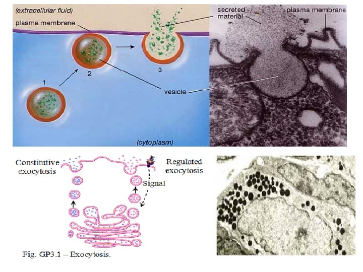
2 - EXOCYTOSIS Exocytosis means the release of cell products into the extracellular compartment. During this process, a vesicle moves from the cytoplasm to the cell membrane, fuses with it and discharges its content. There are 2 general pathways of exocytosis: v Regulated secretion (stimulus- dependent): The secretory products become concentrated and stored forming membrane-bounded secretory granules. As a result of a stimulus (hormonal or neural stimulus), these vesicles move to the surface and fuse with the cell membrane to pour their contents outside the cell. v Regulated exocytosis occurs during release of the digestive enzymes by acinar cells of the pancreas. v Constitutive secretion: The secretory products leave the cell immediately after their synthesis. These cells lack secretory granules. The secretion is released continuously through secretory vesicles that fuse with the plasma membrane. Constitutive exocytosis occurs during release of antibodies by plasma cells. Membrane recycling: During the vesicular transport, the cell membrane is maintained; the excess membrane added to the cell membrane by exocytosis is constantly recycled again into the cytoplasmic compartments by endocytosis through proteins from golgi.

Thank you
- Slides: 15