The Cell Division Cycle Reference Essential cell biology

The Cell Division Cycle Reference Essential cell biology, Alberts 2010

Cell cycle A cell reproduces by carrying out an orderly sequence of events in which it duplicates its contents and then divides in two
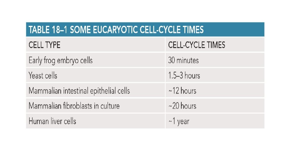

Cell cycle is divided into four phases. the cell grows continuously in interphase, which consists of three phases: G 1, S, and G 2. DNA replication is confined to S phase. G 1 is the gap between M phase and S phase, and G 2 is the gap between S phase and M phase. During M phase, the nucleus divides first, in a process called mitosis; then the cytoplasm divides, in a process called cytokinesis.

Checkpoints in the cell cycle control system ensure that key processes in the cycle occur in the proper sequence. these processes include DNA replication in S phase and the segregation of duplicated chromosomes in mitosis. Feedback from the intracellular events of the cell cycle, as well as signals from the cell’s environment, determine whether the cycle will progress beyond certain checkpoints. three prominent checkpoints are highlighted: the checkpoint in G 1 determines whether the cell proceeds to S phase; the one in G 2 determines whether the cell proceeds to mitosis; and the one in M phase determines whether the cell is ready to pull the duplicated chromosomes apart and segregate them into two new daughter cells

The cell-cycle control system Two types of control systems are involved in cell division: 1 - manufactures the new components of the growing cell, 2 - takes these components into their correct places and partitions them appropriately when the cell divides in two

The protein kinases at the core of the cell-cycle control system are present in proliferating cells throughout the cell cycle. . a Cdk must bind a regulatory protein called a cyclin before it can become enzymatically active.

The accumulation of cyclins regulates the activity of Cdks. the formation of active cyclin–Cdk complexes drives various cell-cycle events, including entry into S phase or M phase. the figure shows the changes in cyclin concentration and Cdk activity responsible for controlling entry into M phase. The increase in the cyclin concentration helps form the active cyclin–Cdk complex that drives entry into M phase. although the enzymatic activity of the cyclin–Cdk complex rises and falls during the course of the cell cycle, the concentration of the Cdk component does not

Distinct Cdks associate with different cyclins to trigger the different events of the cell cycle. For simplicity, only two types of cyclin–Cdk complexes are shown—one that triggers S phase and one that triggers M phase. In both cases, the activation of the Cdk requires phosphorylation and dephosphorylation as well as cyclin binding.

S-Phase • Before a cell divides, it must duplicate its DNA. • This replication must occur with extreme accuracy to minimize the risk of mutations in the next cell generation. • s-cdk initiates DNA replication

-In early G 1: pre-replicative complex is formed: regulatory protein Cdc 6 +ORC -S-Cdk then triggers S-phase by causing the assembly of the protein complexes that initiate DNA synthesis S-Cdk also helps block re-replication by helping to phosphorylate Cdc 6, which dissociates from the origin and is degraded.

M phase it is the most dramatic phase of the cell cycle. During this brief period, the cell reorganizes virtually all of its components and distributes them equally into the two daughter cells. The main problem for a cell in M phase is to accurately segregate its chromosomes, which were replicated in the preceding S phase, so that each new daughter cell receives an identical copy of the genome. Eukaryote cell solve this problem by assembling two specialized cytoskeletal machines: -one pulls the duplicated chromosome -another divides the cytoplasm into two halves (cytokinesis). M phase is conventionally divided into six stages
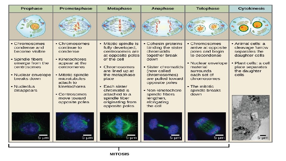

Cytokinesis • , the process by which the cytoplasm is cleaved in two, completes M phase. It usually begins in anaphase but is not completed until the two daughter nuclei have formed in telophase. Whereas mitosis depends on a transient microtubule-based structure, the mitotic spindle, cytokinesis in animal cells depends on a transient structure based on actin and myosin filaments, the contractile ring.

The mitotic spindle determines cytoplasmic cleavage The first visible sign of cytokinesis in animal cells is a puckering and furrowing of the plasma membrane that occurs during anaphase. The contractile ring divides the cell in two

cytokinesis in plant cells involves the formation of a new cell wall • The mechanism of cytokinesis in higher plants is entirely different from that in animal cells, presumably because plant cells are surrounded by a tough cell wall. • The two daughter cells are separated not by the action of a contractile ring at the cell surface but instead by the construction of a new wall that forms inside the dividing cell. • The positioning of this new wall precisely determines the position of the two daughter cells relative to neighboring cells.

Cytokinesis in a plant cell is guided by a specialized microtubule-based structure called the phragmoplast. at the beginning of telophase, after the daughter chromosomes have segregated, a new cell wall starts to assemble inside the cell at the equator of the old spindle (a). the interpolar microtubules of the mitotic spindle remaining at telophase form the phragmoplast and guide the vesicles toward the equator of the spindle. Here, membraneenclosed vesicles, derived from the Golgi apparatus and filled with cell-wall material, fuse to form the growing new cell wall (B), which grows outward to reach the plasma membrane and original cell wall. the plasma membrane and the membrane surrounding the new cell wall (both shown in red) fuse, completely separating the two daughter cells (C).

membrane-enclosed organelles must be distributed to daughter cells • when a cell divides Organelles such as mitochondria and chloroplasts arise only from the growth and division of the pre-existing organelles. • Organelles such as mitochondria and chloroplasts are usually present in large numbers and will be safely inherited if, on average, their numbers simply double once each cell cycle. • The ER in interphase cells is continuous with the nuclear membrane and is organized by the microtubule cytoskeleton. • Upon entry into M phase, the reorganization of the microtubules releases the ER; in most cells, the released ER remains intact during mitosis and is cut in two during cytokinesis. • The Golgi apparatus fragments during mitosis; the fragments associate with the spindle microtubules via motor proteins, thereby linked into the daughter cells as the spindle elongates in anaphase. • Other components of the cell, including all of the soluble proteins, are inherited randomly when the cell divides.

Control of cell number and cell size What adjustment of cell behavior explains the length of an elephant’s trunk or the size of its brain or its liver? Three fundamental processes largely determine organ and body size: cell growth, cell division, and cell death. Each of these processes, in turn, depends on programs intrinsic to the individual cell and is regulated by signals from other cells in the body. Apoptosis helps regulate animal cell numbers: If cells are no longer needed, they can commit suicide by activating an intracellular death program—a process called programmed cell death. In animals, by far the most common form of programmed cell death is called apoptosis.

Animal cells require survival factors to avoid apoptosis Survival factors often suppress apoptosis by regulating bcl 2 family members: The activated receptor activates a transcription regulator in the cytosol. this protein moves to the nucleus, where it activates the gene encoding Bcl 2, a protein that inhibits apoptosis.

Mitogens stimulate cell division • Most mitogens are secreted signal proteins that bind to cell-surface receptors. When activated by mitogen binding, these receptors initiate various intracellular signaling pathways that stimulate cell division. These signaling pathways act mainly by releasing the molecular brakes that block the transition from the G 1 phase of the cell cycle into S phase

Growth factors stimulate cells to Grow The growth of an organism or organ depends on cell growth as much as on cell division. If cells divided without growing, they would get progressively smaller, and there would be no increase in total cell mass. .

Example • Myostatin, for example, is a secreted signal protein that normally inhibits the growth and proliferation of the myoblasts that fuse to form skeletal muscle cells during mammalian development. • When the gene that encodes myostatin is deleted in mice, their muscles grow to be several times larger than normal, because both the number and the size of muscle cells is increased.

Normal cells vs Cancer cells
- Slides: 24