The Cell Cytoplasmic compartments and organelles Prof Dr


- Slides: 41

The Cell Cytoplasmic compartments and organelles Prof. Dr. Özgür Çınar

Histology • Histo- (tissue) + -ology (logos, branch of science) • Marcello Malpighi (10 March 1628 – 29 November 1694) was an Italian biologist and physician, who is referred to as the "Father of microscopical anatomy, histology, physiology and embryology". (wikipedia)

Histology • Histology, also microanatomy, is the branch of biology which studies the tissues of animals and plants using microscopy. • It is commonly studied using a light microscope or electron microscope, the specimen having been sectioned, stained, and mounted on a microscope slide. • Histopathology, the microscopic study of diseased tissue, is an important tool in anatomical pathology, since accurate diagnosis of cancer and other diseases usually requires histopathological examination of samples.

Robert Hooke, cella (latin) = small room

Cytoplasm, Protoplasm, Cytoplasmic matrix • The liquid plasm of the cell • Contains water, ions, proteins etc. • Protoplasm = Cytoplasm + Nucleoplasm • Cytosol = Ground substance = water of the cell

Can cell membrane be seen by a light microscope?

Electron microscope

Modified fluid-mosaic model

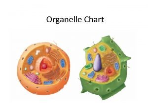
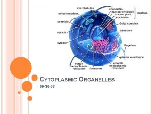
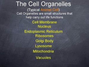
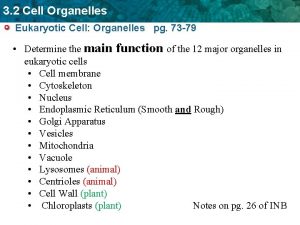
Membranous Organelles • Nucleus • Rough endoplasmic reticulum • Smooth endoplasmic reticulum • Golgi apparatus • Mitochondrion • Peroxisomes • Lysosomes • Endosomes • Secretory vesicles

Nonmembranous Organelles • Proteasome • Ribosome • Centrosome • Inclusions • Cytoskeleton components

Nucleus (karyon) The nucleus is a membrane-limited compartment that contains the genome in eukaryotic cells.

Nucleus • Nuclear envelope • Nucleoplasm • Nucleolus • Chromatin – Euchromatin – Heterochromatin

• The nuclear lamina, a thin, electron-dense intermediate filament network like layer, resides underneath the nuclear membrane. • If the membranous component of the nuclear envelope is disrupted by exposure to detergent, the nuclear lamina remains, and the nucleus retains its shape.

Nuclear lamina • Composed of “Lamin” proteins. • Lamin a and c are structural, lamin b is binding proteins. • Lamin receptors – Emerin: binds both lamin A and lamin B – Nurim: binds lamin A – Lamin B receptor (LBR) • Impairment in nuclear lamina architecture or function is associated with certain genetic diseases (laminopathies) and apoptosis.

Nuclear Pore Complexes

Structure and function • Formed by 50 type of nucleoporin (Nup) proteins • < 40 k. Da (9 nm) passes through • How about the bigger ones!

Karyopherin family members proteins importin x exportin

Nucleolus • Nonmembranous part of the nucleus • The site of r. RNA synthesis and initial ribosomal assembly. • Can be more than one

The nucleolus has three morphologically distinct regions • Fibrillar Center – r. RNA genes (chromosomes– 13, 14, 15, 21, 22 ) – RNA polymerase I – Transcription factors • Fibrillar Material (Pars fibrosa) – Synthesized r. RNA • Granular Material (Pars granulosa) nucleolonema – initial ribosomal assembly – preribosomal particles.

The nucleolus is involved in regulation of the cell cycle. • Nucleostemin – p 53 -binding protein and regulates cell cycle – high in malignant cells – low in differentiated cells • The nucleolus stains intensely with hematoxylin and basic dyes and metachromatically with thionine dyes.

Endoplasmic reticulum Rough ER Ribosome Protein synthesis Smooth ER

Rough endoplasmic reticulum With the TEM, the r. ER appears as a series of interconnected, membrane-limited, flattened sacs called cisternae, with particles located at the exterior surface of the membrane.

Rough endoplasmic reticulum • Basophilic staining (ergastoplasm) • Nissl bodies

Smooth endoplasmic reticulum The s. ER consists of short anastomosing tubules that are not associated with ribosomes.

Smooth endoplasmic reticulum involved in: • lipid and steroid metabolism, • glycogen metabolism and gluconeogenesis • membrane formation and recycling • detoxification • lipoprotein synthesis • storage of calcium – sarcoplasmic reticulum • division of mitochondrion • removing of nonfunctional organelles

Golgi Complex (Apparatus, Body) • Camillo Golgi – osmium-impregnation around nucleus of a nerve cell • Golgi complexes are NOT stained with H&E • Heavy metal impregnation (like. silver, osmium)

Golgi Complex (Apparatus, Body) • In the light microscope, secretory cells that have a large Golgi apparatus typically exhibit a clear area partially surrounded by ergastoplasm. • In EM, it appears as a series of stacked, flattened, membrane-limited sacs or cisternae and tubular extensions embedded in a network of microtubules near the microtubule organizing centers.

Golgi Complex (Apparatus, Body) • It exhibits polarization as cis and trans face. Cis face Trans face

Golgi Complex (Apparatus, Body) • Post-translational modification, • Sorting and packaging of proteins • M-6 -P is added to lysosomal proteins. • Synthesis of sphingomyelin and glycosphingolipid

Cell Secretion and Vesicular Trafficking • Endocytosis is the general term for processes of vesicular transport in which substances enter the cell. • Exocytosis is the general term for processes of vesicular transport in which substances leave the cell.

Coatomers mediate bidirectional traffic between the r. ER and Golgi apparatus. r. ER Golgi

Mitochondrion (mitos + chondros) • 1840, Richard Altman, Bioblast • 1898, Carl Benda, thread (fibre) + granules • 0, 4 – 0, 8 x 4 – 8 um • Circular DNA, • Ability of self-division and protein synthesis • Double membrane Mitochondria are believed to have evolved from an aerobic prokaryote (Eubacterium) that lived symbiotically within primitive eukaryotic cells.

Mitochondria • Present in all cells except red blood cells and terminal keratinocytes. • Abundant in cells that generate and expend large amounts of energy. • Fixed by potassium bichromate or osmium tetroxide • Staining: – Vital: Janus green B – Iron hematoxylin – Acidophilic staining

• Voltage dependent anion channels (Porins) • Several enzymes including phospholipase A 2, monoamine oxidase, and acetyl coenzyme A (Co. A) synthase. • Similar to cytoplasm • Cytochrome C (apoptosis)

• • Crista (folds) Thinner than outer membrane Impermeable to ions -> Cardiolipin Oxidation reactions of the respiratory ETC, ATP synthase (elementary particle), • DNA, RNA, ribosome, • Calcium and other divalent, trivalent cations • TCA cycle enzymes • Fatty acid beta oxidation enzymes

Peroxisomes (Microbodies) • Single membrane-bounded organelles containing oxidative enzymes like catalase and other peroxidases. 2 H O 2 2 Catalase 2 H 2 O + O 2 – Almost all oxidative enzymes produce hydrogen peroxide as a product of the oxidation reaction. • Beta oxidation of fatty acids.

Peroxisomes (Microbodies) • Detoxify alcohol to acetaldehyde in hepatocyte. • Catalyze the first reactions in the formation of plasmalogens, which are the most abundant class of phospholipids in myelin.

Peroxisomes (Microbodies)

• Zellweger syndrome • Adrenoleukodystrophy

Proteasome • are large cytoplasmic or nuclear protein complexes. • destroys abnormal proteins (proteolysis) – nonfunctional – misfolded, denaturated, or contain abnormal amino acids

Inclusions • Endogenous • Exogenous – Melanin – Carbon – Lipid – Carotene – Glycogen – Tattoo – Hemoglobin – Gun powder – Lipofuscin
 Cytoplasmic determinants
 Cytoplasmic streaming
 Internal framework of the cell
 Straw coloured fluid
 During interphase a cell grows duplicates organelles and
 Cell organelle function chart
 Section 4-3 cell organelles and features
 Cells graphic organizer
 Cell organelles graphic organizer
 Label the organelles in the composite cell
 Label the organelles in the composite cell
 Cell organelle song
 White blood cell organelles
 Cell wall function
 What is the organelle of walls and studs
 Mitochondria double membrane function
 Mitochondria house analogy
 Aamfb
Aamfb Cell organelles vocabulary
 Cell organelles song
 Cell organelles game
 Inner life of a cell harvard
 Centrosoma
 Centrosome function
 Robert hooke 1665
 Organelle
 Smallest organelle
 Envelope nuclear
 Titanic watertight compartments diagram
 Nerves of lower limb
 Hand compartments
 Content of popliteal fossa
 Where the stomach located
 Body fluid compartments
 Flexor carpi ulnaris insertion
 For offical use only
 Purpose of community bag
 Third spacing
 Body fluid compartments
 Osmolarity vs tonicity
 Chapter 7 section 3 structures and organelles
 Amoeba organelles and functions
