The Cell Cycle Cell cycle part 1 Exam


 are moved by cell machinery (red) during division of a rat kangaroo")

 Reproduction (b) Growth and development 20 µm (c) Tissue")


")
 can be divided into subphases")
 G 1 M M (M) ITOTIC PHA SE si ito ki")
 Nucleolus Chromosomes (duplicated, uncondensed)")

 Nucleolus Centrosomes (duplicated, uncondensed) Nuclear envelope")


 Kinetochores Microtubules Overlapping nonkinetochore")

 Cleavage of an animal cell (SEM) Cleavage furrow Contractile ring of microfilaments 100")


 reproduce by a type of")


 Diatoms and some yeasts (a) Bacteria")


- Slides: 51
The Cell Cycle Cell cycle part 1 Exam next Friday!
The Key Roles of Cell Division • The ability of organisms to produce more of their own kind best distinguishes living things from nonliving matter • The continuity of life is based on the reproduction of cells, or cell division
Chromosomes (blue) are moved by cell machinery (red) during division of a rat kangaroo cell.
• In unicellular organisms, division of one cell reproduces the entire organism • Multicellular eukaryotes depend on cell division for • Development from a fertilized cell • Growth • Repair • Cell division is an integral part of the cell cycle, the life of a cell from formation to its own division
100 μm 50 μm (a) Reproduction (b) Growth and development 20 µm (c) Tissue renewal
Thing to know #1 Most cell division results in genetically identical daughter cells • Most cell division results in daughter cells with identical genetic information, DNA • The exception is meiosis, a special type of division that can produce sperm and egg cells
Cellular Organization of the Genetic Material • All the DNA in a cell constitutes the cell’s genome • A genome can consist of a single DNA molecule (common in prokaryotic cells) or a number of DNA molecules (common in eukaryotic cells) • DNA molecules in a cell are packaged into chromosomes
20 μm
• Eukaryotic chromosomes consist of chromatin, a complex of DNA and protein that condenses during cell division • Every eukaryotic species has a characteristic number of chromosomes in each cell nucleus • Somatic cells (nonreproductive cells) have two sets of chromosomes • Gametes (reproductive cells: sperm and eggs) have half as many chromosomes as somatic cells
Distribution of Chromosomes During Eukaryotic Cell Division • In preparation for cell division, DNA is replicated and the chromosomes condense • Each duplicated chromosome has two sister chromatids (joined copies of the original chromosome), attached along their lengths by cohesins • The centromere is the narrow “waist” of the duplicated chromosome, where the two chromatids are most closely attached
Sister chromatids Centromere 0. 5 μm
• During cell division, the two sister chromatids of each duplicated chromosome separate and move into two nuclei • Once separate, the chromatids are called chromosomes
Chromosomes 1 Chromosomal DNA molecules Centromere Chromosome arm
Chromosomes 1 Chromosomal DNA molecules Centromere Chromosome arm Chromosome duplication 2 Sister chromatids
Chromosomes 1 Chromosomal DNA molecules Centromere Chromosome arm Chromosome duplication 2 Sister chromatids Separation of sister chromatids 3
• Eukaryotic cell division consists of • Mitosis, the division of the genetic material in the nucleus • Cytokinesis, the division of the cytoplasm • Gametes are produced by a variation of cell division called meiosis • Meiosis yields nonidentical daughter cells that have half as many chromosomes as the parent cell
Thing to know #2 The mitotic phase alternates with interphase in the cell cycle
Phases of the Cell Cycle • The cell cycle consists of • Mitotic (M) phase (mitosis and cytokinesis) • Interphase (cell growth and copying of chromosomes in preparation for cell division)
• Interphase (about 90% of the cell cycle) can be divided into subphases • G 1 phase (“first gap”) • S phase (“synthesis”) • G 2 phase (“second gap”) • The cell grows during all three phases, but chromosomes are duplicated only during the S phase
S (DNA synthesis) G 1 M M (M) ITOTIC PHA SE si ito ki o t y C s is s ne G 2
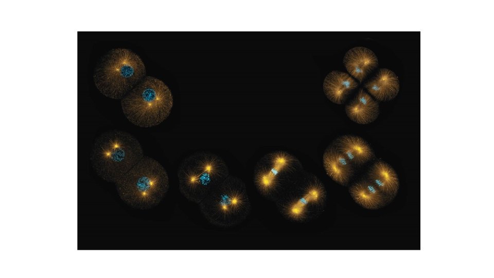
10 μm G 2 of Interphase Centrosomes (with centriole pairs) Nucleolus Chromosomes (duplicated, uncondensed) Nuclear envelope Plasma membrane Prophase Early mitotic spindle Aster Centromere Two sister chromatids of one chromosome Prometaphase Fragments of nuclear envelope Kinetochore Nonkinetochore microtubules Kinetochore microtubule
10 μm Metaphase Anaphase Metaphase plate Cleavage furrow Daughter chromosomes Spindle Telophase and Cytokinesis Centrosome at one spindle pole Nuclear envelope forming Nucleolus forming
G 2 of Interphase Centrosomes (with centriole pairs) Nucleolus Centrosomes (duplicated, uncondensed) Nuclear envelope Plasma membrane Prophase Early mitotic spindle Aster Centromere Two sister chromatids of one chromosome
Prometaphase Fragments of nuclear envelope Kinetochore Metaphase plate Nonkinetochore microtubules Kinetochore microtubule Spindle Centrosome at one spindle pole
Anaphase Telophase and Cytokinesis Cleavage furrow Daughter chromosomes Nuclear envelope forming Nucleolus forming
Bio Flix Mitosis
Sister chromatids Groups 1 -5 Aster Centrosome Metaphase plate (imaginary) Kinetochores Microtubules Overlapping nonkinetochore microtubules Chromosomes Kinetochore microtubules Centrosome 1 µm 0. 5 µm
Cytokinesis: • In animal cells, cytokinesis occurs by a process known as cleavage, forming a cleavage furrow • In plant cells, a cell plate forms during cytokinesis
(a) Cleavage of an animal cell (SEM) Cleavage furrow Contractile ring of microfilaments 100 µm (b) Cell plate formation in a plant cell (TEM) Vesicles forming cell plate Wall of parent cell Cell plate 1 µm New cell wall Daughter cells
Animation: Cytokinesis
Chromosomes 10 µm Nucleus Chromosomes Nucleolus condensing 1 Prophase 2 Prometaphase 3 Metaphase 4 Anaphase Cell plate 5 Telophase
Binary Fission in Bacteria • Prokaryotes (bacteria and archaea) reproduce by a type of cell division called binary fission • In binary fission, the chromosome replicates (beginning at the origin of replication), and the two daughter chromosomes actively move apart • The plasma membrane pinches inward, dividing the cell into two
Origin of replication E. coli cell 1 Chromosome replication begins. Two copies of origin Cell wall Plasma membrane Bacterial chromosome
Origin of replication E. coli cell 1 Chromosome replication begins. 2 One copy of the origin is now at each end of the cell. Two copies of origin Origin Cell wall Plasma membrane Bacterial chromosome Origin
Origin of replication E. coli cell 1 Chromosome replication begins. 2 One copy of the origin is now at each end of the cell. 3 Replication finishes. Two copies of origin Origin Cell wall Plasma membrane Bacterial chromosome Origin
Origin of replication E. coli cell 1 Chromosome replication begins. 2 One copy of the origin is now at each end of the cell. 3 Replication finishes. 4 Two daughter cells result. Two copies of origin Origin Cell wall Plasma membrane Bacterial chromosome Origin
Kinetochore microtubule Bacterial chromosome Intact nuclear envelope (c) Diatoms and some yeasts (a) Bacteria Chromosomes Kinetochore microtubule Microtubules Intact nuclear envelope (b) Dinoflagellates (d) Most eukaryotes Fragments of nuclear envelope
Thing to know #3 The eukaryotic cell cycle is regulated by a molecular control system • The frequency of cell division varies with the type of cell • These differences result from regulation at the molecular level • Cancer cells manage to escape the usual controls on the cell cycle
The Cell Cycle Control System • The cell cycle appears to be driven by specific chemical signals present in the cytoplasm • Some evidence for this hypothesis comes from experiments in which cultured mammalian cells at different phases of the cell cycle were fused to form a single cell with two nuclei
Experiment 1 S G 1 S S Experiment 2 M G 1 Results G 1 nucleus immediately entered S phase and DNA was synthesized. Conclusion M M G 1 nucleus began mitosis without chromosome duplication. Molecules present in the cytoplasm control the progression to S and M phases.
• The sequential events of the cell cycle are directed by a distinct cell cycle control system, which is similar to a clock • The cell cycle control system is regulated by both internal and external controls • The clock has specific checkpoints where the cell cycle stops until a goahead signal is received
G 1 checkpoint Control system G 1 M G 2 M checkpoint G 2 checkpoint S
• For many cells, the G 1 checkpoint seems to be the most important • If a cell receives a go-ahead signal at the G 1 checkpoint, it will usually complete the S, G 2, and M phases and divide • If the cell does not receive the go-ahead signal, it will exit the cycle, switching into a nondividing state called the G 0 phase
G 1 checkpoint G 0 G 1 Without go-ahead signal, cell enters G 0. (a) G 1 checkpoint G 1 With go-ahead signal, cell continues cell cycle.
G 1 M M G 2 M checkpoint Prometaphase Without full chromosome attachment, stop signal is received. (b) M checkpoint Anaphase G 2 checkpoint Metaphase With full chromosome attachment, go-ahead signal is received.
• An example of an internal signal is that cells will not begin anaphase until all chromosomes are properly attached to the spindle at the metaphase plate • This mechanism assures that daughter cells have the correct number of chromosomes
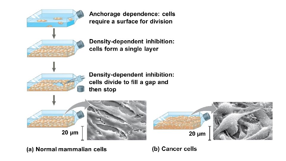
• External factors that influence cell division include specific growth factors • Growth factors are released by certain cells and stimulate other cells to divide • Platelet-derived growth factor (PDGF) is made by blood cell fragments called platelets • In density-dependent inhibition, crowded cells will stop dividing
• Most cells also exhibit anchorage dependence—to divide, they must be attached to a substratum • Density-dependent inhibition and anchorage dependence check the growth of cells at an optimal density • Cancer cells exhibit neither type of regulation of their division
Loss of Cell Cycle Controls in Cancer Cells • Cancer cells do not respond normally to the body’s control mechanisms • Cancer cells may not need growth factors to grow and divide • They make their own growth factor • They may convey a growth factor’s signal without the presence of the growth factor • They may have an abnormal cell cycle control system