THE CELL CYCLE AND MITOSIS Text section 5


 • Cell increases in size and makes")
 • Replication of DNA occurs. Ø The DNA molecule unwinds")
 The chromosomes have duplicated and appear as a jumbled mass")
 • Cell continues to grow and make")


")


- Slides: 27
THE CELL CYCLE AND MITOSIS Text section 5. 1
Why do cells divide? Instead of dividing, why don’t cells just grow larger and larger? If the cell grows too large, it will have trouble moving enough nutrients and wastes across the cell membrane. Ø As the cell grows, the volume of the cell increases much more rapidly than the surface area of the cell membrane. The larger a cell becomes, the more demands the cell places on its DNA.
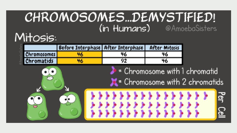
THE CELL CYCLE AND MITOSIS Due to the loss and death of cells, the body must replace them. A good example of this is human skin cells: Each day millions are shed. The life of a cell is divided into three stages known as the cell cycle
The cell cycle consists of three major phases, but often represented as five, since the first phase (interphase) is long: 1. Interphase is normal cell function and growth i. G 1 (first gap) i. S (synthesis) i. G 2 (second gap) 2. Mitosis is the division of the nucleus. 3. Cytokinesis is the division of the cytoplasm. End result: 2 new, identical cells are produced.
Again… let it sink in now
1. INTERPHASE G 1 , S, G 2
INTERPHASE • Nucleus is well-defined and bounded by the nuclear membrane. • Outside of the nucleus are two centrioles. • Their function is to organize the microtubules into a spindle. They will begin to move apart as spindle microtubules grow out of them. centrioles nuclear membrane nucleolus chromosomes
G 1 – 1 st Gap (Interphase) • Cell increases in size and makes the proteins and molecules necessary for the cell to function. • Some organelles begin to duplicate.
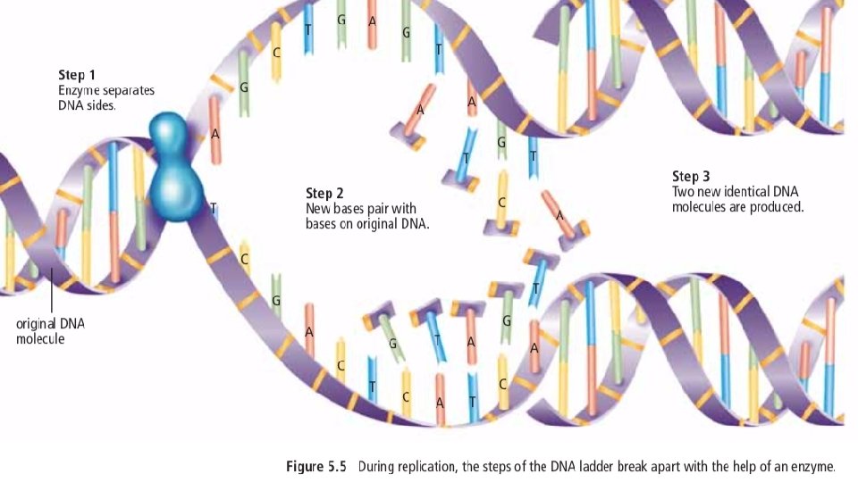
S – synthesis (Interphase) • Replication of DNA occurs. Ø The DNA molecule unwinds with the help of an enzyme. Ø New bases pair with the bases on the original DNA. Ø Two new identical DNA molecules are produced.
S – synthesis (Interphase) The chromosomes have duplicated and appear as a jumbled mass of fibers. They have not yet condensed.
G 2 – 2 nd Gap (Interphase) • Cell continues to grow and make materials such as proteins • The chromatin, which contains the replicated DNA, is in its loosely coiled form • Remaining organelles (such as mitochondria and chloroplasts) are duplicated. Now the cell is ready to divide!
2. MITOSIS
MITOSIS The shortest stage of the cell cycle where the nuclear contents divide, and two daughter nuclei are formed. As the nucleus prepares to divide, replicated DNA in interphase joins to form sister chromatids, joined by a centromere.
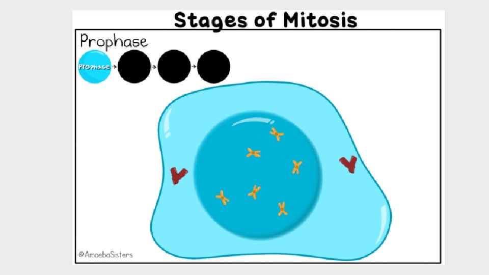
Late Early Prophase Metaphase Anaphase Telophase Mitosis occurs in 4 (well, kind of 5) stages
PMAT EARLY PROPHASE • The chromosomes coil and thicken and become distinct from one another. The chromosomes are now visible. • The nucleolus disappears • The chromosomes are doubled throughout their length. • The centrioles separate and start moving to opposite ends of the cell. A spindle made of microtubules begins to form.
PMAT LATE PROPHASE • The nuclear membrane disappears • The centrioles move to the opposite poles, completing spindle (microtubules form spindle) • The chromosomes attach to the spindle fibres at their centromeres
PMAT METAPHASE • Each chromosome is connected to a spindle fiber at its centromere. • The centrioles are now at opposite sides of the cell. • The spindle fibers will push and pull the chromosomes. • The chromosomes line up at the center of the cell. meta = middle = chromos at the middle of the cell
PMAT ANAPHASE • The microtubules begin to shorten and this pulls the chromatids apart to opposite sides of the cell • Once they separate, each sister chromatid is considered to be a chromosome. • By the end of anaphase, the two ends of the cell have equivalent and complete sets of chromosomes. look at the shape of the chromos. . Little As?
PMAT TELOPHASE • One complete set of chromosomes is now at each pole of the cell • Spindle fibres begin to disappear • Nuclear membrane forms around each set of chromosomes • A nucleolus appears within each nucleus • Now there are two nuclei in one cell, and the cell is ready to divide
CYTOKINESIS • The final stage in the cell cycle, and happens in conjunction with telophase • The two nuclei are separated into two daughter cells • These new cells are identical to the original parent cell
Cytokinesis in ANIMAL cells