The Cardiovascular System Heart The Cardiovascular System A
")


 • Heart muscle cells")


 • Amount of blood pumped by")

- Slides: 36
The Cardiovascular System (Heart)
The Cardiovascular System • A closed system of the heart and blood vessels • The heart pumps blood • Blood vessels allow blood to circulate to all parts of the body • The function of the cardiovascular system is to deliver oxygen and nutrients and to remove carbon dioxide and other waste products
The Heart • The heart is approximately the size of your fist, it’s hollow and cone shaped. Surprisingly, it’s weight is less than a pound. • Located in the mediastinum, the middle cavity of the thorax and flanked by two lungs. • The apex points towards the left hip and rests on the diaphragm. (The muscle that moves to change the volume of your lungs) 3
• The larger portion or the base points towards the right shoulder and lies beneath the second rib. • The heart is enclosed in a double sac membrane called the pericardium. • A slippery fluid called serous fluid allows the heart to beat in a near frictionless environment. 4
The Heart Figure 11. 1
The Heart: Heart Wall • Three layers • Epicardium • Outside layer • Connective tissue layer • Myocardium • Middle layer • Mostly cardiac muscle • Endocardium • Inner layer
External Heart Anatomy Figure 11. 2 a
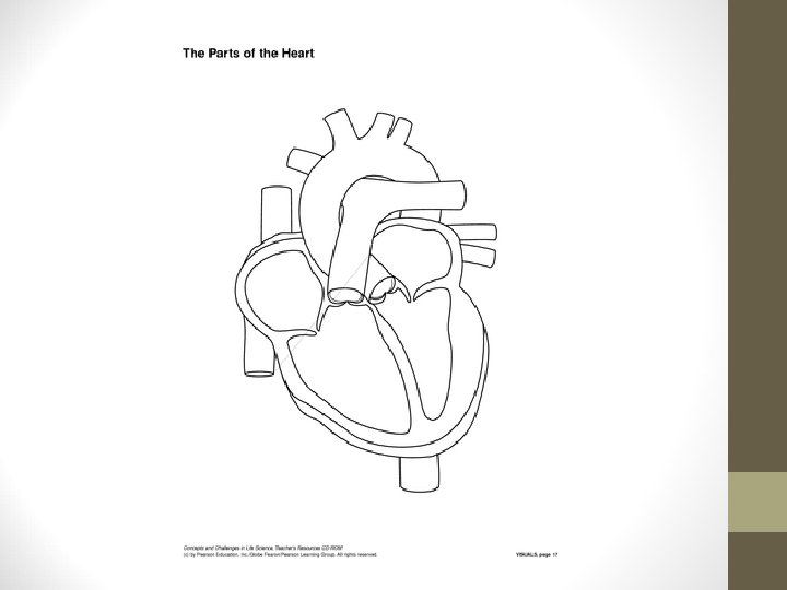
The Heart: Chambers • Right and left side act as separate pumps • Four chambers • Atria • Receiving chambers are supplied • with blood from the veins. • Right atrium • Left atrium • Ventricles • Discharging chambers pump blood out to the body through arteries. • Right ventricle • Left ventricle Figure 11. 2 c
Flow Within The Heart: Valves • Allow blood to flow in only one direction • Four valves • Atrioventricular valves (AV) – between atria and ventricles • Bicuspid valve (left) • Tricuspid valve (right) • Semilunar valves between ventricle and artery • Pulmonary valve • Aortic valve
The Heart: Valves • Valves open as blood is pumped through • Held in place by chordae tendineae (“heart strings”) • Close to prevent backflow
11
Operation of Heart Valves Figure 11. 4
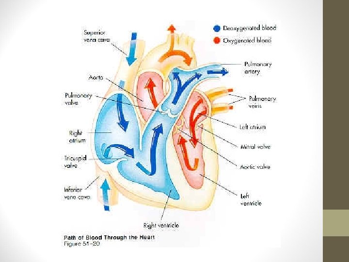
Pulmonary Circulation • Pulmonary Circulation-The flow of blood between the heart and the lungs. • Deoxygenated blood leaves the right ventricle and goes to the lungs via the pulmonary artery. The blood picks up oxygen in the lungs and returns to left atrium of the heart via the pulmonary vein. 13
Systemic Circulation • Systemic Circulation-The oxygenated blood leaves the heart via the aorta and flows throughout the body. The deoxygenated blood returns to the right atrium of the heart via the inferior or superior vena cava. 16
Blood Circulation Figure 11. 3
The Heart: Associated Great Vessels • Aorta • Leaves left ventricle out to the body • Pulmonary arteries • Leave right ventricle out to the lungs • Vena cava (Superior or Inferior) • Enters right atrium from the body • Pulmonary veins (four) • Enter left atrium from the lungs
General Rule-With Exceptions • Arteries carry oxygenated blood out of the heart to the body • EXCEPTION-Pulmonary artery carries deoxygenated blood out of the heart to the lungs. • Veins carry deoxygenated blood back to the heart from the body. • EXEPTION-Pulmonary vein carries oxygenated blood to the heart from the lungs. 19
Coronary Circulation • Blood in the heart chambers does not nourish the myocardium • The heart has its own nourishing circulatory system • 4 major coronary arteries (clogging causes heart attack) • Cardiac veins
4 Major Coronary Arteries 21
22
What is a Heart Attack? • A heart attack occurs when blood flow to a part of your heart is blocked for a long enough time that part of the heart muscle is damaged or dies. The medical term for this is myocardial infarction. 23
Blockage 24
Avoiding Surgery • Patients may experience the effects of a heart attack at any time of day. Some people are lucky enough to get treatment and not need surgery. A stent may be used to open up the clogged artery. 25
Inserting a Stent 26
27
The Heart: Conduction System • Intrinsic conduction system (nodal system) • Heart muscle cells contract, without nerve impulses, in a regular, continuous way • Can be restarted with electrical current
29
The Heart: Conduction System • Special tissue sets the pace • Sinoatrial Node-Located in the right atrium and initiate the contraction • Pacemaker • Atrioventricular Node-Located between the atria and the ventricles • Purkinje Fibers-Spread within the muscles of the ventricle walls.
Heart Contractions Figure 11. 5
Filling of Heart Chambers – The Cardiac Cycle Figure 11. 6
The Cardiac Cycle • The events in one complete cycle of heartbeats. • The average heart beats about 75 beats per minute. • Atria contract simultaneously • Atria relax, then ventricles contract • Systole = contraction • Diastole = relaxation 33
The Heart: Cardiac Cycle • Cardiac cycle – events of one complete heart beat • Mid-to-late diastole – blood flows into ventricles • Ventricular systole – blood pressure builds before ventricle contracts, pushing out blood • Early diastole – atria finish re-filling, ventricular pressure is low
The Heart: Cardiac Output • Cardiac output (CO) • Amount of blood pumped by each side of the heart in one minute • CO = (heart rate [HR]) x (stroke volume [SV]) • Stroke volume • Volume of blood pumped by each ventricle in one contraction
Cardiac Output Regulation Figure 11. 7