The Cardiovascular System Anatomy and Physiology UPA What


 Platelets (Clotting")
 which transport O 2 This")


 They are not cells; they are fragments of HUGE cells called megakaryoctes")


 at their external surfaces.")

 Antibodies in plasma Blood that can be")

 Originally")


- Slides: 44
The Cardiovascular System Anatomy and Physiology UPA
What is the Circulatory System? A closed system of the heart and blood vessels The Heart pumps blood Blood vessels allow blood to circulate The function is to deliver oxygen and nutrients to the body and to remove carbon dioxide and other wastes First we will focus on the blood.
Physical Characteristic and Volume Blood is sticky, opaque, metallic in taste Blood is heavier than water and more viscous Has a temperature around 1 oo. 4°C Accounts for 8% body weight Healthy average adults have 5 -6 liters Equivalent to 1. 5 gallons or 12 pints
Functions of the Blood Distribution Delivers oxygen and nutrients while eliminating wastes Delivers hormones, adrenaline, medicine, etc. Regulation Maintains body temperatures by distributing heat Maintains p. H to neutralize acid/base built-up Protection Prevents further blood loss by clotting wounds Prevents infection by bringing antibodies and white blood cells to
Components of the Blood is unique because it is the only fluid tissue Blood is considered connective tissue because of the liquid matrix Made up of 2 parts… Formed Elements: living blood cells Plasma: nonliving fluid matrix
Blood Plasma Straw-colored fluid that is 90% water Contains over 100 different dissolved solutes Gasses, nutrients, hormones, wastes, ions, proteins, etc. Albumin accounts for 60% of plasma proteins It serves to keep water in the bloodstream These proteins serve to make dozens of daily adjustments to sustain life. Example: When blood becomes too acidic (from diet) both three respiratory and kidneys are called change the plasma p. H back to normal.
Formed Elements Components are Erythrocytes (Red Blood Cells Leukocytes (White Blood Cells) Platelets (Clotting Proteins) Most of these cells only survive a few days These cells don’t divide instead are replaced in the bone marrow
Erythrocytes 45% of blood is Erythrocytes (Red Blood Cells) which transport O 2 This part is also called the Hematocrit “Blood Fraction” Healthy males are 47% and females are 42% They have no true nuclei, organelles and are little “bags” of hemoglobin RBC pick up oxygen in the capillary bed of the lungs and release it to other cells in the body They don’t consume any of the oxygen for themselves and are very efficient!
Blood Cell Formation Blood Cell formation is called hematopoiesis In adults, red marrow is found in the bones of the axial skeleton, humerus, and femur On average, the marrow turns out an ounce of new blood (100 billion new cells) EVERY DAY!
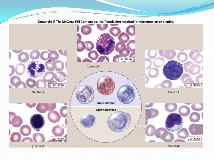
Leukocytes Also called white blood cells Only 1% of total blood volume WBC are able to slip out of the blood and transport themselves to areas that need them (whereas RBC are only in the bloodstream) Whenever WBC are mobilized for action, the body speeds up their production and 2 x as many appear in the blood within a few hours A WBC Count over 11, 000 cells/mm 3 is sign of bacteria/viral infection
Categories of WBC The Following phrase will help you: (Listed from most to least abundant) Never Let Monkeys Eat Bananas Neutrophils Multi-lobed nucleus, granules; “eat” bacteria, lives a few days Lymphocytes Large nucleus, pale blue, no granules; Mount immune response via antibodies, lifespan is months-years
Categories of WBC Monocytes Nucleus U-shaped, largest, no granules; wander to fight infection, highly phagocytic Eosinophil Nucleus lobed, many granules, red; prevents allergic reactions, defend against parasitic worms Basophil Nucleus lobed, blue, granules; release histamines in response to allergens, anti-inflammatory response
Types of White Blood Cells
Platelets (Thrombocytes) They are not cells; they are fragments of HUGE cells called megakaryoctes Essential for blood clotting that occurs in plasma when blood vessels are ruptured Platelet formation is hormone driven
Process of blood clotting 1. Vascular spasms Immediately following injury, blood vessels constrict (called Vascular Spasm) 2. Platelet plug formation A “plug” is created for temporary seal Platelets become sticky and swell, adhere to collagen Sends chemicals through blood to increase production of more platelets (positive feedback mechanism) 3. Coagulation/Blood clot Blood turns from a liquid to a gel in the region Within 30 -60 minutes the clot is stabilized
The Process of Blood Clotting
Bleeding Disorders Thrombocytopenia Number of circulating platelets is too low Causes spontaneous bleeding all over the body Whole blood transfusions are required to temporally stop bleeding
Bleeding Disorders… Hemophilia Refers to several different hereditary bleeding disorders They are sex-linked (primarily males) Symptoms begin early in life; minor tissue damage (scrapes, etc. ) can be life threatening Helped with transfusions or injections of purified clotting factors Both are expensive and inconvient
Transfusions and Blood Replacements Loss of 15 -30%: Weakness Loss of +30%: Severe shock (fatality) The shelf life for collected blood is only 35 days!
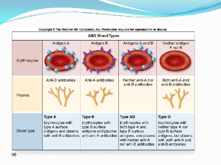
Human Blood Groups RBC plasma membranes contain specific proteins (antigens) at their external surfaces. The presence or absence of these allows each person’s blood to be categorized
ABO Groups Based upon the presence or absence of 2 antigens called type A and type B Blood types are either A, B, AB, or O. Unique to ABO blood types are preformed antibodies called agglutinins. They act to attack RBC that are not normal to the individual.
ABO Blood Groups Blood Group Antigen (Protein) Antibodies in plasma Blood that can be received AB A B None All *Universal recipient B B Anti-A B, O A A Anti-B A, O O None Anti-A Anti-B O *Universal donor
Easy way to remember…. Type O Type AB Type B
Rh Factor Blood Groups Rh factor is named for the Rh antigen (protein) Originally named after the rhesus monkeys in which it was discovered You can be Rh+ or Rh-
Interesting to Know Blood Circulation & Respiration One complete round trip will take, on average only 30 to 45 seconds. And, even less during exercise. The human body has so many miles of blood vessels inside of it that they could encircle the earth twice, then a little bit more
The Heart
The Location of the Heart The heart is located in the mediastinum, the medial section of the thoracic cavity It rests superior to the diaphragm and 2/3 of its mass lies to the left of the sternum Its pointed apex points towards the left hip It weighs less than 1 pound.
Coverings of the Heart: The Fibrous Pericardium The external, double-walled sac is called the pericardium Its function is to… Protect the heart Anchor it to the diaphragm and vessels Prevent overfilling of the heart
Coverings of the Heart: The Serous Pericardium It is a thin, slippery membrane of 2 layers Parietal layer: Attaches the arteries leaving the heart Visceral layer: Also called the epicardium
Coverings of the Heart: Pericardial Cavity Between the 2 layers of the pericardium Contains a film of serous fluid Fluid keeps the heart lubricated for a friction-free environment
Layers of the Heart Wall Epicardium Outer layer covered in fat Also called the visceral layer of the pericardium Myocardium Forms the bulk of the heart Cardiac muscle cells Endocardium Inner most layer White sheet of endothelium (squamous)
Chambers and Internal Anatomy Humans have a 4 chambered heart 2 atria and 2 ventricles Left and right sides divided by the septum
The Atria The top chambers of the heart They are the receiving chambers of blood returning from circulation They are smaller since they only push blood to the ventricles “next door”
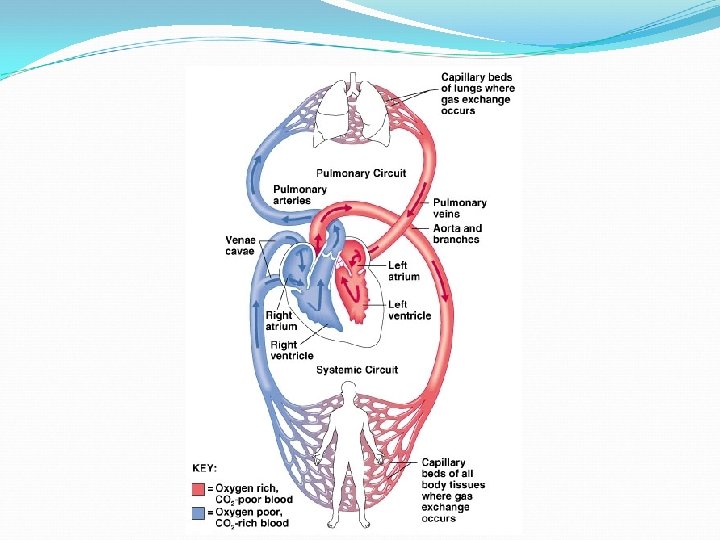
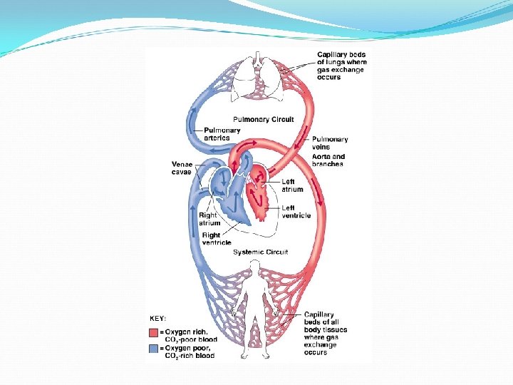
The Ventricles They are larger and make up most of the mass of the heart They are the lower chambers in the heart They are the discharging chambers that actually pump blood to the body The right pumps to the pulmonary circuit (lungs) The left pumps to the systemic circuit (all body tissue)
The Pathway of Blood: Pulmonary Circuit Blood is returned to the body through the superior and inferior vena cava (Low O 2) Blood enters right atrium Pumps into the right ventricle through the tricuspid valve Blood is pumped out of the r. ventricle and into the pulmonary arteries (one to each lung) Blood fills lungs to collect O 2 The blood returns from the lungs via the pulmonary veins
The pathway of blood: Systemic Circuit Blood returned from lungs enters left atrium with O 2 Blood passes to left ventricle through the mitral valve Blood leaves the heart via the aorta From there the blood is transported to smaller arteries in body tissue Gases are exchanged in capillary beds of all body tissues
Electrocardiography Reading a ECG The electrical currents generated and transmitted are spread throughout the whole body These currents can be monitored and ampliphied with a electrocardiograph
The graphic recording of the heart activity is calles an elecctrocardiogram (EKG or ECG) Electrodes are placed at various sites; 12 standards leads are used
A ECG consists of 3 distinguishable waves, called deflection waves The P Wave The first small wave Lasts 0. 08 seconds Results from movement of the depolarization wave from the SA node through the atria In other words, the contraction of the atria.
The QRS Wave Contraction of the ventricles Average duration is 0. 08 seconds The Tw