THE BRAIN Tools of Discovery Older Brain Structures

THE BRAIN Tools of Discovery Older Brain Structures The Limbic System The Cerebral Cortex

EEG ELECTROENCEPHALOGRA M

MRI MAGNETIC RESONANCE IMAGING

CT SCAN COMPUTED TOMOGRAPHY

PET SCAN POSITRON EMISSION TOMOGRAPHY

MEG MAGNETOENCEPHALOGRAPHY

FM RI FUNTIONAL M AG NE TI C RESO NANCE I MA GI NG
 Electrodes placed on the scalp measure electrical")
Name How does it work? Electroencephalogram (EEG) Electrodes placed on the scalp measure electrical activity in neurons Symptoms of depression and anxiety correlate with increased activity in the right frontal lobe (associated with negative emotion) Magnetic resonance imaging (MRI) People sit or lie down in a chamber that uses magnetic fields and radio waves to provide a map of brain structure People with history of violence tend to have smaller frontal lobes (associated with moral judgement and self control) Computed tomography (CT) X-rays of the head generate images that may locate brain damage Positron emission tomography (PET) Tracks where a temporarily radioactive form of glucose goes while the brain of the person given it performs a given task Magnetoencephalography (MEG) A head coil records magnetic fields from the brain’s natural electrical currents Soldiers with PTSD show stronger magnetic fields in the visual cortex when they view trauma-related images Funtional magnetic resonance imaging (f. MRI) Measures blood flow to brain regions by comparing continuous MRI scans
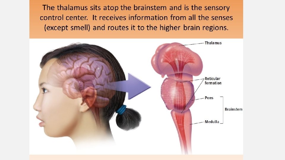

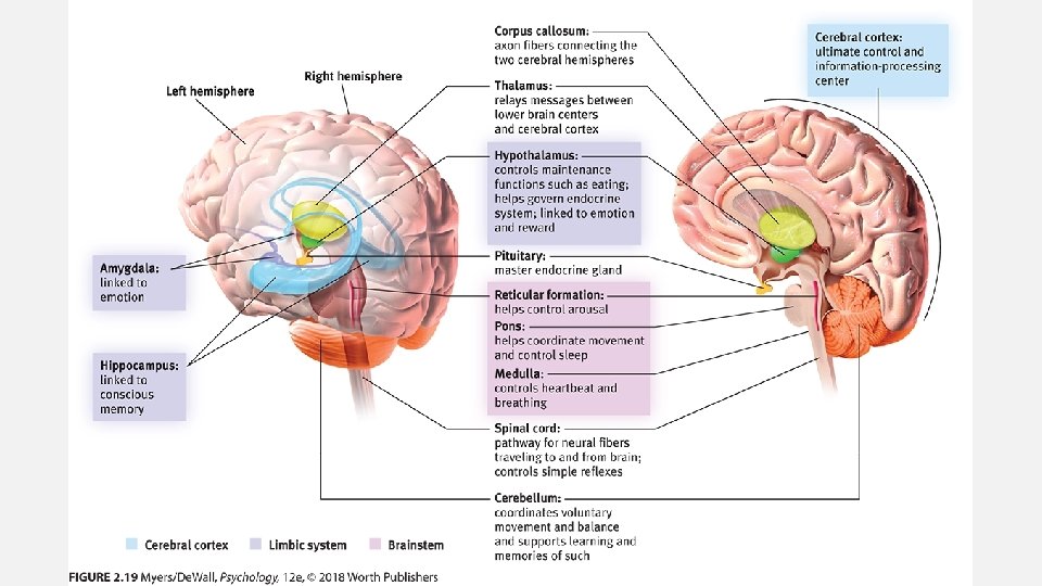
Limbic System

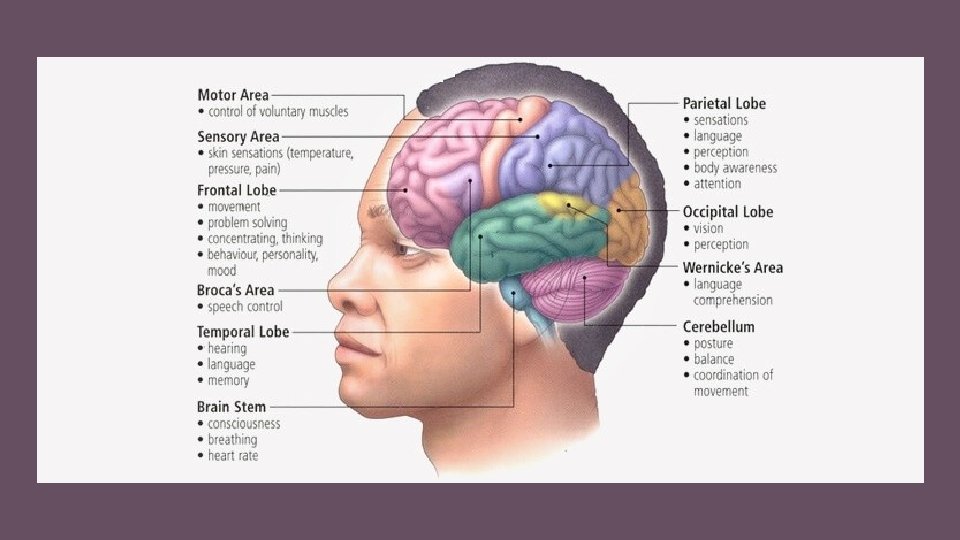
Cerebral Cortex

FUSIFORM GYRUS • • ANGULAR GYRUS In temporal lobe Processing colors Face recognition Synesthesia • In parietal lobe • Aids in abstractions and metaphors https: //www. bing. com/videos/search? q=brain+structure+a nd+function&&view=detail&mid=F 348 BD 098 F 270 CE 3 AB 5 DF 348 BD 098 F 270 CE 3 AB 5 D&&FORM=VRDGAR

Play-Doh Brains

• https: //www. youtube. com/watch? v=v. Hrmiy 4 W 9 C 0
- Slides: 16