The Brain subdivided into Forebrain Midbrain Hindbrain Anatomical

The Brain: § subdivided into § Forebrain § Midbrain § Hindbrain

Anatomical classification § Cerebral hemispheres § Diencephalon § Thalamus § Hypothalamus § Brain stem § Midbrain § Pons § Medulla

Usual pattern of gray/white in CNS § White exterior to gray _________ § Gray surrounds hollow central cavity______________ § Two regions with additional gray called “cortex”_______________ § Cerebrum: “cerebral cortex” § Cerebellum: “cerebellar cortex”

Gray and White Matter § Like spinal cord but with another layer of gray outside the white § Called cortex § Cerebrum and cerebellum have § Inner gray: “brain nuclei” (not cell nuclei) § Clusters of cell bodies Remember, in PNS clusters of cell bodies were called “ganglia”
 § Lined by")
Ventricles § Central cavities expanded § Filled with CSF (cerebrospinal fluid) § Lined by ependymal cells (these cells lining the choroid plexus make the CSF) § Continuous with each other and central canal of spinal cord
 § Elevated ridges § Entire surface §")
Surface anatomy § Gyri (plural of gyrus) § Elevated ridges § Entire surface § Grooves separate gyri § A sulcus is a shallow groove (plural, sulci) § Deeper grooves are fissures

Parts of Brain Cerebrum Diencephalon Brainstem Cerebellum

simplified… § Back of brain: perception § Top of brain: movement § Front of brain: thinking

Cerebral hemispheres § Lobes: under bones of same name § Frontal § Parietal § Temporal § Occipital

Cerebral hemispheres: note lobes § Divided by longitudinal fissure into right & left sides § Central sulcus divides frontal from parietal lobes

§ Lateral sulcus separates temporal lobe from parietal lobe § Parieto-occipital sulcus divides occipital and parietal lobes (not seen from outside) § Transverse cerebral fissure separates cerebral hemispheres from cerebellum

coronal section § Note: longitudinal fissure, lateral sulcus, insula

Cerebral cortex § Executive functioning capability § Gray matter: neuron cell bodies, dendrites, short unmyelinated axons § 100 billion neurons § No fiber tracts (would be white) § 2 -4 mm thick (about 1/8 inch) § Brodmann areas (historical: 50 structurally different areas given #s) § Neuroimaging: functional organization (example later)

Cerebral cortex § All the neurons are interneurons § By definition confined to the CNS § They have to synapse somewhere before the info passes to the peripheral nerves § Three kinds of functional areas § Motor areas: movement § Sensory areas: perception § Association areas: integrate diverse information to enable purposeful action

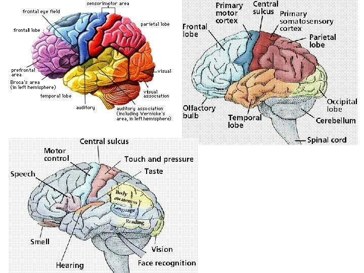
Sensory areas Posterior to central sulcus § Primary somatosensory cortex: postcentral gyrus of parietal lobe (allows conscious awareness of sensation and the ability to localize it: where the sensation is from) § Somatosensory association area: behind it (understanding of what is being felt: the meaning of it)
 §")
From special sense organs § Sight: occipital lobe § Primary visual cortex (17) § Handles info from contralateral retina (right ½ of visual field is on left side) § Map of visual space § If damaged: functionally blind because no conscious awareness of sight § Visual association area (18 & 19) § Face recognition is usually on the right side § Hearing: temporal lobe § Primary auditory area (41) § Auditory association area (22)

Refer back to this labeled version as needed
: uncus § Deep in temporal lobe along medial surface")
§ Smell (olfactory sense): uncus § Deep in temporal lobe along medial surface

§ f. MRI: functional magnetic resonance imaging § Cerebral cortex of person speaking & hearing § Activity (blood flow) in posterior frontal and superior temporal lobes respectively

Motor areas Anterior to central sulcus § Primary motor area § Precentral gyrus of frontal lobe (4)

§ Primary motor area continued § Precise, conscious or voluntary movement of skeletal muscles § Large neurons called pyramidal cells § Their axons: form massive pyramidal or corticospinal tracts § Decend through brain stem and spinal cord § Cross to contralateral (the other) side in brainstem § Therefore: right side of the brain controls the left side of the body, and the left side of the brain controls the right side of the body
: specialized motor speech area § Base")
Motor areas – continued § Broca’s area (44): specialized motor speech area § Base of precentral gyrus just above lateral sulcus in only one hemisphere, usually left § Word articulation: the movements necessary for speech § Damage: can understand but can’t speak; or if can still speak, words are right but difficult to understand
: complex movements asociated with highly processed")
Motor areas – continued § Premotor cortex (6): complex movements asociated with highly processed sensory info; also planning of movements § Frontal eye fields (inferior 8): voluntary movements of eyes

Homunculus – “little man” § Body map: human body spatially represented § Where on cortex; upside down
 1. Motor areas: movement")
Association Areas Remember… § Three kinds of functional areas (cerebrum) 1. Motor areas: movement 2. Sensory areas: perception 3. Association areas: everything else

Association Areas § Tie together different kinds of sensory input § Associate new input with memories § Is to be renamed “higher-order processing“ areas

Prefrontal cortex: cognition This area coordinates the brain/body and inter-personal world as a whole Intellect Abstract ideas Judgment Personality Impulse control Persistence Complex Reasoning Long-term planning Social skills Appreciating humor Conscience Mood Mental flexibility Empathy Executive functioning e. g. multiple step problem solving requiring temporary storage of info (working memory)

Wernicke’s area Region involved in recognizing and understanding spoken words § § Junction of parietal and temporal lobes One hemisphere only, usually left (Outlined by dashes) Pathology: comprehension impaired for written and spoken language: output fluent and voluminous but incoherent (words understandable but don’t make sense; as opposed to the opposite with Broca’s area)

Cerebral white matter § Extensive communication § Areas of cortex with each other § Areas of cortex with brain stem and spinal cord § Via (mostly) myelinated axon fibers bundled into tracts § Commissures § Association fibers § Projection fibers

§ Commissures: interconnect right and left hemispheres so can act as a whole § Corpus callosum is largest § Association fibers: connect different parts of the same hemisphere; can be long or short
 §")
§ Projection fibers: run vertically § Cerebral cortex running down (with motor instructions) § Or ascend to cerebral cortex from below (sensory info to cortex)
 gyrus § Combines")
§ Corona radiata: spray of projection fibers § From precentral (motor) gyrus § Combines with sensory fibers traveling to sensory cortex § Form a band of fibers called internal capsule* Motor output from brain_____ * ______Sensory input to brain

§ Projection fibers § Corona radiata: _________ fanning out of the fibers § Internal capsule: __________ bundled, pass down § Commisure § Corpus callosum: ________ connects right and left hemispheres § Decussation: crossing of ___________ pyramidal tracts
- Slides: 34