The Brain Sections 9 11 and 9 13

The Brain Sections 9. 11 and 9. 13 * Contains approximately 100 Billion neurons * Weighs about 3 pounds
 �")
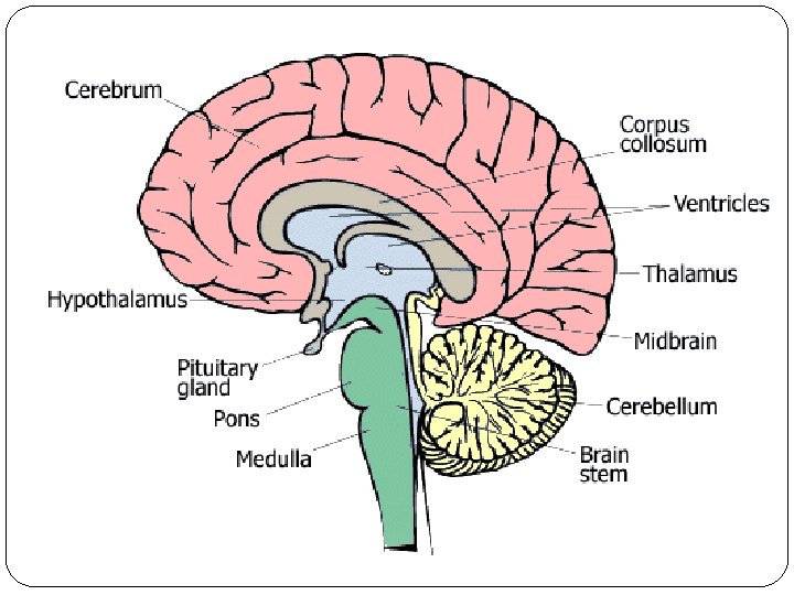
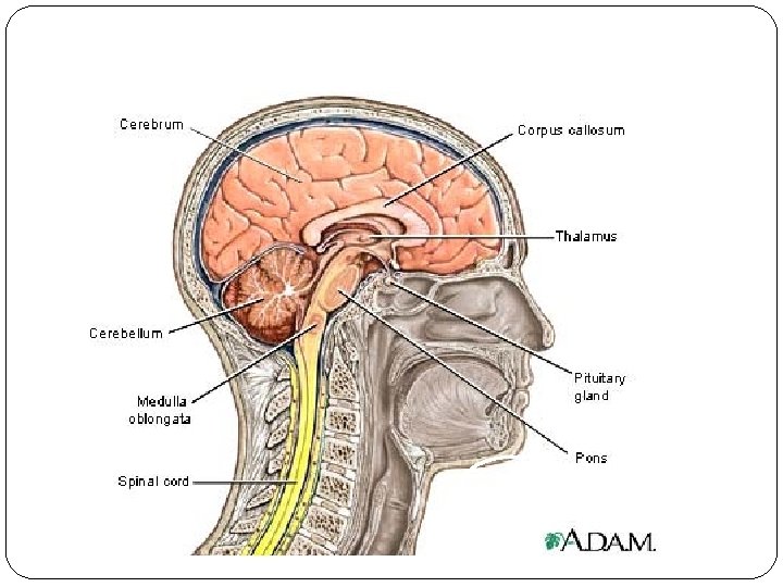
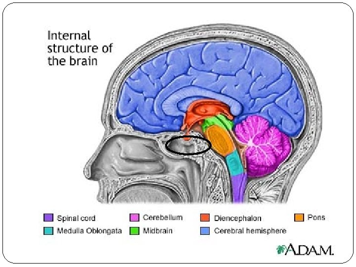
Major Brain Structures � Cerebrum- Divided into 4 lobes: (frontal, parietal, occipital, temporal) � Diencephalon: (thalamus, hypothalamus, epithalamus) � Brain Stem: (midbrain, pons, medulla oblongata) Cerebellum **(HINT IMPORTANT –KNOW FOR YOUR TEST)** � �

Brain Blood Supply �Oxygen – The brain requires 20% of the body’s oxygen supply. Neurons that are without oxygen for 4 or more minutes may be permanently damaged

Glucose nervous system �Glucose in - theour glucose in the blood is the source of energy for the brain’s cells. Very little glucose is stored in the brain, so if there isn’t enough glucose in the blood a person can experience confusion, dizziness, convulsions and loss of consciousness.

Cerebrum � Two cerebral hemispheres � Longitudinal fissure separates hemispheres. � Surface area increased with Gyri (ridges) and Sulci (creases) or Fissures (deep grooves). � Connected by the Corpus Callosum � Function: Intelligence, memory, learning � Cerebral cortex – gray matter, outermost, all conscious thinking occurs here � Olfactory bulb – sense of smell
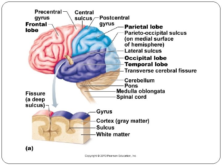

Lobes �Frontal �Parietal �Temporal �Occipital

Functions of the Cerebrum
")
Diencephalon Main structures: 1. Thalamus – main relay station for sensory impulses (except smell) to the cerebral cortex. 2. Hypothalamus – regulates visceral movement (BP, GI tract, HR), body temperature, water and electrolytes, hunger, thirst, stimulate pituitary, maintains sleep and wake patterns. 3. Epithalamus – contains the Pineal gland which regulates biological clock.

Cerebellum � 2 nd largest part of brain �Controls muscular coordination �Maintains posture �Allows for smooth, refined movements �Involuntary once learned

Check point � 1. What part of the brain helps you play a musical instrument? � 2. What part of the brain has 4 lobes, what are the four lobes? � 3. This part of your brain allows you regulates body temperature, electrolytes, and thirst, which part of the brain allow you to do this?
 �Protects the brain from harmful substances and pathogens by")
The Blood Brain Barrier (BBB) �Protects the brain from harmful substances and pathogens by blocking passage of many substances from the blood into brain tissue. Does NOT protect from lipid-soluble substances such as oxygen, carbon dioxide, alcohol, and most anesthetic agents.
 �A clear, colorless liquid that carries oxygen, glucose, and other needed")
Cerebrospinal Fluid (CSF) �A clear, colorless liquid that carries oxygen, glucose, and other needed chemicals from the blood to the neurons. It removes wastes and toxic substances produced by the brain and spinal cord cells

Brain Stem Three sections: 1. Midbrain – visual and auditory reflex centers, main motor pathway 2. Pons – “bridge”, relays impulses between: a. medulla/cerebrum b. cerebrum/cerebellum 3. Medulla Oblongata –regulates heart rate, blood pressure, respiration, coughing, sneezing, vomiting, swallowing

Activity � 140 -141 in nervous packet �Coloring parts of the brain in nervous system packet �LAB: Mouse party!

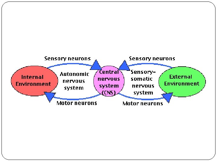
Central Nervous System �Consists of Brain and Spinal Cord

The Spinal Cord

Spinal Cord � Functions – to conduct nerve impulses and serve as the center of spinal reflexes. � Anatomy of the spinal cord: 1. Ascending tracts – from sensory to brain 2. Descending tracts – from brain to motor 3. Composed of gray and white matter. 4. Central canal contains the CSF. 5. Conus medullaris – end of cord at L 1 6. Cauda Equina – cord fans out into nerves.

Cross-Section of Spinal Cord Afferent impulses travel through dorsal root to cord. Efferent impulses travel through ventral root to effector.

Spinal Cord Protection and Covering �Vertebral Colum �Ring of bone formed by vertebral foramina protect the spinal cord

Spinal Cord Protection and Covering �Meninges – connective tissue covering that protect the spinal cord. They are found in 3 layers

Meninges: The Coverings � Three Layers: 1. Dura mater – outermost, tough, white 2. Arachnoid mater – middle, web-like, CSF in subarachnoid space 3. Pia mater – innermost, very thin on top of brain tissue Meningitis: Inflammation of the meninges and CSF; typical causes are bacteria or virus; spinal tap needed to diagnose

Meninges �Dura Mater – outer layer of meninges �Composed of tough connective tissue

Meninges �Arachnoid Mater – middle layer of meninges �Composed of collagen and elastic fibers that resemble a spider’s web

Meninges �Pia Mater – inner layer of meninges �Composed of transparent collagen and elastic fibers that adhere to the spinal cord and brain

Cross-Section of Spinal Cord Afferent impulses travel through dorsal root to cord. Efferent impulses travel through ventral root to effector.

Cross-section of Spinal Cord Orientation of grey and white matter is opposite of the brain. Grey matter = cell bodies White matter = myelinated axons

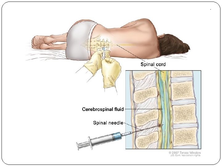
Subarachnoid Space �Area between the arachnoid mater and pia mater containing CSF �Spinal Tap (lumbar puncture) – a long needle is inserted into the subarachnoid space (generally between L 3 & L 4 or L 4 & L 5) to withdraw CSF for diagnostic purposes or to introduce medication.

Epidural given outside of the dura mater.

External Structure �The adult spinal cord is only about 16 -18 inches long…it ends around L 2

Cauda Equina �Nerves on the lower portion of the cord angled downward through the vertebral canal.

Enlargements �Cervical Enlargement – contains nerves that supply the upper limbs �Lumbar Enlargement – contains nerves that supply the lower limbs

Indentations/divisions �Anterior Median Fissure & Posterior Median Sulcus – divide the cord into right and left halves

Central Canal �Space down the center of the spinal cord containing CSF

Spinal Nerves �Paths of communication between the spinal cord and the nerves that serve the specific regions of the body
 root –")
Roots �Connect each spinal nerve segment to the spinal cord �Posterior (dorsal) root – contains only sensory axons �Dorsal Root Ganglion – swollen area containing sensory cell bodies �Anterior (ventral) root – contains both somatic motor and autonomic motor neurons

Reflexes � � � � Rapid, automatic responses to specific stimuli. Their purpose is to preserve homeostasis. Little variability in the responses. Only a few neurons are needed. “Wiring” of a single reflex = “Reflex Arc” Testing somatic reflexes can be used for diagnostic purposes. Examples: swallowing, sneezing, vomiting, and knee jerk.

Reflex Classification

Stretch Reflex: “Patellar reflex”

Muscle spindles = sensory receptors involved in stretch reflexes

Innate Reflex: “Withdrawal reflex”

Peripheral Nervous System Sections 14 and 15 READ TONIGHT!!!
 Sensory (Afferent) Somatic Skin and special senses Somatic")
Peripheral Nervous System PNS Motor (Efferent) Sensory (Afferent) Somatic Skin and special senses Somatic (Voluntary) Visceral Skeletal Muscles Autonomic (Involuntary) Sympathetic Parasympathetic

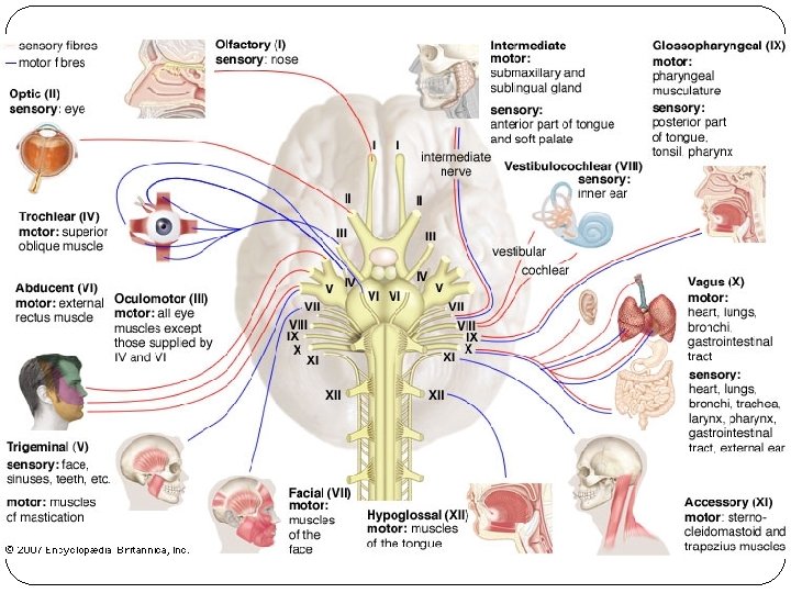
General Info All nerves that branch off the CNS and connect to other body parts. � Cranial nerves – 12 pairs � Spinal nerves – 31 pairs Functions: 1. To receive stimulus input and send to CNS. 2. To relay the response from the CNS to the appropriate effector organ.

Nerve Anatomy

What is this?
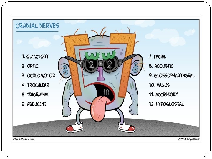

How to remember the Cranial Nerves �https: //youtu. be/Ft. Jt. YMRVw 7 A

Internal Structure �Gray Matter �Anterior Horns – contain cell bodies of somatic motor neurons �Posterior Horns – contain cell bodies of somatic and autonomic sensory neurons �Lateral Gray Horns – present only in lower portion of the spinal cord. Contains cell bodies of autonomic nervous system

Ventral Horn Lateral Horn Dorsal Horn

White Matter �Organized into columns containing myelinated and unmyelinated axons of sensory and motor neurons Posterior White Column Lateral White Column Anterior White Column

White Matter �Tracts – bundles of axons having a common origin or destination carrying similar information �Sensory (ascending) tracts – consist of axons that conduct nerve impulses toward the brain �Motor (descending) tracts – consists of axons that carry nerve impulses down the spinal cord

Tracts

Spinal Nerves �Paths of communication between the spinal cord and the nerves that serve the specific regions of the body

Afferent or Sensory Nerves 1. 2. 3. Picks up stimuli from: a. internal environment – visceral nerves b. external environment – somatic nerves Sends to CNS for interpretation Typically unipolar or bipolar neurons

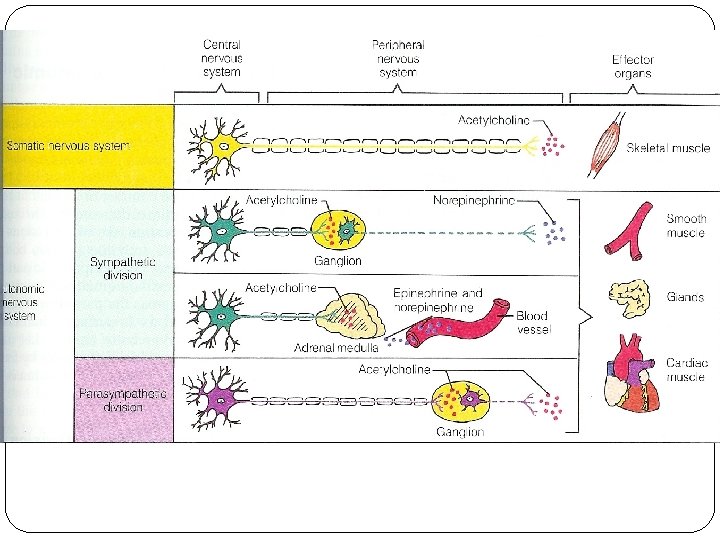
Somatic nervous system � All voluntary or conscious activities � Nerves connect to skeletal muscles � Pathways have one motor neuron to muscle cells. � Neurotransmitter: Acetylcholine

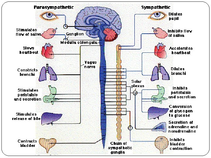
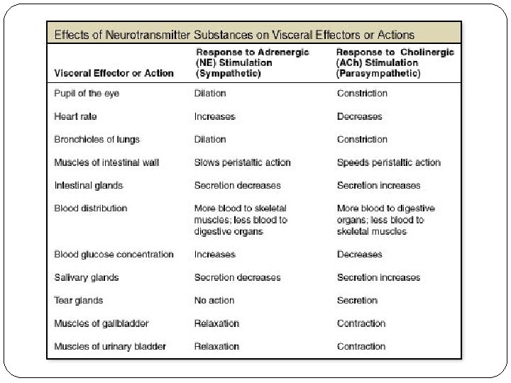
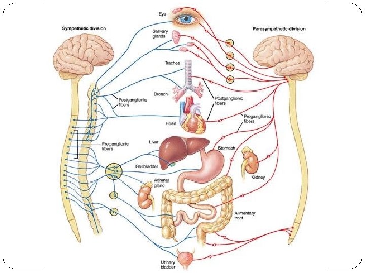
Autonomic nervous system - All involuntary or unconscious activities. - Maintains internal environment - Nerves connect to cardiac, smooth muscle or glands - Pathways have two neurons synapsing at a ganglia before effector. - Neurotransmitters: Acetylcholine or norepinephrine - Two divisions counterbalance each other: Sympathetic and Parasympathetic

Comparing Motor Neurons

Sympathetic � prepares for energy-expenditure, excitement or stressful situations � "fight" or take "flight“. Nerve fibers originate from the thoracic and lumbar regions of spinal cord. � Short Pre and Long postganglionic fibers �

Parasympathetic � Stimulated during calm and relaxing situations � "rest" and "digest". Nerve fibers originate from the brain and sacral region. � Long preganglionic fibers and short post �

Which pathway is responsible here? Sympathetic or Parasympathetic

Division of the ANS are distinguished by: 1. Unique origin sites a. Sacrocranial vs. Thoracolumbar 2. Different lengths of their fibers a. Preganglionic (long – Para, short – S) b. Postganglionic (long – S, short – Para) 3. Location of their ganglia a. P – close to effector b. S – close to spinal cord

Brain Disorders and Diseases

Common Brain Injuries � Concussion – abrupt, but temporary loss of consciousness from a blow to the head. Symptoms: headache, confusion, memory loss, lack of concentration � Contusion – bruising of the brain due to trauma, leaking capillaries, commonly follows a concussion. Pia mater torn. Signs: Immediate loss of consciousness, loss of reflexes, decreased blood pressure, cessation of respiration. � Laceration – tear of the brain, large vessel rupture, cerebral hematoma, increased intercranial pressure.

Brain Tumors

Brain Aneurism Cerebral thrombosis video

Strokes

Injury Effects

Phineas Gage Man who survived a terrible brain injury in the 1800’s. First opportunity for scientists to study the frontal lobe and limbic system connection. Limbic system is the emotional region and frontal keeps the limbic region in control. Injury re-enactment video

CAN YOU READ THIS? cdnuolt blveiee taht I cluod aulaclty uesdnatnrd waht I was rdanieg. The phaonmneal pweor of the hmuan mnid, aoccdrnig to a rscheearch at Cmabrigde Uinervtisy, it deosn't mttaer in waht oredr the ltteers in a wrod are, the olny iprmoatnt tihng is taht the frist and lsat ltteer be in the rghit pclae. The rset can be a taotl mses and you can sitll raed it wouthit a porbelm. Tihs is bcuseae the huamn mnid deos not raed ervey lteter by istlef, but the wrod as a wlohe. Amzanig huh? yaeh and yuo Iawlyas tghuhot slpeling was ipmorantt!
- Slides: 80