The Brain LowerLevel Brain Structures The Brainstem Brainstem

The Brain

Lower-Level Brain Structures: The Brainstem

Brainstem • The oldest part of the brain • Is responsible for automatic survival functions • Located where the spinal cord swells and the brain just begins

Medulla • The base of the brainstem • Controls life-supporting functions like heartbeat and breathing • Damage to this area can lead to death.

Lower-Level Brain Structures: The Thalamus

Thalamus • Sits atop the brainstem • The brain’s sensory switchboard -directs messages to the sensory receiving areas in the cortex • Thalamus is Greek for “inner chamber. ”

Module 8: The Brain Lower-Level Brain Structures: The Cerebellum

Cerebellum • Latin for the “little brain” • Located in the rear of the brain • Helps coordinate voluntary movements and balance • If damaged, the person could perform basic movements but would lose fine coordination skills.

Module 8: The Brain Lower-Level Brain Structures: The Limbic System

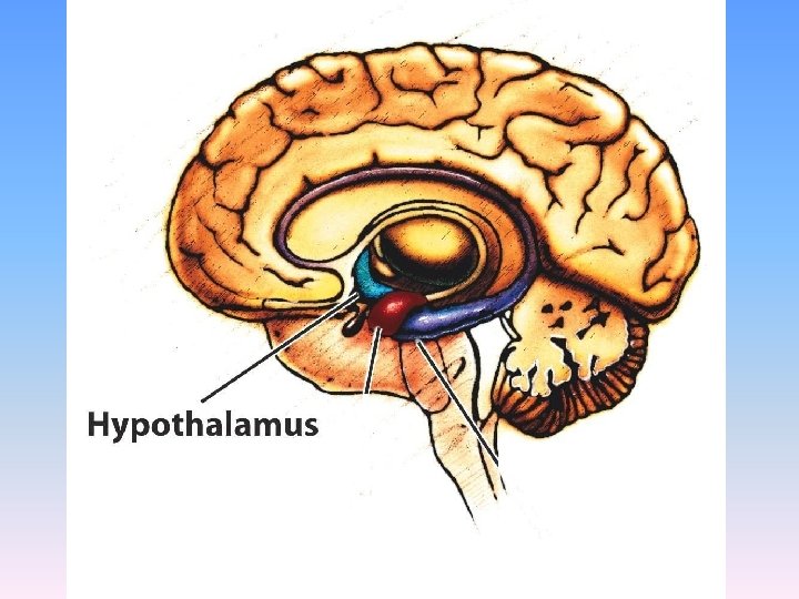
Limbic System • A ring of structures around the thalamus; at the border of the brainstem and cerebral cortex • Helps regulate memory, aggression, fear, hunger, and thirst • Includes the hypothalamus, hippocampus, and amygdala

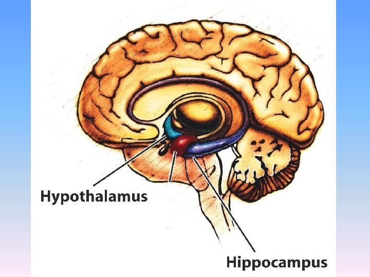
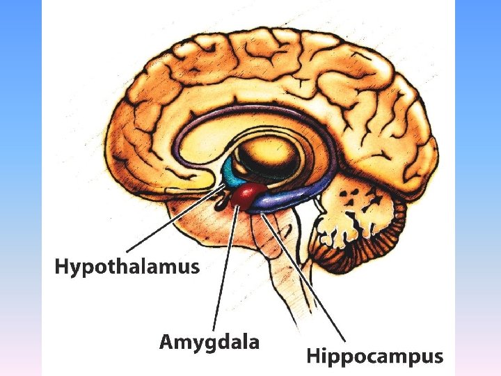
Hypothalamus • Located directly under the front of the thalamus • Regulates eating, drinking, body temperature, and the fight or flight reactions to stress • Plays a role in emotions, pleasure, and sexual function

Hippocampus • Wraps around the back of the thalamus • Plays a role in processing new memories for permanent storage • Looks something like a seahorse – Hippo is Greek for “horse. ”

Amygdala • Two almond shaped structures • Controls emotional responses such as fear and anger

Module 8: The Brain The Cerebral Cortex

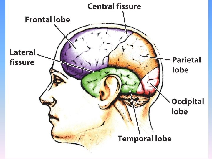
Cerebral Cortex • The body’s ultimate control and information processing center • Covers the brain’s lower level structures • Contains an estimated 30 billion nerve cells • Divided into four lobes

Corpus Callosum • The large band of neural fibers that connects the two brain hemispheres and carries messages between them • Is sometimes cut to prevent seizures

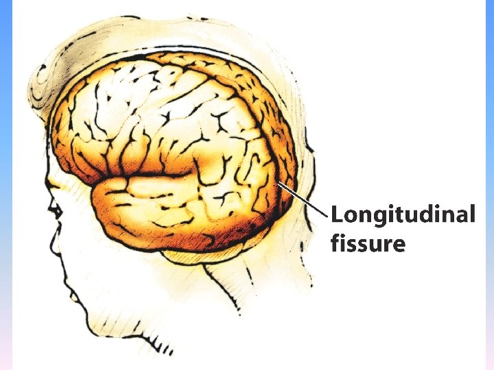
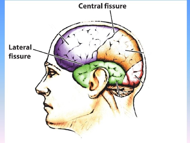
Longitudinal Fissure • The crevice that divides the brain into two halves or hemispheres • This and other fissures in the brain create major divisions in the brain called lobes

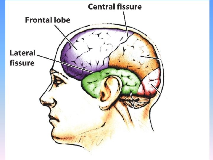
Frontal Lobes • The portion of the cerebral cortex lying just behind the forehead • Is involved in making plans and judgments

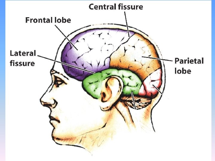
Parietal Lobes • Regions available for general processing, including mathematical reasoning • Designated as the association lobes • Behind the frontal lobes

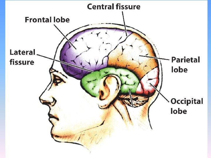
Occipital Lobe • The primary visual processing area • Located in the back of the head

Temporal Lobes • Includes the auditory cortex where sound information is processed • Located roughly above the ears

Cerebral Cortex

Cerebral Cortex

Cerebral Cortex

Cerebral Cortex

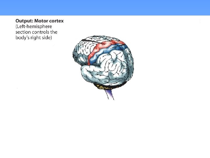
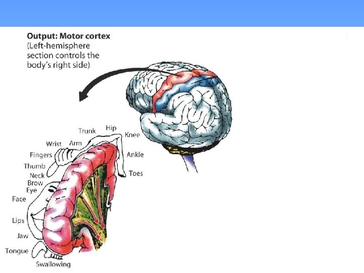
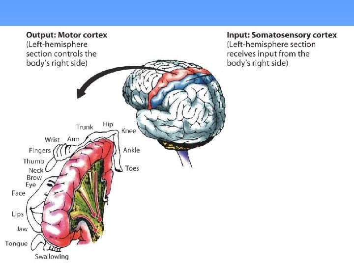
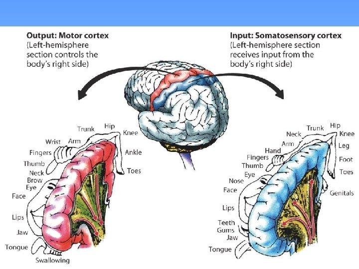
Motor Cortex • Area at the rear of the frontal lobes • Controls voluntary movement • Different parts of the cortex control different parts of the body. • The motor cortex in the left hemisphere controls the right side of the body and visa versa.

Somatosensory Cortex • Located in the front of the parietal lobes • Registers and processes body senses • Soma is Greek for “body. ”

Module 8: The Brain Hemispheric Differences

Hemispheric Differences • “Left-brained” and “right-brained” debunked • Brain is divided into two hemispheres but works as a single entity. • Both sides continually communicate via the corpus callosum, except in those with split brains.

Module 8: The Brain Hemispheric Differences: Language and Spatial Abilities

The Brain’s Left Hemisphere • For most people, language functions are in the left hemisphere. • For a small percentage of people, language functions are in the right hemisphere.

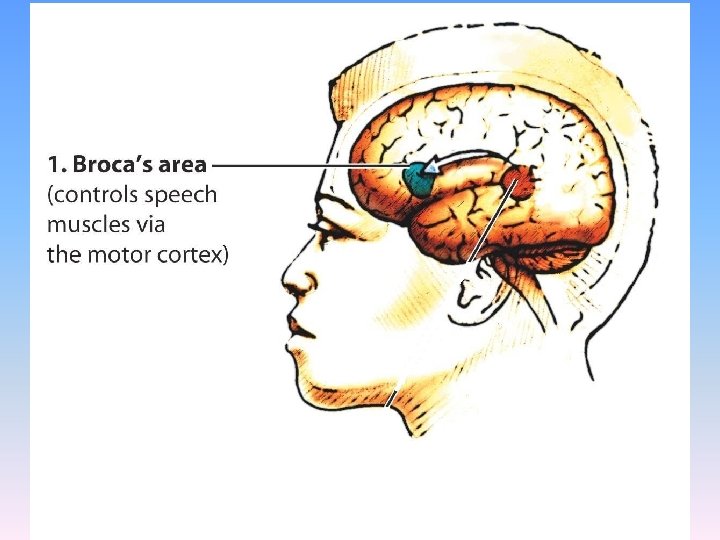
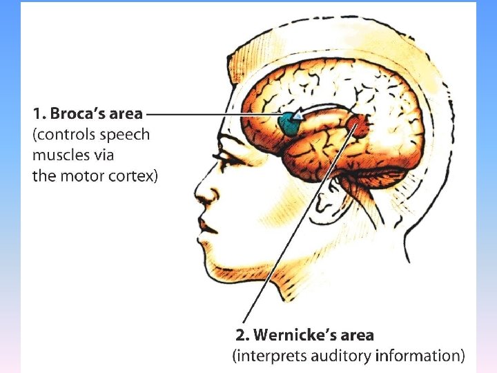
Broca’s Area • Located in the frontal lobe and usually in the left hemisphere • Responsible for the muscle movements of speech • If damaged the person can form the ideas but cannot express them as speech

PET Scan of Broca’s Area

Broca’s Area This is the brain of “Tal” from whom Broca discovered the area for speech. Note the damage to Broca’s Area.

Wernicke’s Area • Located in the temporal lobe • Involved in language comprehension and expression; our ability to understand what is said to us • Usually in the left temporal lobe

PET Scan of Wernicke’s Area

The Brain’s Right Hemisphere • Houses the brain’s spatial abilities • Our spatial ability allows us to perceive or organize things in a given space, judge distance, etc. • Helps in making connections between words

Module 8: The Brain Plasticity

Plasticity • The ability of the brain tissue to take on new functions • Greatest in childhood • Important if parts of the brain are damaged or destroyed

The End

Name of Concept • Use this slide to add a concept to the presentation

Name of Concept Use this slide to add a table, chart, clip art, picture, diagram, or video clip. Delete this box when finished
- Slides: 57