The Brain ChaoYing Wang 102 9 12 Outline

The Brain Chao-Ying Wang 102. 9. 12

Outline n n n n n The Organization of brain Meninges Ventricular System: Ventricles and Cisterns Cerebrum: Lobes and Basal ganglion Diencephalon: Thalamus, Epithalamus, Hypothalamus , Pituitary gland Limbic System Brain Stem: Midbrain, Pons and Medulla Oblongata Cerebellum Cerebral Vascular system: Arterial and Venous System
 Sagittal Anterior (Ventral) Posterior (Dorsal) (Transverse, tranaxial) (Coronal) Inferior (Caudal)")
Superior (Cranial) Sagittal Anterior (Ventral) Posterior (Dorsal) (Transverse, tranaxial) (Coronal) Inferior (Caudal)

Imaging Planes Transverse Axial Sagittal Coronal

The organization of brain n An adult brain weighs between 1. 35 and 1. 4 kilograms (kg) (around 3 pounds) and has a volume of about 1200 cubic centimeters (cc). Cerebrum is 83% of brain volume; cerebellum contains 50% of the neurons Brain size is not directly correlated with intelligence It is not the physical size of the brain that determines intelligence—it is the number of active synapses.
 n n n Mes-encephalon(midbrain) n n Telencephalon: cerebrum")
Embryonic development: neural tube n Pros-encephalon(forebrain) n n n Mes-encephalon(midbrain) n n Telencephalon: cerebrum Diencephalon: epithalamus, hypothalamus Mesencephalon: cerebral peduncles, colliculi Rhomb-encephalon(hindbrain) n n Metencephalon: pons, cerebellum Myelencephalon: medulla oblongata
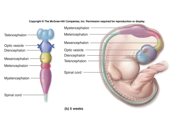

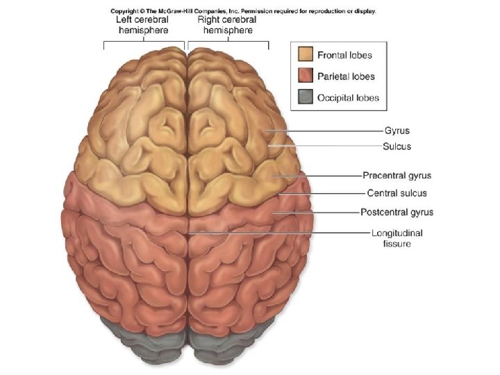
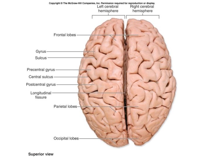
The Brain’s 4 Major Regions Cerebrum, diencephalon, brainstem, and cerebellum. n The cerebrum is divided into two halves, called the left and right cerebral hemispheres. n Each hemisphere is subdivided into five functional areas called lobes. n Outer surface of an adult brain exhibits folds called gyri (gyrus) and shallow depressions between those folds called sulci (sulcus). n The brain is associated with 12 pairs of cranial nerves.

Support and Protection of the Brain n The brain is protected and isolated by multiple structures: n Bony cranium n Meninges: n Protective connective tissue membranes n surround and partition portions of the brain. n Cerebrospinal fluid (CSF) n acts as a cushioning fluid. n Blood-brain barrier: n prevents entry of harmful materials from the bloodstream.

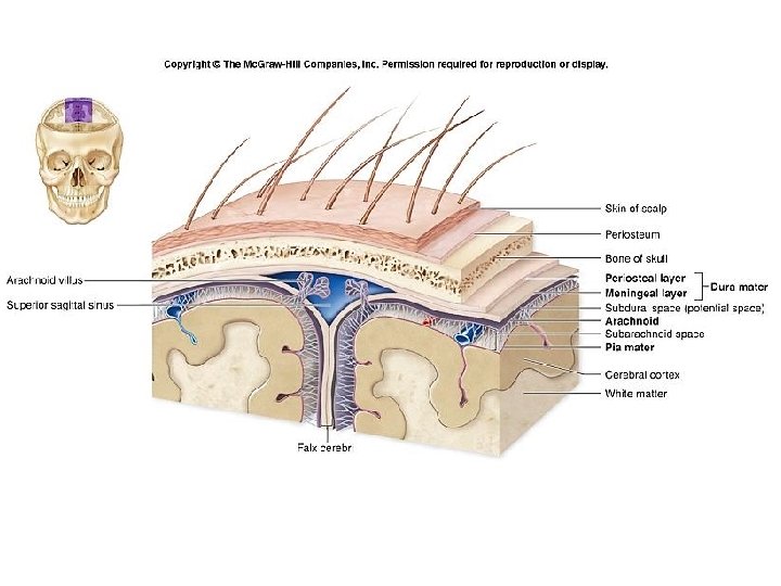
Brian Cushing Brain Cushing n n Three dense regular connective tissue layers: n Separate the soft tissue of the brain from the bones of the cranium. n Enclose and protect blood vessels that supply the brain. n Contain and circulate cerebrospinal fluid(CSF). n Parts of the cranial meninges form some of the veins that drain blood from the brain. From superficial to deep, the cranial meninges are the dura mater, the arachnoid, and the pia mater.

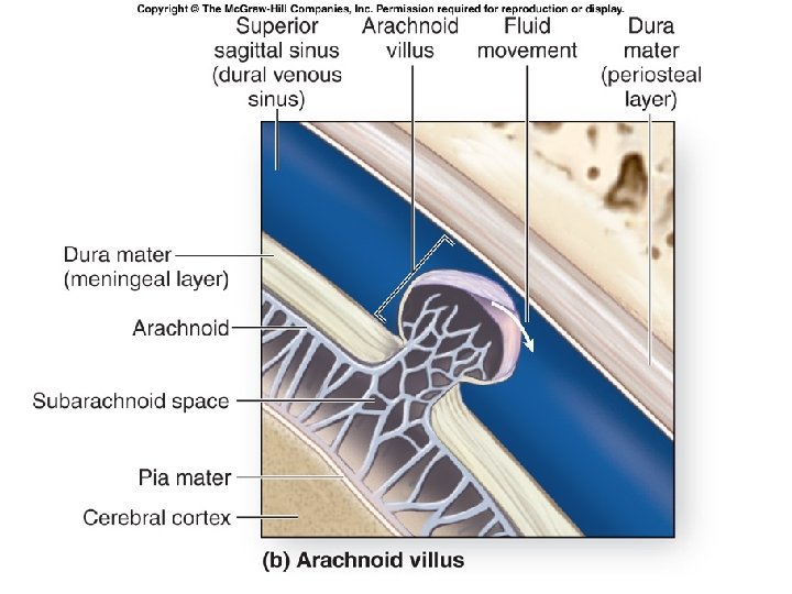
Cranial Meninges Dura mater n Strongest of the meninges. n Dura mater is composed of two layers. n periosteal layer, attaches to the periosteum of the cranial bones n meningeal layer lies deep to the periosteal layer Arachnoid n Lies immediately internal to the dura mater. n Composed of a delicate web of collagen and elastic fibers(arachnoid trabeculae). n Between the arachnoid and the overlying dura mater is the subdural space. n Immediately deep to the arachnoid is the subarachnoid space. Pia mater n The innermost of the cranial meninges. n Thin layer of delicate connective tissue that tightly adheres to the brain

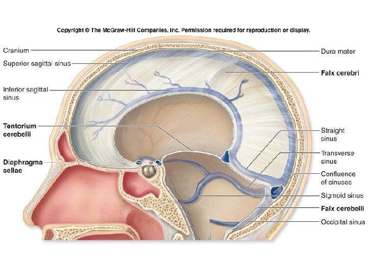
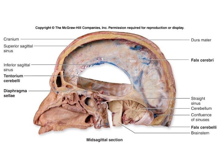
Cranial Dural Septa n n The meningeal layer of the dura mater extends as flat partitions (septa) deep into the cranial cavity; n at four locations n called cranial dural septa. Membranous partitions separate specific parts of the brain and provide additional stabilization and support to the entire brain. 1 1. falx cerebri 2. tentorium cerebelli 4 3 3. falx cerebelli 4. diaphragma sellae 2

MRI of the brain, T 1 -weighted coronal cut. 1, Sulcus. 2, Gyrus. 3, Falx cerebri. 4, Temporal lobe (Left side). 5, Temporal lobe (Right side). 1, Tentorium cerebelli. 2, Gyrus. 3, Sulcus. 4, Cerebellum. MRI of the brain, T 1 -weighted sagittal cut. 1, Occipital lobe. 2, Tentorium cerebelli. 3, Cerebellum. 4, Midbrain.

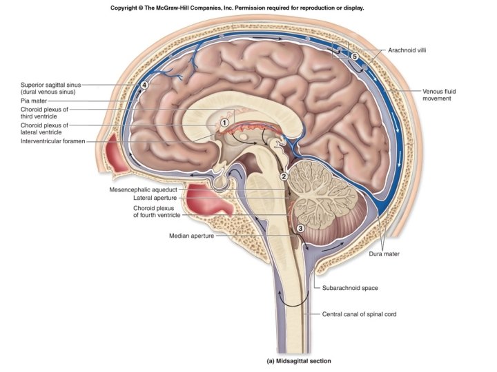
Brain Ventricles n n Cavities or expansions within the brain that are derived from the lumen (opening) of the embryonic neural tube. Continuous with one another as well as with the central canal of the spinal cord. Four ventricles in the brain. n two lateral ventricles are in the cerebrum, separated by a thin medial partition called the septum pellucidum n within the diencephalon is a smaller ventricle called the third ventricle n each lateral ventricle communicates with the third ventricle through an opening called the interventricular foramen The fourth ventricle is located within the pons and cerebellum.
 Frontal(anterior) horn Occipital(Post. ) horn Temporal(Inferior) horn (Foramen of Luschka) (Foramen")
(Foramen of Monro) Frontal(anterior) horn Occipital(Post. ) horn Temporal(Inferior) horn (Foramen of Luschka) (Foramen of Magendie) Cistern Magna

Cerebellopontine angle cistern

Cerebrospinal Fluid n n n A clear, colorless liquid that circulates in the ventricles and subarachnoid space. Is similar to blood plasma. Made in choroid plexuses (roofs of ventricles) n Filtration of plasma from capillaries through ependymal cells (electrolytes, glucose) 500 ml/d; total volume 100 -160 ml (1/2 c) Performs several important functions. n buoyancy n protection n environmental stability n nourishes brain Assayed in diagnosing meningitis, bleeds, MS Hydrocephalus: excessive accumulation

Subarachnoid cisterns • Areas within the subarachnoid space where the pia mater and arachnoid membrane are not in close approximation. • Subarachnoid space and CSF gathers to form pools or cisterns. • Some major subarachnoid cisterns: 1. cisterna magna (cerebellomedullary cistern): the largest of the subarachnoid cisterns 2. pontine cistern 3. suprasellar cistern 4. interpeduncular cistern 5. quadrigeminal cisterns (superior cistern of the great cerebral vein) 6. ambient cistern 7. Cerebellopontine angle(CPA) cistern

Subarachnoid cisterns

Cerebral hemispheres: note lobes • Divided by longitudinal fissure into right & left sides • Central sulcus divides frontal from parietal lobes • Lateral sulcus separates temporal lobe from parietal lobe • Parieto-occipital sulcus divides occipital and parietal lobes (not seen from outside) • Transverse cerebral fissure separates cerebral hemispheres from cerebellum • Insula – deep within the lateral sulcus

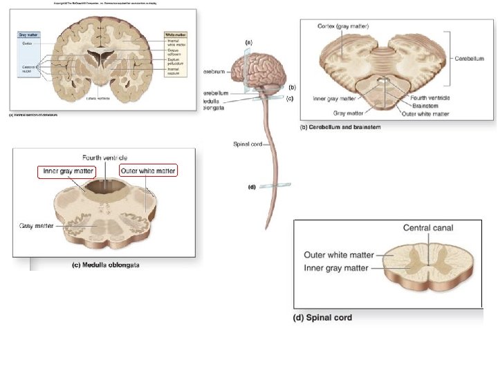
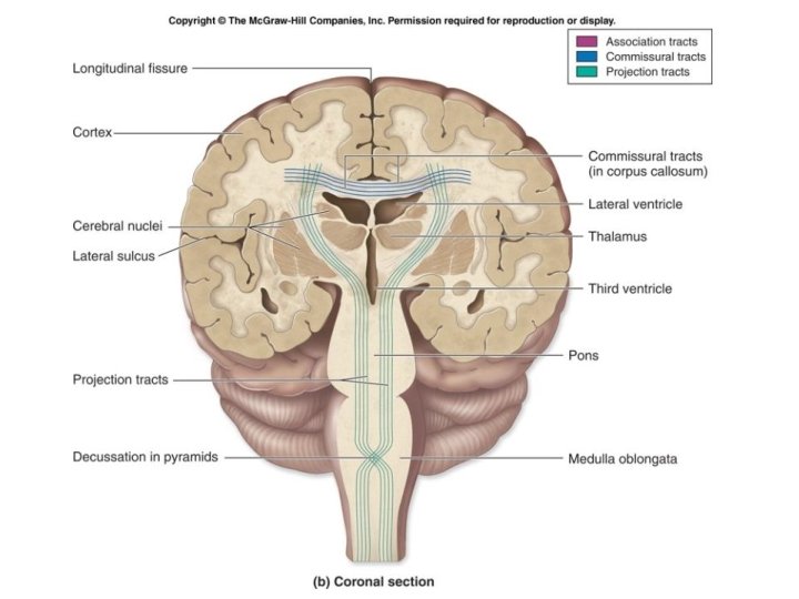
Organization of Brain Tissue • During brain development, an outer, superficial region of gray matter forms from migrating peripheral neurons. • External sheets of gray matter, called the cortex, cover the surface of most of the adult brain (the cerebrum and the cerebellum). • Gray matter: outer – motor neuron and interneuron cell bodies, dendrites, axon terminals – unmyelinated axons. • White matter: inner – composed primarily of myelinated axons. • Within the white matter: – discrete innermost clusters of gray matter called cerebral nuclei (or basal nuclei). – are oval, spherical, or sometimes irregularly shaped clusters of neuron cell bodies.

Cerebral cortex n n n Composed of gray matter-- 3 mm thick n Neuronal cell bodies, dendrites, and short axons Folds in cortex – triples its size 2 types of cells n stellate cells n have dendrites projecting in all directions n pyramidal cells n have an axon that passes out of the area

Functional areas of the cortex n Three kinds of functional areas n n n Motor areas Sensory areas Association areas

Motor areas Anterior to central sulcus • Primary motor area – – Precentral gyrus of frontal lobe (4) Conscious or voluntary movement of skeletal muscles Large neurons called pyramidal cells Their axons: form massive pyramidal or corticospinal tracts • Decend through brain stem and spinal cord • Cross to contralateral (the other) side in brainstem • Therefore: right side of the brain controls the left side of the body, and the left side of the brain controls the right side of the body
: specialized motor speech area – Base")
Motor areas – continued • Broca’s area (44): specialized motor speech area – Base of precentral gyrus just above lateral sulcus in only one hemisphere, usually left – Word articulation: the movements necessary for speech – Damage: can understand but can’t speak; or if can still speak, words are right but difficult to understand
: complex movements associated with highly processed")
Motor areas – continued • Premotor cortex (6): complex movements associated with highly processed sensory info; also planning of movements • Frontal eye fields (inferior 8): voluntary movements of eyes

Sensory areas Posterior to central sulcus • Primary somatosensory cortex: postcentral gyrus of parietal lobe (allows conscious awareness of sensation and the ability to localize it: where the sensation is from) • Somatosensory association area: behind it (understanding of what is being felt: the meaning of it)
 –")
From special sense organs • Occipital lobe: visual – Primary visual cortex (17) – Visual association area (18 & 19) • Face recognition is usually on the right side • Hearing: temporal lobe – Primary auditory area (41) – Auditory association area (22)
: uncus – Deep in temporal lobe along medial surface")
• Smell (olfactory sense): uncus – Deep in temporal lobe along medial surface

Homunculus – “little man” • Body map: human body spatially represented – Where on cortex; upside down

Refer back to this labeled version as needed

Auditory Association Area • The auditory association area contains two special regions. • BROCA'S AREA is a region of the brain that allows for speech. • WERNICKE’S AREA is the region of the brain that allows understanding of words. • Arcuate Fasciculus - A white matter tract that connects Broca’s Area and Wernicke’s Area through the temporal, parietal and frontal Lobes. Allows for coordinated, comprehensible speech. • Damage may result in Conduction Aphasia - Where auditory comprehension and speech articulation are preserved, but people find it difficult to repeat heard speech.

Cerebral White Matter n n Extensive communication n Areas of cortex with each other n Areas of cortex with brain stem and spinal cord Via (mostly) myelinated axon fibers bundled into tracts n Commissures n Association fibers n Projection fibers

Cerebral White Matter n Types of tracts or fibers n Commissures – composed of commissural fibers n Allows communication between cerebral hemispheres n Corpus callosum – the largest commissure n Association fibers n Connect different parts of the same hemisphere


44

Projection tracts n Projection fibers run vertically n Cerebral cortex running down (with motor instructions) n Or ascend to cerebral cortex from below (sensory info to cortex) n Internal capsule – projection fibers form a compact bundle n Passes between the thalamus and basal nuclei n Corona radiata – superior to the internal capsule n Fibers run to and from the cerebral cortex

• Projection fibers _________ – Corona radiata: fanning out of the fibers – Internal capsule: bundled, __________ pass down • Commisure – Corpus callosum: connects right and left hemispheres • Decussation: crossing of pyramidal tracts _____________________

White Matter-Fractional Anisotropy map of DTI

White Matter Fiber Tracts Association Projection Callosum Wakana S, Jiang H, Nagae-Poetscher LM, van Zijl PC, Mori S. Fiber tract-based atlas of human white matter anatomy. Radiology. 2004 Jan; 230(1): 77 -87. Epub 2003 Nov 26.

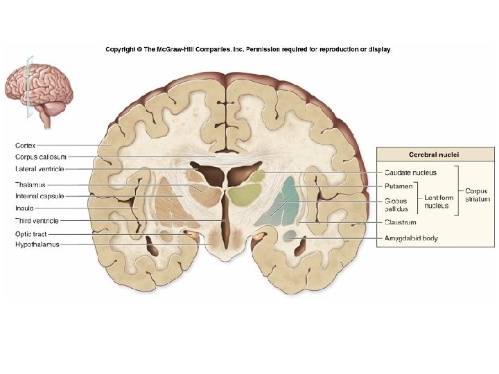
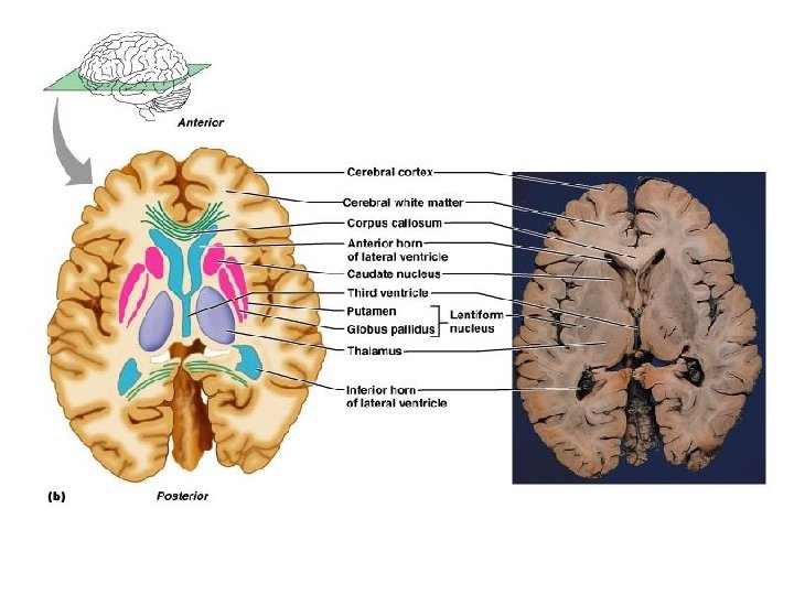
Basal nuclei n n A group of nuclei deep within the cerebral white matter n Caudate nucleus – arches over the thalamus(head, body and tail) n Lentiform nucleus – “lens shaped” n Amygdala – sits on tail of the caudate nucleus n Functionally belongs with the limbic system Lentiform nucleus n Divided into two parts n Globus pallidus n Putamen

Basal nuclei n n Cooperate with the cerebral cortex in controlling movements Receive input from cortical areas, send most of output back to motor cortex through thalamus Involved with stopping/starting & intensity of movements “Dyskinesias”-bad movements n Parkinson’s disease: loss of inhibition from substantia nigra of midbrain – everything slows down n Huntington disease: overstimulation (“choreoathetosis”) degeneration of corpus striatum which inhibits; eventual degeneration of cerebral cortex

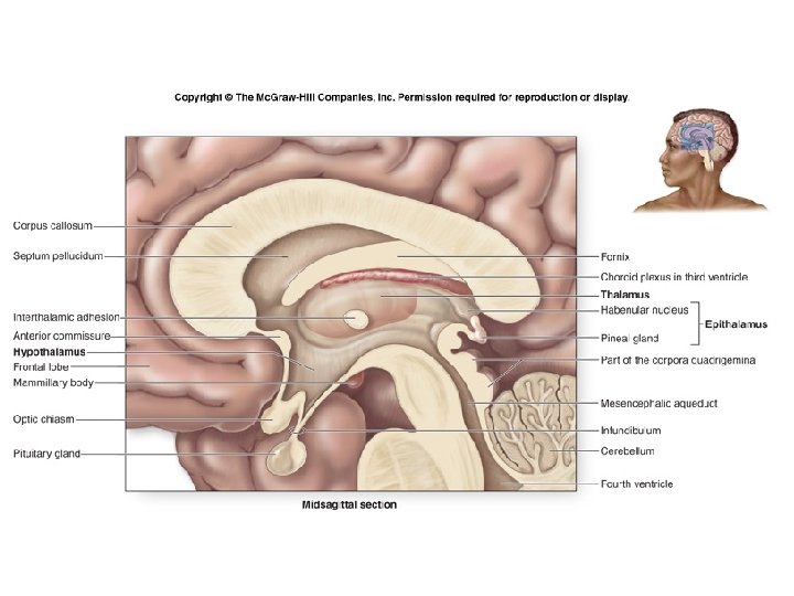
The Diencephalon n n Border the third ventricle Surrounded by the cerebral hemispheres Primarily composed of gray matter Forms the center core of the forebrain Composed of three paired structures: n Thalamus, hypothalamus, and epithalamus

The Thalamus n n Two large lobes of gray matter Laterally enclose the 3 rd ventricle Gateway to cerebral cortex: every part of brain that communicates with cerebral cortex relays signals through a nucleus in the thalamus Processing (editing) occurs also in thalamus
")
(Mass intermedia)

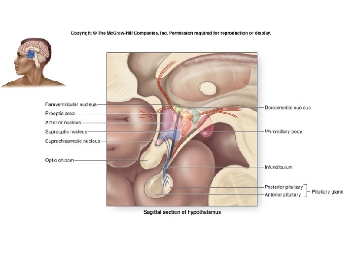
The Diencephalon – The Hypothalamus n n Lies between the optic chiasm and the mammillary bodies Pituitary gland projects inferiorly Contains approximately a dozen nuclei Main visceral control center of the body

The Hypothalamus n Functions include the following: n n n n Control of the autonomic nervous system Control of emotional responses Regulation of body temperature Regulation of hunger and thirst sensations Control of behavior Regulation of sleep-wake cycles Control of the endocrine system Formation of memory

Mammillary Bodies • A pair of small round bodies at the anterior end of the fornix • Part of the diencephalon; they form part of the limbic system. • They relay information (recognition memory) from the hippocampus. They also add the element of smell to memories. • Damage to the mammillary bodies due to thiamine deficiency (vit B 1) or alcohol causes Wernicke-Korsakoff syndrome (anterograde amnesia)

The Diencephalon – The Epithalamus n n n Forms part of the ”roof” of the third ventricle Consists of a tiny group of nuclei Includes the pineal gland (pineal body) n n Secretes the hormone melatonin Under influence of the hypothalamus


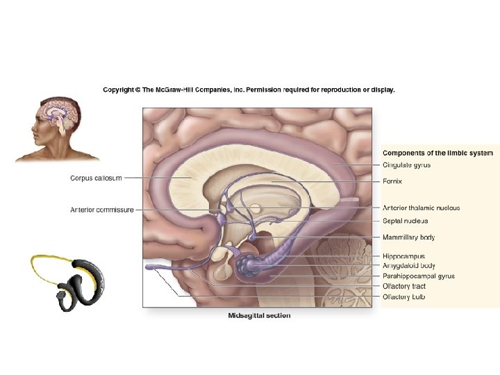
Functional Brain Systems – The Limbic System n n n Location n Medial aspect of cerebral hemispheres n Also within the diencephalon Composed of: n Septal nuclei, cingulate gyrus, and hippocampal formation n Part of the amygdala The fornix and other tracts link the limbic system together The “emotional brain” n Cingulate gyrus n Allows us to shift between thoughts n Interprets pain as unpleasant Hippocampal formation n Hippocampus and the parahippocampal gyrus

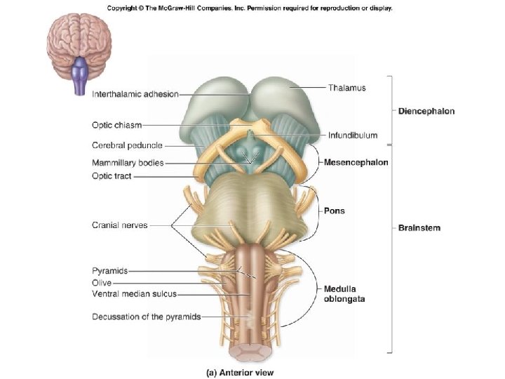
The Brain Stem n n Includes the midbrain, pons, and medulla oblongata Several general functions n Produces automatic behaviors necessary for survival n Passageway for all fiber tracts running between the cerebrum and spinal cord n 10 of the 12 pairs of cranial nerves attach to it

The Brain Stem – The Midbrain n n Lies between the diencephalon and the pons Central cavity – the cerebral aqueduct Cerebral peduncles located on the ventral surface of the brain n Contain pyramidal (corticospinal) tracts Superior cerebellar peduncles n Connect midbrain to the cerebellum

The Brain Stem – The Midbrain n Corpora quadrigemina – the largest nuclei n Divided into the superior and inferior colliculi n Superior colliculi – nuclei that act in visual reflexes n Inferior colliculi – nuclei that act in reflexive response to sound

70

The Brain Stem – The Midbrain n Two pigmented nuclei n Substantia nigra – neuronal cell bodies contain melanin n Functionally linked to the basal nuclei n Red nucleus – lies deep to the substantia nigra n Largest nucleus of the reticular formation

The Brain Stem – The Pons n n n Located between the midbrain and medulla oblongata Contains the nuclei of cranial nerves V, VI, and VII Two general groups of cranial nerve nuclei n Motor nuclei n Sensory nuclei

73

The Brain Stem – The Medulla Oblongata n Most caudal level of the brain stem n Continuous with the spinal cord n Choroid plexus lies in the roof of the fourth ventricle n Pyramids of the medulla – lie on its ventral surface n Cranial nerves VIII–XII attach to the medulla

75

76

Functional Brain Systems – The Reticular Formation n n Runs through the central core of the medulla, pons, and midbrain Forms three columns n n n Midline raphe nuclei Medial nuclear group Lateral nuclear group

Functional Brain Systems – The Reticular Formation

The Cerebellum n n n Located dorsal to the pons and medulla n Smoothes and coordinates body movements n Helps maintain equilibrium Consists of two cerebellar hemispheres Surface folded into ridges called folia Hemispheres each subdivided into anterior and posterior Thick tracts connecting the cerebellum to the brain stem n Superior cerebellar peduncles n Middle cerebellar peduncles n Inferior cerebellar peduncles

80

The Cerebellum n n Composed of three regions n Cortex – gray matter n Internal white matter n Deep cerebellar nuclei – deeply situated gray matter Cerebellum must receive information n On equilibrium n On current movements of limbs, neck, and trunk n From the cerebral cortex

82

Outline • • • The Organization of brain Meninges Ventricular System: Ventricles and Cisterns Cerebrum: Lobes and Basal ganglion Diencephalon: Thalamus, Epithalamus, Hypothalamus , Pituitary gland Limbic System Brain Stem: Midbrain, Pons and Medulla Oblongata Cerebellum Cerebral Vascular system: Arterial and Venous System

Principle Brain Arteries
 The segments of the internal carotid artery are as follows:")
Internal Carotid Artery( ICA) The segments of the internal carotid artery are as follows: 1. Cervical segment or portion, or C 1 2. Petrous segment, or C 2 3. Lacerum segment, or C 3 • C 2 and C 3 compose the commonly termed Petrous portion 4. Cavernous segment, or C 4, commonly used Cavernous portion 5. Clinoid segment, or C 5, lies between the commonly used Cavernous portion and Cerebral or Supraclinoid portion 6. Ophthalmic, or supraclinoid segment, or C 6 7. Communicating, or terminal segment, or C 7

Anterior circulation -Internal carotid arteries C 6 C 4 C 2 C 1
 • The vertebral-basilar arteries supply about 20% of the blood flow")
Vertebral Arteries (VA) • The vertebral-basilar arteries supply about 20% of the blood flow to the brain. • Derived from subclavian arteries in the thoracic cavity. • When they get to the atlas, they exit the spine and migrate backwards toward the atlantooccipital membrane. • Shortly after entering the cranial vault the vertebral arteries unite and become the basilar artery. • Vetebral-Basilar system include: • Posterior inferior cerebellar artery (PICA) • Anterior inferior cerebellar artery (AICA) • Pontine artery • Superior cerebellar artery(SCA)

Posterior circulation -Vertebral arteries

Circle of Willis The Circle of Willis is composed of the following arteries: 1. Anterior cerebral artery (ACA) 2. Anterior communicating artery(ACo. A) 3. Internal carotid artery (l. CA) 4. Posterior cerebral artery (PCA) 5. Posterior communicating artery (PCo. A) The basilar artery and middle cerebral arteries, supplying the brain, are not considered part of the circle

Circle of Willis 2 1 3 5 4

Lenticulostriate Arteries Supply the Basal Ganglia and Internal Capsule

Anatomy and Vascular Territories of the 3 Main Cerebral Arteries: Middle Cerebral Artery M 3 M 2 M 1

Anterior Cerebral Artery and Posterior Cerebral Artery

Superficial and Deep Arterial Supply to the Cerebral Hemispheres Coronal Plane

Superficial and Deep Arterial Supply to the Cerebral Hemisphere Axial Plane
- Slides: 95