THE BRAIN APPROACHES OVER TIME BRAIN VERSUS HEART

THE BRAIN APPROACHES OVER TIME
, they didn’t even preserve")
BRAIN VERSUS HEART When Tutankhamen was mummified (3300 years ago), they didn’t even preserve the brain or the heart.

ANCIENT GREEK PHILOSOPHERS The brain hypothesis • Alcmaeon • Located mental processes in the brain • Discovered the optic nerve connecting the eye to the brain The heart debate • Empedocles • Located mental processes in the heart • Believed every living thing is made from earth, fire, air and water. • Reasoned that the heart was the centre of the body’s bleed vessels & so our thoughts must be located in the blood.

Loosing your mind

MIND-BODY PROBLEM Are our mind and our body two separate entities or are they the same thing?
 Body: fleshy")
TH 7 C - DESCARTES DUALISM Mind: non-physical, spiritual entity (a soul) Body: fleshy structure Come into contact through the pineal gland enables them to interact. Located in the centre of the brain. They could affect each other.

PHRENOLOGY – GALL 1758 -1828 Different parts of the brain have different functions Personality characteristics and mental abilities were controlled by brain organs, located in the outer surface The size indicated how developed it was strength influence Development would push the skull out (bumps)
 Within")
PHRENOLOGY – GALL 1758 -1828 Researched on many different people Faculties (mental abilities/traits/behaviours) Within that there were affective (feelings) and intellectual faculties.

FIRST EXPERIMENTS

BRAIN ABLATION Disabling, destroying or removing selected brain tissue followed by an assessment of subsequent changes in behaviour.
 He found that")
PIERRE FLOURENS Introduced ablation 1820 s with animals (rabbits and pigeons) He found that removing different areas had different effects on movement, breathing etc. He believed that you could fully recover though as other areas would take over

KARL LASHLEY 1920 s used ablation to find the location of learning and memory on rats and monkeys He failed to produce amnesia by removing small areas so concluded that memory is throughout the entire brain.

KARL LASHLEY Mass action Equipotentialit y • The large areas of the brain function as a whole in complex functions • If a part is destroyed then loss of function will depend on the amount of cortex that is destroyed • Any healthy part of the cortex can take over the function of an injured part.

ELECTRICAL STIMULATION A small electrified fine wire or disc that is inserted or placed onto a specific area of the brain. If a particular response occurs (or stops), then that is believed to be linked to that area of the brain.

FRITSCH AND HITZIG – THE MOTOR CORTEX 1870 They found 5 sites that triggered distinctive movements in dogs – on the opposite side of the body Demonstrated contralateral function of limb movements

PENFIELD – MAPS THE CEREBRAL CORTEX Humans 1940 s Mapped the cerebral cortex Removed parts of the brain believed to be responsible for epileptic fits (patient had to be awake)

SPLIT-BRAIN SURGERY Surgical cutting of the corpus callosum and sometimes other connecting nerves to separate the two cerebral hemispheres.

Severed Corpus Collosum
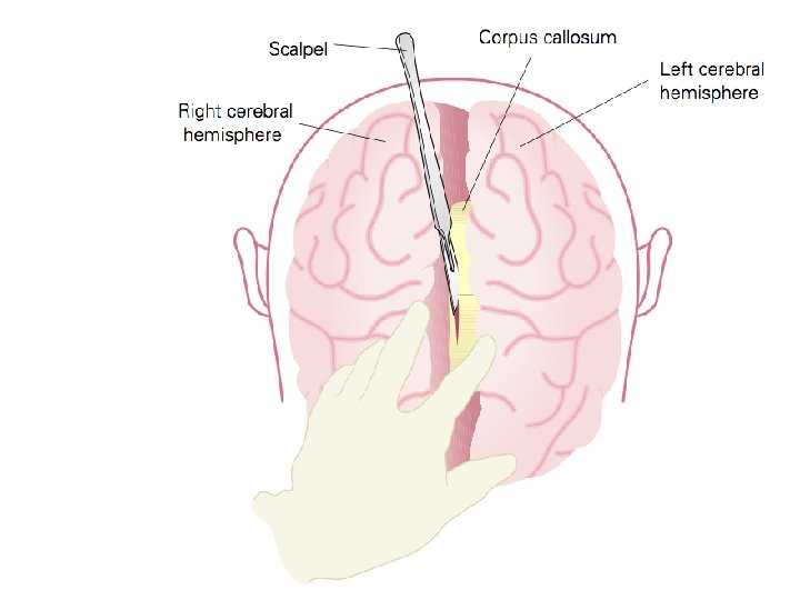

SPLIT-BRAIN SURGERY But how can the brain communicate if the corpus callosum is cut?

SPLIT-BRAIN SURGERY Severed Corpus Callosum Video

SPLIT-BRAIN SURGERY SPERRY AND GAZZANIGA Right visual field Sent to left hemisphere Participant named the object.

SPLIT-BRAIN SURGERY SPERRY AND GAZZANIGA Left visual field Sent to right hemisphere Denied they saw anything.

SPLIT-BRAIN SURGERY SPERRY AND GAZZANIGA Denied they saw anything. Why?

SPLIT-BRAIN SURGERY SPERRY AND GAZZANIGA Left visual field Sent to right hemisphere Participant picked up the object.

SPLIT-BRAIN SURGERY SPERRY AND GAZZANIGA Participant picked up the object. WHY?

NEUROIMAGING TECHNIQUE

NEUROIMAGING A technique that captures a picture of the brain

NEUROIMAGING Neuroimagin g Structural CT Functional MRI PET f. MRI
 Functional • Images")
NEUROIMAGING TECHNIQUES Structural • Images that shown brain structure (CT, MRI) Functional • Images that provide views of some particular aspect of brain function by showing images of the brain at work (PET, f. MRI)

Why is this an important technique?
Computerised technology b)Computerised tomography c) Computerised telepathy")
STRUCTURAL IMAGING CT DOES CT STAND FOR: a)Computerised technology b)Computerised tomography c) Computerised telepathy
 Patient")
COMPUTERISED TOMOGRAPHY X-ray equipment Scans the brain at different angles Horizontal cross-section (slices) Patient takes a substance called contrast Highlights brain’s blood vessels Helpful for tumours/ stroke/mental illness Only shows brain structure and not tissue

STRUCTURAL IMAGING MRI Uses magnetic fields to vibrate atoms in the brain’s neurons and generate a computer image of the brain.

MRI More sensitive than a CT, colour Distinguish between tissue that is benign or malignant, structural abnormalities etc.

Video

FUNCTIONAL NEUROIMAGING - PET Positron emission tomography Produces colour images showing brain structure, activity and function. Patient does a task and level of activity is recorded. Image of the ‘working brain’

FUNCTIONAL NEUROIMAGING - PET Tracks a glucose solution containing a short-lived radioactive tracer Assumed that brain areas that require increased blood flow have increased neuronal activity Colour code areas of high and low activity

Video

FUNCTIONAL NEUROIMAGING - FMRI Detects and records brain activity by measuring oxygen consumption across the brain. Uses radioactive tracers.

FUNCTIONAL NEUROIMAGING - FMRI Blood is more oxygenated in highly active parts of the brain Brain areas that are more or less active during a given task are identified by detecting changes in oxygen levels in the blood as it flows. Takes rapid pictures Highly detailed and precise
- Slides: 45