The Bodys Defenses Nonspecific defense mechanisms not selective

The Body’s Defenses Nonspecific defense mechanisms • not selective in their response + fight off everything the same way Specific Defense Mechanisms • Immune System + generates efficient and selective response

Nonspecific Defenses Against Infection 1 st Line of Defense • physical and chemical barriers + skin and mucous membrane + acid and lysozyme 10�m

2 nd Line of Defense Part I: Non-specific Killers Figure 43. 1 3�m WBC = Generic Term for Phagocytes: 1 -Monocytes macrophages: Ingest pathogens and serve as APC to B & T 2 -Neutrophils: “kamkazes” that release “neutralizer” chemicals-kill pathogens and themselves! 3 -Eosinophils: combat multicellular pathogens, inflammatory response 4 -Natural Killer (NK) cells: Attack infected body cells NOT microbes. Release perforin, triggering apoptosis Watch Video Clips of WBC in in infected cell. action

Macrophages A MACROPHAGE OVERCOMES AND EATS A CANCER CELL – secrete cytokine proteins which help stimulate other
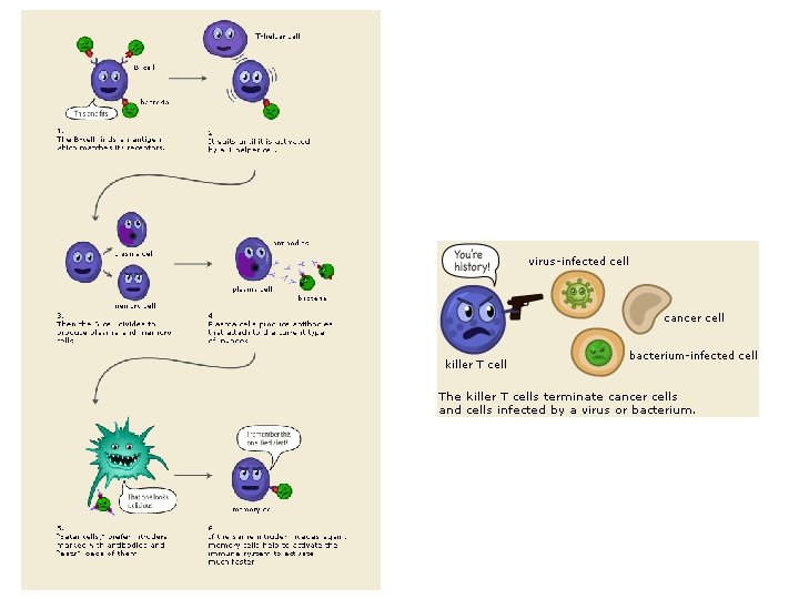
 Cells Our first line of defense NK cells are interferon activated.")
Natural Killers (NK) Cells Our first line of defense NK cells are interferon activated. ‘Venom’ is perforin & granzyme protein.

2 nd Line Part II: Inflammatory & Temperature Response • localized response to tissue damage (cut) or entry of microorganism - increased blood supply (redness/swelling/warmth) - histamine and prostaglandins = chemical alarm system triggering vasodilation + aid in delivering clotting elements and immune cells Pyrogens: FEVER inhibits microbial growth but > 103 F dangerous & 105 F often fatal( WHY? )

Specific Immunity Immune System Response • lymphocytes + B cells and T cells - come from stem cells in bone marrow + mature in different locations before moving on to lymphoid tissue (lymph nodes, spleen, blood, lymph) - respond to specific antigens + clonal selection - effector cells and memory cells + primary and secondary immune response

• Newly formed lymphocytes are all alike – But they later develop into B cells or T cells, depending on where they continue their maturation Bone marrow Lymphoid stem cell Thymus T cell B cell Figure 43. 10 Blood, lymph, and lymphoid tissues (lymph nodes, spleen, and others)

B & T Lymphocytes secrete perforin protein which punctures tumor cells when activated by Ag Activated by Ag & produce cytokine proteins (interferons & interleukins) which help activate immune response Antibody (Ab): Proteins that attach to Antigens & to signal destruction

• An antigen is any foreign molecule – That is specifically recognized by lymphocytes and elicits a response from them • A lymphocyte actually recognizes and binds – To just a small, accessible portion of the antigen called an epitope Antigenbinding sites Antibody A Antigen Antibody B Figure 43. 7 Antibody C Epitopes (antigenic determinants)

Cell-Mediated Immune Response T cells • kill cells that have been infected, or parasites • response initiated through contact with cell or macrophage + divides into four cell lines - T c TH TS TM T memory cells cytotoxic “killer” T cells T 4 helper cells + core of immune system; infected by HIV + “messenger” T suppressor cells + protect our own tissues

• The activated cytotoxic T cell – Secretes proteins that destroy the infected target cell 1 A specific cytotoxic T cell binds to a class I MHC–antigen complex on a target cell via its TCR with the aid of CD 8. This interaction, along with cytokines from helper T cells, leads to the activation of the cytotoxic cell. 2 The activated T cell releases perforin molecules, which form pores in the target cell membrane, and proteolytic enzymes (granzymes), which enter the target cell by endocytosis. Cytotoxic T cell Released cytotoxic T cell Perforin Cancer cell Granzymes 1 TCR Class I MHC molecule Target cell 3 The granzymes initiate apoptosis within the target cells, leading to fragmentation of the nucleus, release of small apoptotic bodies, and eventual cell death. The released cytotoxic T cell can attack other target cells. 3 CD 8 2 Apoptotic target cell Pore Peptide antigen Cytotoxic T cell Figure 43. 16 Video
 • “antibody-mediated")
Humoral Immune Response B cells • fights infections of plasma (generally bacteria) • “antibody-mediated response” + antibody is quaternary protein (multiple polypeptide chains) - Y-shaped - can bind to variety of antigens + response to foreign antigen - divides into two different cell lines + memory cells + plasma cell - antibody factory - makes antigens easier to locate by phagocytes

• Clonal selection of B cells – Generates a clone of short-lived activated effector cells and a clone of long-lived memory cells – Activated by Ag itself or Th cell Antigen molecules B cells that differ in antigen specificity Antigen receptor Antigen molecules bind to the antigen receptors of only one of the three B cells shown. The selected B cell proliferates, forming a clone of identical cells bearing receptors for the selecting antigen. VIDEO Some proliferating cells develop into long-lived memory cells that can respond rapidly upon subsequent exposure to the same antigen. Antibody molecules Clone of memory cells Figure 43. 12 Clone of plasma cells Some proliferating cells develop into short-lived plasma cells that secrete antibodies specific for the antigen.

1 After a macrophage engulfs and degrades 2 a bacterium, it displays a peptide antigen complexed with a class II MHC molecule. A helper T cell that recognizes the displayed complex is activated with the aid of cytokines secreted from the macrophage, forming a clone of activated helper T cells (not shown). A B cell that has taken up and degraded the same bacterium displays class II MHC–peptide antigen complexes. An activated helper T cell bearing receptors specific for the displayed antigen binds to the B cell. This interaction, with the aid of cytokines from the T cell, activates the B cell. 3 The activated B cell proliferates and differentiates into memory B cells and antibody-secreting plasma cells. The secreted antibodies are specific for the same bacterial antigen that initiated the response. Bacterium Macrophage Peptide antigen Class II MHC molecule B cell 2 3 1 TCR Clone of plasma cells Endoplasmic reticulum of plasma cell CD 4 Cytokines Helper T cell Activated helper T cell Clone of memory B cells Figure 43. 17 Secreted antibody molecules

• In the secondary immune response – Memory cells facilitate a faster, more efficient response 1 Day 1: First exposure to antigen A 2 Primary response to antigen A produces antibodies to A 3 Day 28: Second exposure to antigen A; first exposure to antigen B 4 Secondary response to antigen A produces antibodies to A; primary response to antigen B produces antibodies to B Antibody concentration (arbitrary units) 104 103 102 Antibodies to A Antibodies to B 101 100 Figure 43. 13 0 7 14 21 28 35 Time (days) 42 49 56

• The roles of the major participants in the acquired immune response Cell-mediated immune response Humoral immune response First exposure to antigen Intact antigens WBC engulf & display Antigens engulfed and displayed by. Ag dendritic cells Antigens displayed by infected cells Activate B cell Helper T cell Figure 43. 14 Memory B cells Active and memory helper T cells Secrete antibodies that defend against pathogens and toxins in extracellular fluid Cytotoxic T cell Gives rise to Plasma cells Secreted cytokines activate Memory cytotoxic T cells Active cytotoxic T cells Defend against infected cells, cancer cells, and transplanted tissues

Active vs. Passive Immunization • Active immunity – Develops naturally in response to an infection – Can also develop following immunization, also called vaccination • Passive immunity, which provides immediate, short-term protection – Is conferred naturally when antibodies pass from mother to infant in breast milk – Can be conferred artificially by injecting antibodies into a non-immune person

MHC: Self vs. Non-self • The immune system – Can wage war against cells from other individuals • Transplanted tissues – Are usually destroyed by the recipient’s immune system MHC: Major Histocompatibility Complex which is unique set of surface proteins on an individual’s cells Class I MHC molecules, found on almost all nucleated cells of the body = “self” Class II MHC molecules, located mainly on WBC’s, macrophages, and B cells – allow for communication/coordination between animal’s immune
 responses – To typically harmless antigens called allergens")
Allergies • Allergies are exaggerated (hypersensitive) responses – To typically harmless antigens called allergens – DUST MITES & ITCHY SKIN VIDEO Ig. E Allergen Histamine 1 3 2 Granule Mast cell 1 Ig. E antibodies produced in response to initial exposure to an allergen bind to receptors or mast cells. 2 On subsequent exposure to the same allergen, Ig. E molecules attached to a mast cell recognize and bind the allergen. Figure 43. 20 3 Degranulation of the cell, triggered by cross-linking of adjacent Ig. E molecules, releases histamine and other chemicals, leading to allergy symptoms.

Autoimmune Diseases – The immune system loses tolerance for self and turns against certain molecules of the body

• Rheumatoid arthritis – Is an autoimmune disease that leads to damage and painful inflammation of the cartilage and bone of joints Figure 43. 21

• Other examples of autoimmune diseases include – Systemic lupus erythematosus – Multiple sclerosis – Insulin-dependent diabetes
 • Human Immunodeficiency Virus (HIV) –")
HIV & AIDS • Acquired Immunodeficiency Syndrome (AIDS) • Human Immunodeficiency Virus (HIV) – retrovirus (RNA) that causes AIDS by infected Th cells • People with AIDS – Are highly susceptible to opportunistic infections and cancers that take advantage of an immune system in collapse – HIV Infection Overview

Review: Specific Defenses Th then helps to activates B & Tc cells

Immune Quiz • Click SELF QUIZ and follow the link for ‘quiz on immunology. • When you have completed the quiz you can re -click the link and new set of questions will appear.
- Slides: 27