
The blood vessels The Urinary System Prepared by

The blood vessels & The Urinary System Prepared by : Amal Awad Al-Harbi
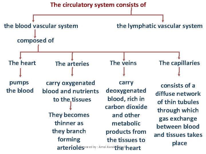
The circulatory system consists of the blood vascular system the lymphatic vascular system composed of The heart pumps the blood The arteries The veins The capillaries carry consists of a deoxygenated diffuse network blood, rich in of thin tubules carbon dioxide through which They becomes and other gas exchange thinner as metabolic between blood they branch products from and tissues takes forming the tissues to place Prepared by : Amal Awad Al-Harbi arterioles the heart carry oxygenated blood and nutrients to the tissues
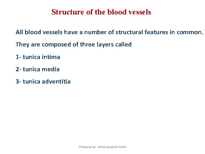
Structure of the blood vessels All blood vessels have a number of structural features in common. They are composed of three layers called 1 - tunica intima 2 - tunica media 3 - tunica adventitia Prepared by : Amal Awad Al-Harbi
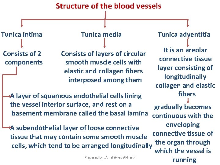
Structure of the blood vessels Tunica intima Tunica media Tunica adventitia Consists of 2 components Consists of layers of circular smooth muscle cells with elastic and collagen fibers interposed among them It is an areolar connective tissue layer consisting of longitudinally collagen and elastic fibers A layer of squamous endothelial cells lining the vessel interior surface, and rest on a gradually becomes basement membrane called the basal lamina continuous with the enveloping A subendothelial layer of loose connective tissue that may contain some smooth muscle connective tissue of cells, which tend to be arranged longitudinally the organ through which the vessel is Prepared by : Amal Awad Al-Harbi running
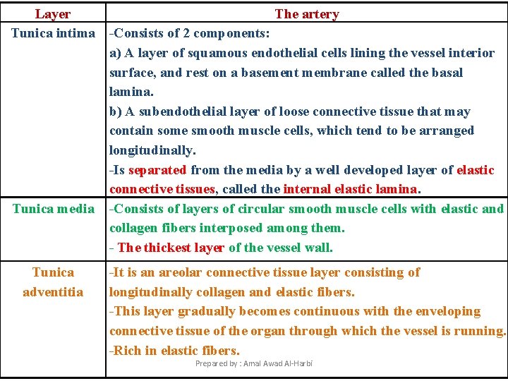
Layer The artery Tunica intima -Consists of 2 components: a) A layer of squamous endothelial cells lining the vessel interior surface, and rest on a basement membrane called the basal lamina. b) A subendothelial layer of loose connective tissue that may contain some smooth muscle cells, which tend to be arranged longitudinally. -Is separated from the media by a well developed layer of elastic connective tissues, called the internal elastic lamina. Tunica media -Consists of layers of circular smooth muscle cells with elastic and collagen fibers interposed among them. - The thickest layer of the vessel wall. Tunica adventitia -It is an areolar connective tissue layer consisting of longitudinally collagen and elastic fibers. -This layer gradually becomes continuous with the enveloping connective tissue of the organ through which the vessel is running. -Rich in elastic fibers. Prepared by : Amal Awad Al-Harbi
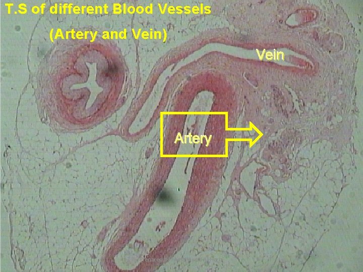
T. S of different Blood Vessels (Artery and Vein) Vein Artery Prepared by : Amal Awad Al-Harbi

Sector of the wall of an artery Tunica adventitia Tunica media Tunica intima Lumen Prepared by : Amal Awad Al-Harbi
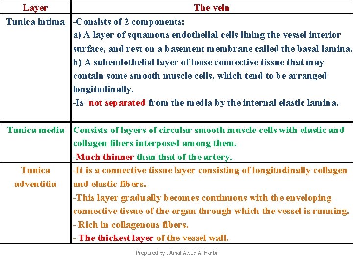
Layer The vein Tunica intima -Consists of 2 components: a) A layer of squamous endothelial cells lining the vessel interior surface, and rest on a basement membrane called the basal lamina. b) A subendothelial layer of loose connective tissue that may contain some smooth muscle cells, which tend to be arranged longitudinally. -Is not separated from the media by the internal elastic lamina. Tunica media Consists of layers of circular smooth muscle cells with elastic and collagen fibers interposed among them. -Much thinner than that of the artery. Tunica -It is a connective tissue layer consisting of longitudinally collagen adventitia and elastic fibers. -This layer gradually becomes continuous with the enveloping connective tissue of the organ through which the vessel is running. - Rich in collagenous fibers. - The thickest layer of the vessel wall. Prepared by : Amal Awad Al-Harbi
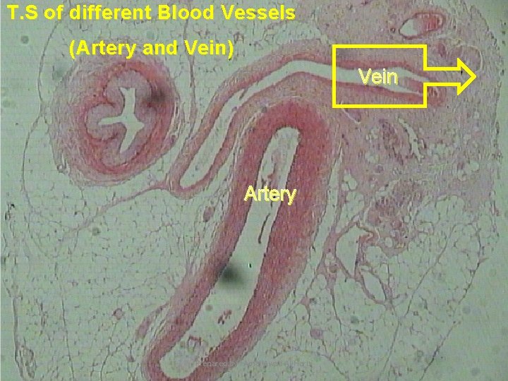
T. S of different Blood Vessels (Artery and Vein) Vein Artery Prepared by : Amal Awad Al-Harbi

Sector of the wall of a vein Tunica adventitia Tunica intima Tunica media Lumen Prepared by : Amal Awad Al-Harbi
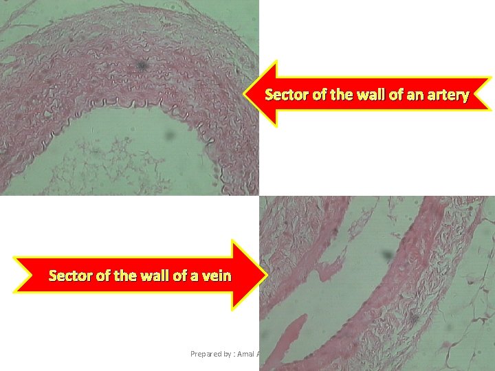
Sector of the wall of an artery Sector of the wall of a vein Prepared by : Amal Awad Al-Harbi
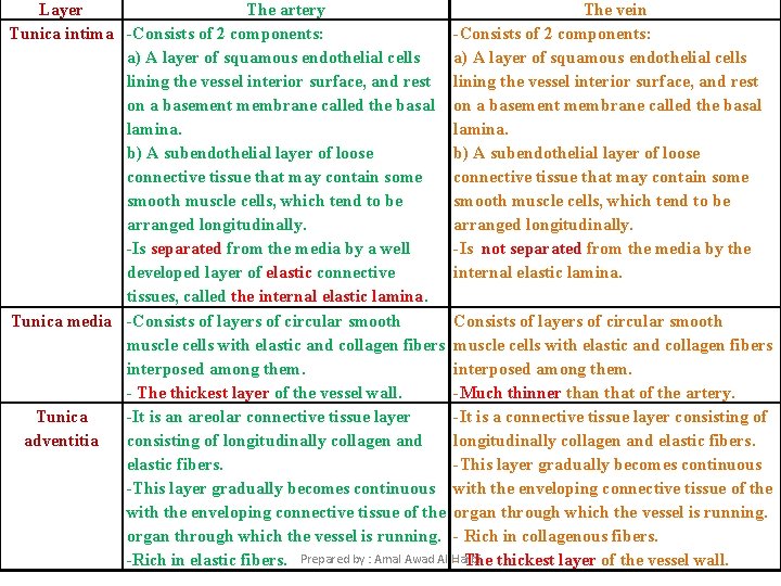
Layer The artery The vein Tunica intima -Consists of 2 components: a) A layer of squamous endothelial cells lining the vessel interior surface, and rest on a basement membrane called the basal lamina. b) A subendothelial layer of loose connective tissue that may contain some smooth muscle cells, which tend to be arranged longitudinally. -Is separated from the media by a well -Is not separated from the media by the developed layer of elastic connective internal elastic lamina. tissues, called the internal elastic lamina. Tunica media -Consists of layers of circular smooth muscle cells with elastic and collagen fibers interposed among them. - The thickest layer of the vessel wall. -Much thinner than that of the artery. Tunica -It is an areolar connective tissue layer -It is a connective tissue layer consisting of adventitia consisting of longitudinally collagen and elastic fibers. -This layer gradually becomes continuous with the enveloping connective tissue of the organ through which the vessel is running. - Rich in collagenous fibers. -Rich in elastic fibers. Prepared by : Amal Awad Al-Harbi - The thickest layer of the vessel wall.
The Urinary System Prepared by : Amal Awad Al-Harbi

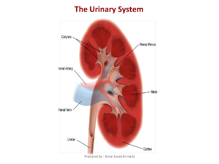
*The kidney is bean-shaped with a concave medial border , the hilum (where nerves enter, blood and lymph vessels enter and exit and the ureter exits) and a convex lateral surface. *The kidney is surrounded by a mass of adipose tissue. *The kidney tissue surrounds a cavity, the renal pelvis. This pelvis is the expanded upper end of the ureter. *The functional unit of the kidney is the uriniferous tubule which consists of the nephron and the collecting duct into which the renal tubule empties its contents. Prepared by : Amal Awad Al-Harbi
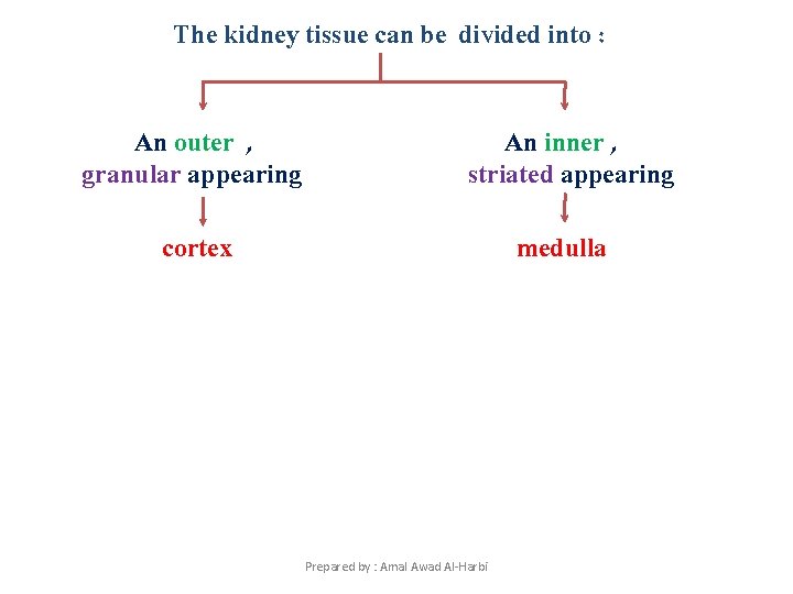
The kidney tissue can be divided into : An outer , granular appearing An inner , striated appearing cortex medulla Prepared by : Amal Awad Al-Harbi
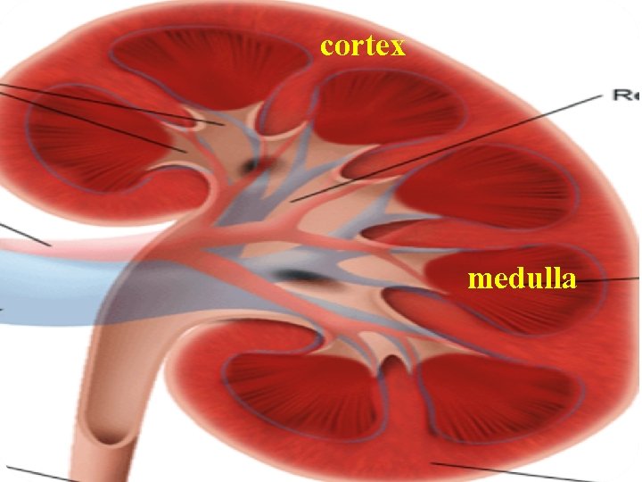
cortex medulla Prepared by : Amal Awad Al-Harbi
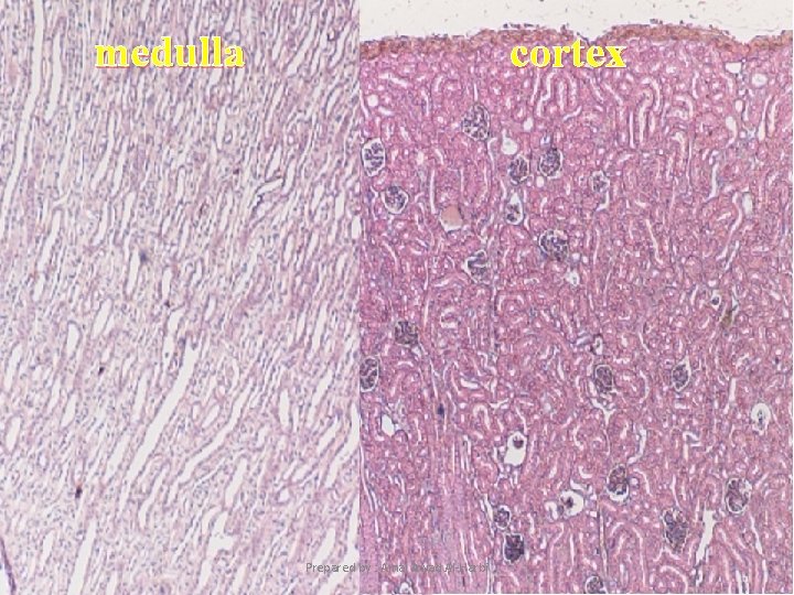
medulla cortex Prepared by : Amal Awad Al-Harbi
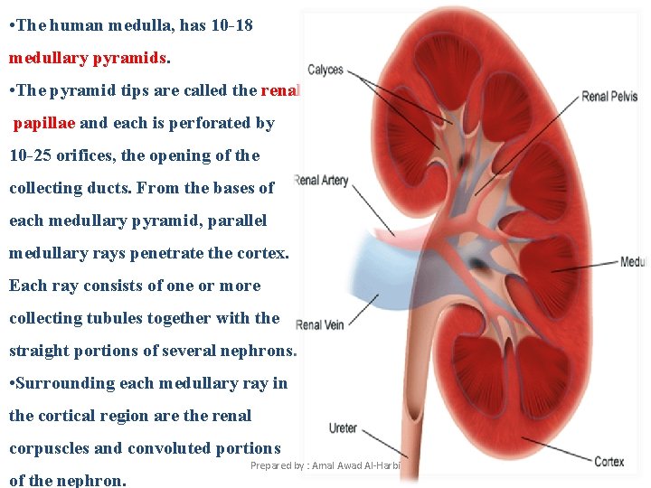
• The human medulla, has 10 -18 medullary pyramids. • The pyramid tips are called the renal papillae and each is perforated by 10 -25 orifices, the opening of the collecting ducts. From the bases of each medullary pyramid, parallel medullary rays penetrate the cortex. Each ray consists of one or more collecting tubules together with the straight portions of several nephrons. • Surrounding each medullary ray in the cortical region are the renal corpuscles and convoluted portions of the nephron. Prepared by : Amal Awad Al-Harbi
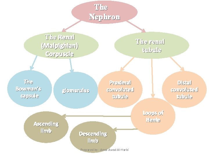
The Nephron The Renal (Malpighian) Corpuscle The Bowman’s capsule Ascending limb The renal tubule glomerulus Proximal convoluted tubule Distal convoluted tubule Loops of Henle Descending limb Prepared by : Amal Awad Al-Harbi
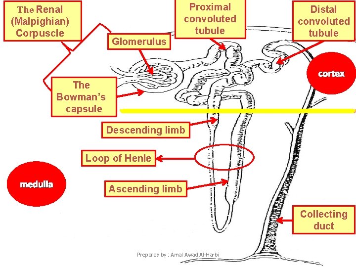
Proximal convoluted tubule The Renal (Malpighian) Corpuscle Glomerulus Distal convoluted tubule cortex The Bowman’s capsule Descending limb Loop of Henle medulla Ascending limb Collecting duct Prepared by : Amal Awad Al-Harbi
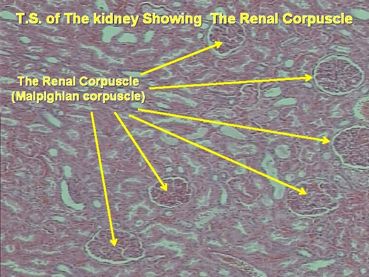
T. S. of The kidney Showing The Renal Corpuscle (Malpighian corpuscle) Prepared by : Amal Awad Al-Harbi
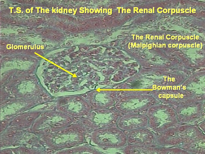
T. S. of The kidney Showing The Renal Corpuscle (Malpighian corpuscle) Glomerulus The Bowman’s capsule Prepared by : Amal Awad Al-Harbi
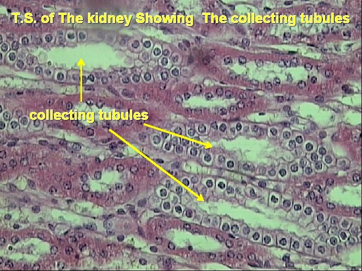
T. S. of The kidney Showing The collecting tubules Prepared by : Amal Awad Al-Harbi

Prepared by : Amal Awad Al-Harbi
- Slides: 24