The biochemistry of connective tissues Connective tissue CT

The biochemistry of connective tissues
 is a kind of biological tissue that supports, connects, or separates")
Connective tissue (CT) is a kind of biological tissue that supports, connects, or separates different types of tissues and organs of the body. All CT has three main components: cells, fibers, and extracellular matrix, all immersed in the body fluids. -The extracellular portions are composed of fibers imbedded in an amorphous ground substance. -Cells are spread through an extracellular fluid. -Ground substance - A clear, colorless, and viscous fluid containing glycosaminoglycans (are long unbranched polysaccharides consisting of a repeating disaccharide unit) and proteoglycans to fix the body water and the collagen fibers in the intercellular spaces. Ground substance slows the spread of pathogens. -The fibers of connective tissue are probably of two chemical types, collagenous and elastic.

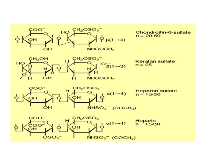
Proteoglycans • Proteoglycans are proteins modified by glycosaminoglycans. • Glycosaminoglycans are long-chain compounds made up of hundreds repeating disaccharide units. One of the sugars in each disaccharide unit is a hexosamine (glucosamine). • Many proteoglycans contain a core protein which links them to the cellular membrane.

Connective tissue can be subdivided into: 1 - connective tissue proper 2 - special connective tissue 3 - less classifiable types of connective tissues. -Connective tissue proper consists of loose connective tissue and dense connective tissue (dense regular and dense irregular connective tissues. ) -Special connective tissue consists of reticular connective tissue, adipose tissue, cartilage, bone, and blood. -Other kinds of connective tissues include fibrous, elastic, and lymphoid connective tissues. Functions of connective tissue • • • Transport Immunological defense Mechanical support Growth and repair Hematopoiesis

Collagen is the major structural protein of vertebrate extracellular matrix. Structure of collagen Collagen is made up of three polypeptides (referred to as "α-chains") are twisted around one another (tropocollagen) in a rope-like triple-helix and are held together by hydrogen bonds. Collagen is formed from tropocollagen subunits. The triple helix in tropocollagen is highly extended and strong. Features: (1) Three separate polypeptide chains (2) 3. 3 residues per turn (3) Each chain forms hydrogen bonds with the other two
The fibers have diameter between 80 to 160")
Types of collagen Collagen type I i)The fibers have diameter between 80 to 160 nm. ii)Found in bone, dentin, skin, tendon, muscles and walls of blood vessels. Collagen type II i)have a diameter <80 nm ii)found in invertiberal discs and hyaline cartilage. Collagen type III i)Found in spleen, muscle, and aorta. Collagen type IV Found around different types in the basement membranes and muscles. Collagen type V It is found in embryonic cell cultures and the basement membranes. Collagen type VI It is found in muscle and skin.

Collagen Amino Acid Composition • Nearly one residue out of three is Gly • Proline content is unusually high • Proline facilitates the formation of the helical conformation of each α-chain because its ring structure causes "kinks" in the peptide chain. • Many modified amino acids are present: – 4 -hydroxyproline – 3 -hydroxyproline – 5 -hydroxylysine The hierarchical design of collagen. The structural features of collagen ranges from the amino acid sequence, tropocollagenmolecules, collagen fibrils to collagen fibers.

Elastin is a protein in connective tissue that is elastic and allows many tissues in the body to resume their shape after stretching or contracting. ü Elastin serves an important function in arteries and is particularly abundant in large elastic blood vessels such as the aorta. Elastin is also very important in the lungs, elastic ligaments, the skin, the bladder, elastic cartilage. ü Elastin is primarily composed of the amino acids glycine, valine, alanine, and proline. ü Elastin polypeptide chains are crosslinked together to form rubberlike, elastic fibers. ü Each elastin molecule uncoils into a more extended conformation when the fiber is stretched and recoils spontaneously as soon as the stretching force is relaxed.

Metabolic muscle diseases

What Causes Metabolic Diseases? The metabolic diseases of the muscle results from problems that occur when certain fuel molecules are processed before they enter the mitochondria, or by the cell's inability to get fuel molecules into the mitochondria. Each of the metabolic disorders of the muscle are caused by different genetic defects that impair the body's ability to process chemical reactions that occur within cells during normal functioning. Under normal conditions, fuel molecules broken down from the food, and then are broken down further inside of each cell before they can be used by the mitochondria of the cell to produce ATP drives all muscle activity in the body, and when ATP levels are low, muscle weakness, pain or cramps may occur.
")
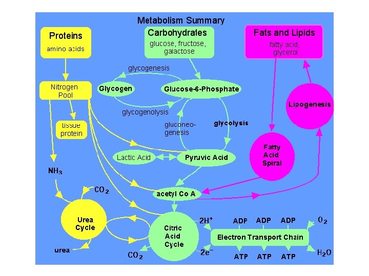
-In normal metabolism, food provides fuel that's processed inside the cells, producing energy (ATP) for muscle contraction and other cellular functions. -In metabolic myopathies, missing enzymes prevent mitochondria from properly processing fuel, and no energy is produced for muscle function.

Glycolysis
; also called glycogen storage disease type II Acid Maltase Deficiency")
Acid Maltase Deficiency (AMD); also called glycogen storage disease type II Acid Maltase Deficiency is a rare, autosomal recessive metabolic disorder that is a result of a lack in the enzyme acid maltase, which is necessary for the break down of glycogen. Inheritance In is an autosomal recessive metabolic disorder, meaning that two copies of the mutated gene (one from each parent) must be inherited to have this condition. Cause The disease is caused by a mutation in a gene (acid alpha-glucosidase: also known as acid maltase) on long arm of chromosome 17 at 17 q 25. 2 -q 25. 3 Description, Signs and Symptoms The excessive accumulation of glycogen due to this condition results in progressive muscle weakness (myopathy) all throughout the body and affecting major body tissues in the heart, skeletal muscles, liver and nervous system.

Carnitine deficiency is an inborn error of fatty acid transport caused by a defect in the transporter responsible for moving carnitine across the plasma membrane Carnitine is involved in the oxidation of fatty acids Genetics is an autosomal recessive condition. The gene responsible for the OCTN 2 carnitine transporter is SLC 22 A 5, located at 5 q 31. 1 -32. Patients who present clinically with this would fall into two categories: 1 -metabolic presentation with hypoglycemia 2 - cardiac presentation characterized by cardiomyopathy. Muscle weakness can be found with either presentation.
 is a mitochondrial")
Carnitine palmitoyl transferase deficiency Carnitine palmitoyl transferase II precursor (CPT 2) is a mitochondrial membrane protein which is transported to the mitochondrial inner membrane. CPT 2 together with carnitine palmitoyltransferase I oxidizes long-chain fatty acids in the mitochondria. Carnitine palmitoyl transferase II deficiency (CPT-II) is a metabolic disorder characterized by an enzymatic defect that prevents long-chain fatty acids from being transported into the mitochondria for utilization as an energy source. Inheritance CPT II deficiency has an autosomal recessive pattern of inheritance.

Debrancher enzyme deficiency This disease is a metabolic muscle disorder, a group of diseases that interferes with the processing of food (in this case, carbohydrates) for energy production. Genetics The disorder is caused by a defect in the debrancher enzyme gene, which interferes with the breakdown of glycogen in the muscles and liver. This disease principally affects the liver. It causes swelling of the liver, slowing of growth, low blood sugar level.

Lactate dehydrogenase deficiency This disease is a metabolic muscle disorder, a group of diseases that interferes with the processing of food (in this case, carbohydrates) for energy production. What causes lactate dehydrogenase deficiency? The disease is caused by a genetic defect in the lactate dehydrogenase enzyme, which normally recycles byproducts of carbohydrate metabolism

Myoadenylate deaminase deficiency is a metabolic muscle disease that interferes with the muscle cell's processing of adenosine triphosphate (ATP), the major energy molecule of the cell. Symptoms of myoadenylate deaminase deficiency The disease may cause exercise intolerance, cramps and muscle pain; although, in many cases, people with deficiencies in this enzyme may experience no symptoms. What causes myoadenylate deaminase deficiency? Myoadenylate deaminase deficiency is caused by a genetic defect in the myoadenylate deaminase enzyme, which affects the cell's ability to process ATP
 This disease is one of a group of metabolic muscle")
Phosphofructokinase deficiency (Tarui disease) This disease is one of a group of metabolic muscle disorders that interferes with the processing of food (in this case, carbohydrates) for energy production. What causes phosphofructokinase deficiency? Phosphofructokinase deficiency is caused by a genetic defect in the phosphofructokinase enzyme, which affects the breakdown of glucose

Phosphoglycerate kinase deficiency is one of a group of metabolic muscle diseases that interferes with the processing of food (in this case, carbohydrates) for energy production. What causes phosphoglycerate kinase deficiency? The condition is caused by a genetic defect in the phosphoglycerate kinase enzyme, which normally breaks down glucose for energy production
- Slides: 23