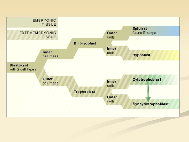
The bilaminar embryo Formation of ectoderm and endoderm

The bilaminar embryo. Formation of ectoderm and endoderm. Mark Kozsurek, M. D. , Ph. D. mark@kozsurek. hu ED I. , 10/11/2018

N O TI A L B A L U ST Note, that implantation runs simultaneously with the formation of girm layers!

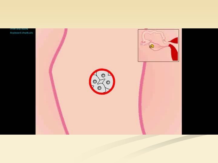
Apposition first and adhesion somewhat later is mediated by numerous receptor-ligand interactions. Among others this is responsible for the differentiation of trophoblasts blastocoel ENDOMETRIUM (secretory phase - high progesteron, low estrogen) embryoblasts (inner cell mass) BLASTOCYST (day 5, 200 -250 cells) embryoblasts (30 -34 cells) blastocoel trophoblasts (170 -220 cells) (blastocystic cavity) The blastocoel fluid cavity contains amino acids, growth factors, and other molecules necessary for cellular differentiation

ENDOMETRIUM Syncytiotrophoblast destroys the endometrium locally and errode the endometrial vessels using digestive enzymes. Lacunae filled with maternal blood appear within the syncytiotrophoblast. epiblast hypoblast

EMBRYOBLAST amnion cavity amnioblast epiblast hypoblast Cavity forms within the inner cell mass and is bounded by amnioblasts and epiblasts.

Dividing hypoblasts emigrate and attach to the interior of trophoblasts. amnion cavity
 yolk sac is formed. amnion cavity primitive yolk sac")
Primitive (or primary) yolk sac is formed. amnion cavity primitive yolk sac

Former hypoblasts giving the wall of the primitive yolk sac create a new layer: the extraembryonic mesoderm. amnion cavity extraembryonic mesoderm primitive yolk sac

In the loose tissue ot extraembryonic mesoderm cavities appear which later unite. amnion cavity primitive yolk sac extraembryonic mesoderm

Extraembryonic mesoderm is condensed and persist on the internal surface of trophoblasts and encloses the amnionic cavity (somatic layer) as well as the yolk sac (splanchnic layer). somatic layer connecting stalk amnion cavity primitive yolk sac extraembryonic coelom splanchnic layer

Formation of secondary yolk sac has started. amnion cavity secondary yolk sac

Primitive yolk sac is replaced by the secondary one and the exocoelomic cyst. amnion cavity secondary yolk sac exocoelomic cyst

Second week of pregnancy: „the week of twos” 2 nd week Trophoblast divide into syncytiotrophoblast and cytotrophoblasts. Embryonic disk is formed by two layers (epiblasts, hypoblasts). Two cavities exist on the two sides of the embryonic disk: the amnionic cavity and the yolk sac.

Importance and fate of the amnion and the yolk sac 2 nd week 4 th week During its growth embryo moves into the amnionic cavity and the amnion (blue) fuses with the chorion made of the extraembryonic mesoderm (red) and the trophoblast (black). Amnionic epithelium covers the umbilical cord and the fetal surface of the placenta.

amniotic epithelium chorionic plate
 in umbilical")
• • • Amniotic epithelium, mesenchymal cells Endothelium (simple squamous epithelium) in umbilical vessels RBCs, lymphocytes and neutrophils persisting in vessels Smooth muscle cells in the wall of arteries Simple cuboidal epithelium lining the allantois

Importance and fate of the amnion and the yolk sac 2 nd week 4 th week During its growth embryo moves into the amnionic cavity and the amnion (blue) fuses with the chorion made of the extraembryonic mesoderm (red) and the trophoblast (black). Amnionic epithelium covers the umbilical cord and the fetal surface of the placenta. Amnionic fluid contains exfoliated epithelial cells of the fetal skin, so if sample is taken from the amniotic fluid, DNA analysis of these cells might be performed.

Amniocentesis 15 -17 th week

The yolk sac is a membranous sac formed by hypoblasts on the ventral surface of the embryo. Nutrients stored in the yolk sac are utilized by the vitelline circulation, the most primitive circulatory system of the embryo. During folding of the embryo the midgut is getting separated from the remnant of the yolk sac, but a connection called omphalomesenteric (or vitelline) duct persists for a while. Blood formation is first observable in the extraembryonic mesoderm attached onto the external surface of the yolk sac and persists until the 6 th week, when liver becomes the principal site of hemopoiesis. Furthermore, primordial germ cells that later immigrate into the sex glands appear first in the wall of the yolk sac, too.

Further proliferation of epiblast is observable: on one end of the embryonic disk epiblasts proliferate quickly, daughter cells migrate toward the midline resulting in the appearance of the primitive streak and primitive node.
 primitive streak What happens to the sinking epiblasts?")
primitive node primitive pit (Hensen’s node) primitive streak What happens to the sinking epiblasts?

Junctions among epiblasts become looser and due to the collision at the primitive node and primitive streak epiblasts sink below their original layer. (1) Some of them replace the hypoblasts and differentiate into the ultimate endoderm, while others, the (2) mesoblasts, form a third, middle layer, the intraembryonic mesoderm. Epiblast remaining in their original layer (3) will form the ectoderm.

ectoderm epiblasts primitive streak hypoblasts wall of yolk sac intraembryonic mesoderm ? In fact all the tissues and cells of the human are derived from the epiblasts! endoderm extraembryonic mesoderm

Proliferation of the epiblasts in the anterolateral area slows down. The primitive node with the primitive pit moves caudally, the notochord elongates simultaneously, while the primitive streak is getting shorter and shorter. Finally, the primitive nod, pit and streak disappear. If the process is not arrested properly, saccrococcygeal terratoma may develop. In teratomas usually derivatives of all the germ layers are represented, they usually contain skin, hair, cartilage, bone, fat (sebum) and sometimes tooth or teeth! Prognosis is quite good!

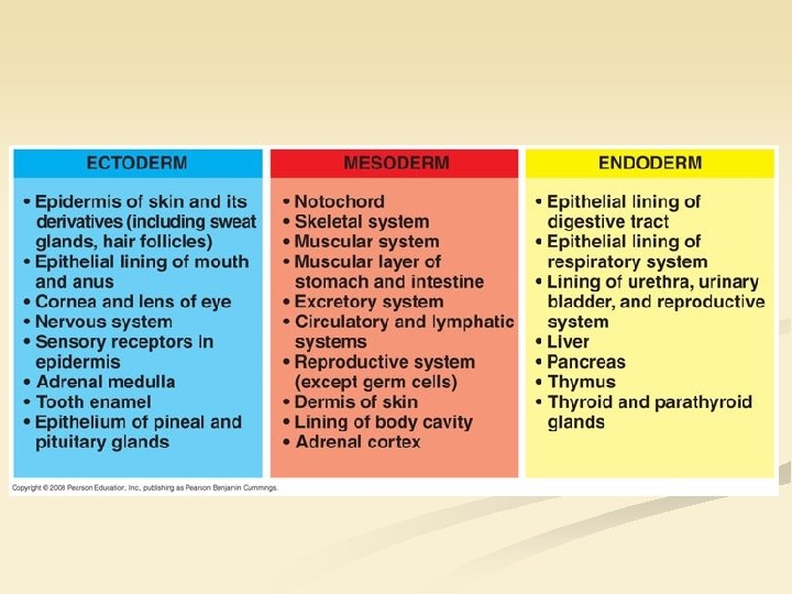
ECTODERM Ectoderm will be formed by those epiblasts, which persist in their original layer. neural plate neural crest placodes 1. Neuroectoderm neurons and glial cells of the peripheral and central nervous system 2. Surface ectoderm surface and glandular epithelium of the skin (but not the connective tissue!)
")
AZAN: reddish structures – ectodermal, bluish – mesodermal (with some simplification)

ENDODERM Epiblasts derived from the primitive streak and replacing original hypoblasts at the roof of the yolk sac form the endoderm. Epithelium (surface and glandular) of the GI tract and the airways, the urinary bladder

A brief summary


Thank you for your attention!
- Slides: 33