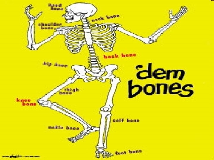
The Axial Skeleton forms the longitudinal axis of


 – Pectoral girdle")
 Girdle is composed of two bones, which allow the upper limb")


 – Carpals—wrist – Metacarpals—palm")

- Slides: 22
The Axial Skeleton forms the longitudinal axis of the body. It is divided into three parts – Skull – Vertebral column – Bony thorax
The skull consists of two sets of bones, which are joined by sutures. Only the mandible is attached by a freely movable joint. – Cranium (eight large flat bones) • Frontal, Parietal(2), Temporal(2), Occipital, Sphenoid, Ethmoid – Facial bones (all paired except mandible and vomer) • Paired Maxilla, Palatine, Zygomatic, Lacrimal, Nasal, and Inferior Nasal Conchae and Single Mandible and Vomer(plow)
The Fetal Skull • The fetal skull is large compared to the infant’s total body length • Fontanels—fibrous membranes connecting the cranial bones – Allow the brain to grow – Convert to bone within 24 months after birth
Skeletal Changes Throughout Life Figure 5. 33 a
Paranasal Sinuses • The Paranasal Sinuses are hollow portions of bones surrounding the nasal cavity • Functions of paranasal sinuses – Lighten the skull – Give resonance and amplification to voice
The Hyoid bone is the only bone that does not articulate with another bone-it is held into place by thyroid ligaments. • Serves as a moveable base for the tongue • Aids in swallowing and speech Notice the bruising around the neck area. The appearance of finger indentations indicate manual strangulation. The impression was confirmed by finding
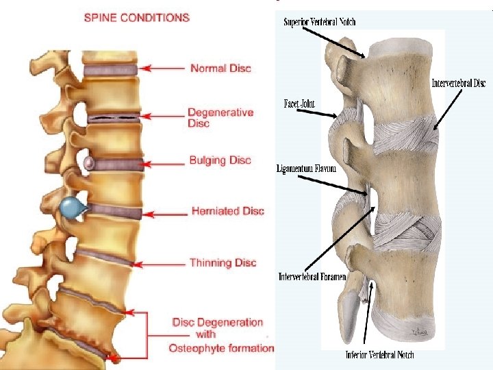
The Vertebral Column • Each vertebrae is given a name according to its location – There are 24 single vertebral bones separated by intervertebral discs • Seven cervical vertebrae are in the neck (C 1 -C 7) • Twelve thoracic vertebrae are in the chest region (T 1 -T 12) • Five lumbar vertebrae are associated with the lower back (L 1 -L 5)
The Vertebral Column • The remaining nine vertebrae fuse to form two composite bones – Sacrum – Coccyx (tailbone)
Sacrum and Coccyx • Sacrum – Formed by the fusion of five vertebrae • Coccyx – Formed from the fusion of three to five vertebrae – “Tailbone, ” or remnant of a tail that other vertebrates have
• The spine has a normal curvature – Primary curvatures are the spinal curvatures of the thoracic and sacral regions • Present from birth – Secondary curvatures are the spinal curvatures of the cervical and lumbar regions • Develop after birth (cervical with head raising and lumbar with walking)
Abnormal Spinal Curvatures of the Vertebral Column Scoliosis- “crooked”-side to side curvature-causes multifactorial Kyphosis-”hunchback” causes include trauma “slouching”, osteoporosis with compression fractures. Lordosis “swayback” an inward curvature of the vertebrae usually caused by differing anterior and posterior disc thickness
The Bony Thorax forms a cage to protect major organs. It consists of three parts – Sternum – Ribs • True ribs (pairs 1– 7 attached to sternum) • False ribs (pairs 8– 12) • Floating ribs (pairs 11– 12) – Thoracic vertebrae
The Appendicular Skeleton is composed of 126 bones – Limbs (appendages) – Pectoral girdle – Pelvic girdle
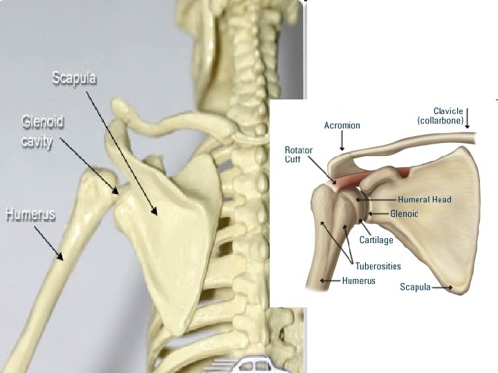
The Pectoral (shoulder) Girdle is composed of two bones, which allow the upper limb to have exceptionally free movement – Clavicle—collarbone (acts as a brace to hold arm away from thorax and prevents shoulder dislocation) – Scapula—shoulder blade (provides exceptional range of motion for the arm)
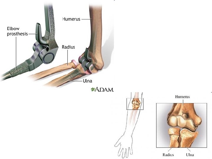
Bones of the Upper Limbs • Humerus – Forms the upper arm – Single bone
Bones of the Upper Limbs • The forearm has two bones – Ulna • Medial bone in anatomical position (pinky side) – Radius • Lateral bone in anatomical position (thumb side)
Bones of the Upper Limbs • The hand (27 bones) – Carpals—wrist – Metacarpals—palm – Phalanges—fingers
http: //www. anatomyarcade. com/ games/games. Skeletal. html