The Avian Egg Structure Production Function By Akrum

The Avian Egg Structure, Production, Function By Akrum Hamdy

Topics • Anatomy of the Egg • Anatomy of Avian Female Reproductive Tract • Process of Egg Formation • Aspects of Incubation and Hatching • Dystocias

Egg Types • Eight Basic Shapes • Egg sizes range from 10 mm to 145 mm in length

Egg Colors • Multitude of colors, but all formed from two pigments derived from porphyrins • Color and mottling serves to camouflage the eggs in the nest • Cavity nesting birds have colorless eggs • Colors are added in the uterus during shell formation (below)

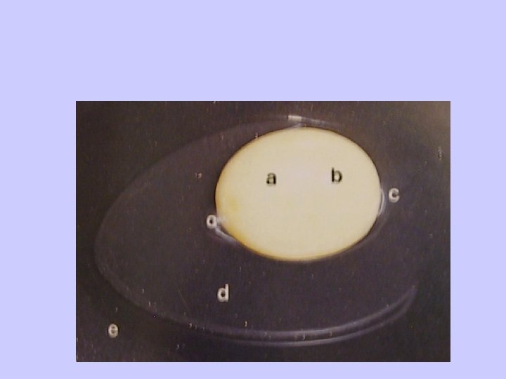
Anatomy of the Egg Seven Components • • • Yolk Albumin Membranes Chelazia Chorioallantoic Membrane • Air Cell • Shell

Anatomy of the Egg Seven Components • • • Yolk Albumin Membranes Chelazia Chorioallantoic Membrane • Air Cell • Shell

Yolk • Formed in the liver, transported to ovarian follicle • 33% lipid • 19% protein • 48% water • Layed in concentric layers

Albumin • Four distinct layers – Chalaziferous - inner thick – Inner thin layer – Outer thick layer – Outer thin layer • Protects yolk from invasion by microorganisms and provides water, protein and minerals to the embryo
: source of amino acids • Ovotransferrin (13%):")
Proteins found in Albumin • Ovalbumin (54%): source of amino acids • Ovotransferrin (13%): iron chelator -prevents bacterial growth • Ovomucoid, ovoglobulins, avidin comprise the remainder – Avidin is a biotin inhibitor - reduces bacterial growth

Chelaziae • Twisted fiber-like structures at each pole • Hold yolk in place inside the egg – permit limited rotation – inhibit lateral displacement

Shell Membranes – Inner and Outer Membranes – Envelope yolk and albumin – Contiguous with each other except at one end where they separate to form air cell – Mammilary cores are embedded in the outer membrane -- initial site of calcification

Shell Matrix • Organic mucopolysaccharide matrix • Becomes calcified to form hard outer shell • Hens egg weighs 50 - 60 grams at time of lay • 2. 5 grams of calcium in the egg • A hen laying 280 eggs per year transports 30 times the calcium content of her entire body for shell formation • Cuticle: waxy layer with pigments

Cross-section of Egg Shell • Membranes • Pores • Gas Exchange – Oxygen – Carbon Dioxide – 15% weight loss during incubation – Related to incubation time (see tables)

Female Reproductive Tract • Ovary • Infundibulum - site of fertilization • Magnum - albumin addition • Isthmus - membranes • Uterus - shell gland • Vagina - transport to exterior • Sperm storage occurs at various sites in tract in some species

Female Reproductive Tract • Ovary • Infundibulum - site of fertilization • Magnum - albumin addition • Isthmus - membranes • Uterus - shell gland • Vagina - transport to exterior • Sperm storage occurs at various sites in tract in some species

Cloacal Structure Oviduct

Incubation Issues • Temperature – Regulates rate of development – Tolerance for lowered temperature decreases as embryo grows – Less tolerance for increase temps: 46 -47 C is lethal for more than 60 -90 min depending on stage of embryo development • Humidity: regulates water loss – Determined by internal egg temp (humidity is 100%) and ambient humidity and gradient between the two • Turning

Incubation Times

Humidity and Shell Conductance

Incubation Issues • Turning – Most critical from day 3 to day 7 – Required for: • Proper incorporation of albumin into amnion • Failure to incorporate leads to water loss from albumin, increased viscosity and setttling between chorioallantoic membrane and inner shell membrane • This results in decreased oxygen diffusion – Ideal turning rate: • Minimum of 3 x/d • More than 24 x/d is not necessary

Managing Water Loss by the 14 16 % Principle • Weigh egg at time of lay • Calculate projected weight at hatch by subtracting 15 -18% of weight • Plot laid weight on day 1; and pip weight at appropriate point for incubation time (e. g. 21 days) • Connect with a straight line • Weigh eggs periodically during incubation

Altering Weight Loss • Too much loss: – Place in incubator with higher humidity – Cover part of the egg with white glue • Too little loss: – Place in incubator with lower humidity – Thin part of the egg-shell by sandpapering

Appearance of Fertile Egg at Lay and during Early Development

Stages of Embryonic Development

Stages of Embryonic Development

Assessing Eggs during Incubation: Candling • Blood Vessels of Chorioallantoic layer • • Position

Assessing Eggs during Incubation: Candling • Blood Vessels of Chorioallantoic layer • Embryonic Position and Condition

Assessing Eggs during Incubation: Radiology • Embryonic Position and Condition Note head down position

Hatching Process • Membrane Drawdown - due to water-loss • Air Cell Formation - initiation of airbreathing by developing chick • Pipping • Rotation - counterclock-wise • Assisted Hatching - do not initiate until chorioallantoic blood vessels shut down.

Appearance of Healthy Chick at Hatch • Color • Hydration Status

Malpositions and Hatching Problems • Malpositions 1 - 6 – Head at small end of egg is most common (mp 2) - reduced hatchability – Head under left wing (mp 3) - lethal • Oversize Embryos • Unretracted Yolk Sacs

Malpositions and Hatching Problems • Unretracted Yolk Sacs • Idiopathic

Malpositions and Hatching Problems • Malpositions 1 - 6 – Head at small end of egg is most common (mp 2) - reduced hatchability – Head under left wing (mp 3) - lethal

Malpositions and Hatching Problems • Oversized Embryos
 and Other Problems Internal Laying")
Dystocias (Egg-Binding) and Other Problems Internal Laying

Radiographic Appearance of Egg -bound Cockatiel

Management of Dystocias • • • Correct dehydration Provide Warmth Correct Calcium Depletion Prostaglandin application to cloaca Forced expulsion/removal

Management of Dystocias Lubricate and Apply Pressure Crush and remove fragments

Management of Dystocias

Management of Dystocias Impaction in Oviduct

Management of Chronic Egg-Laying • Photoperiod Control • Removal of stimuli • Chemical – Lupron – HCG • Surgical

Summary • Egg is self-contained external development encasement for embryo • Formation and laying is a significant physiological and metabolic factor for the hen • Embryonic development can be monitored through the egg shell • There are six classical malpositions

Summary • Hatching can be assisted after draw-down of chorioallantoic membranes • Dystocias can be treated through a variety of removal techniques • Chronic egg-laying is a significant problem for owners of some birds; management methods vary.
- Slides: 44