THE APPENDICULAR SKELETON The Appendicular Skeleton Composed of

 �Pectoral girdle �Pelvic girdle")

 Girdle � Composed of two bones �Clavicle — collarbone �Scapula —")


 � 2")


 � Patella (2) � Tibia (2) � Fibula")


- Slides: 31
THE APPENDICULAR SKELETON
The Appendicular Skeleton � Composed of 126 bones �Limbs (appendages) �Pectoral girdle �Pelvic girdle
The Appendicular Skeleton Figure 5. 6 a
The Pectoral (Shoulder) Girdle � Composed of two bones �Clavicle — collarbone �Scapula — shoulder blade � These bones allow the upper limb to have exceptionally free movement
Bones of the Shoulder Girdle Figure 5. 21 c–d
Clavicles � � � Slender, rodlike bones with elongated S shapes Located at base of the neck and run horizontally between the sternum and the shoulders Sternal ends – articulate with the manubrium Acromial ends – articulate with the scapulae Brace the scapulae, holding the shoulders in place Structurally weak
Scapulae � � � Broad, triangular bones located on either side of the upper back Spine – divides posterior surface 2 processes at the head: � Acromion process – forms tip of the shoulder and articulates with the clavicle � Coracoid process – curves anteriorly and inferiorly to the clavicle � � Glenoid cavity – between the acromion and coracoid processes; articulates with the head of the humerus Suprascapular notch – passage way for nerves
Upper Limb Bones � � � Bones form the framework of the arm, forearm, and hand Bones function as levers for muscle contraction Includes: � Humerus (2) � Radius (2) � Ulna (2) � Carpals (16) � Metacarpals (10) � Phalanges (28)
Humerus � � � Long bone that extends from scapula to the elbow Head fits into glenoid cavity of scapula Greater tubercle – on lateral side Lesser tubercle – on anterior side Surgical neck – tapering region below head and tubercles (common fracture site) Deltoid tuberosity – rough area near the middle of the shaft on the lateral side � attachment site for the deltoid muscle
Humerus Bone Features continued… � � � Coronoid fossa – process where the elbow bends: receives the ulna Capitulum – articulates with the radius Olecranon fossa – on posterior surface, receives the olecranon process of the ulna when the elbow straightens Trochlea – articulates with the ulna Epicondyles – attachments for elbow muscles and ligaments
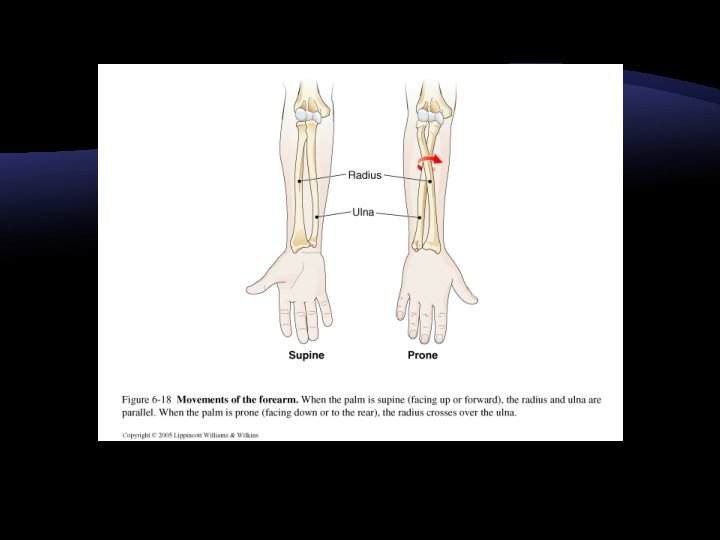
Radius � � � On thumb side of forearm Shorter than the ulna Extends from the elbow to the wrist and crosses over the ulna when hand is turned over at the wrist Radial tuberosity – process just below the head; attachment for the biceps Styloid process – attachment for wrist ligaments at the distal end
Ulna � � � Longer than the radius Trochlear notch – at proximal end, wrench-like opening that articulates with the trochlea of the humerus Olecranon process – above the trochlear notch; attachment for triceps that straightens the upper limb at the elbow; fits into olecranon fossa Coronoid process – below trochlear notch, fits into coronoid fossa when elbow bends Styloid process – at distal end provides attachment for wrist ligaments
Wrist � � � Wrist consists of carpals bound in 2 rows of 4 bones each Articulate with radius and ulna proximally and metacarpals distally Carpal bones are: � Pisiform � Triquetrum � Lunate � Scaphoid � Hamate � Capitate � Trapezoid � Trapezium � She Left Town. Please Take The Cat Home
Metacarpals � � � Form the palm of the hand 5 per hand Long bones with rounded distal ends (knuckles) Articulate with carpals and phalanges Lateral metacarpal is the most freely moveable Numbered 1 -5, starting at the thumb
Phalanges � Finger bones � 3 per finger (proximal, middle, and distal) � 2 in thumb – no middle phalanx
Bones of the Pelvic Girdle � The total weight of the upper body rests on the pelvis � It protects several organs �Reproductive organs �Urinary bladder �Part of the large intestine
Coxal Bones � Each coxa develops from 3 parts: � Ilium � Ishium � Pubis � Acetabulum – cup-shaped cavity where the 3 parts of coxa fuse, making the socket for the femur
Ilium � � � Largest and most superior portion of the coxa Flares outward and forms the prominence of the hip Iliac crest – margin of the ilium Sacroiliac joint – where ilium and sacrum join Anterior superior iliac spine – found lateral to the groin, provides attachments for ligaments and muscles Posterior superior iliac spine – on posterior border
Ischium � � Forms lowest portion of the coxa L-shaped Ischial tuberosity – rough surface that points down and back; supports body weight when sitting Ischial spine – sharp projection above ischial tuberosity, near the junction between the ilium and the ischium � Creates the narrowest part of the pelvis
Pubis � � Anterior portion of coxa Pubic symphysis – fibrocartilage joint between the 2 pubic bones Pubic arch – angle between pubic bones Obturator foramen – largest opening in the body � Formed between ischium and pubis � Covered and nearly closed by obturator membrane
Male vs. Female Pelvis � Male Pelvis: �Heavier bone �More evidence of muscle attachments � Female Pelvis �Iliac bones are more flared �Broader hips �Greater angle of pubic arch �Greater distance between ischial spines and tuberosities �Shorter, flatter sacral curvature �More delicate bones
Lower Limb Bones � Femur (2) � Patella (2) � Tibia (2) � Fibula (2) � Tarsals (7/foot) � Metatarsals (5/foot) � Phalanges (14/foot)
Total Knee Replacement Surgery
Femur Bone Features � � � Thigh bone Longest bone in body Extends from hip to knee Head of femur – large and rounded; projects medially into acetabulum of coxal bone Greater trochanter and lesser trochanter – attachments for muscles of buttocks and lower limbs Lateral and medial condyles – articulate with tibia
Patella � � � Articulates with the femur on distal anterior surface Kneecap Flat sesamoid bone located in a tendon that passes anteriorly over the knee
Tibia Bone Features � � � Shin bone Larger of 2 leg bones; located on the medial side Medial and lateral condyles – on proximal end, articulate with condyles of femur Tibial tuberosity – below condyles on anterior surface; attachment of patellar ligament Anterior crest – extends downward from tuberosity; Medial malleolus – inner ankle
Fibula Bone Features � Long, slender bone located on the lateral side of the tibia � Articulates with the tibia just below the lateral condyle � Lateral malleolus – distal end that forms the outer ankle
Bones of the Foot � � Tarsus – consists of 7 tarsal bones Talus – tarsal bone that can move freely where it joins the tibia and fibula � Forms the ankle � � Other tarsals are bound firmly together to support the talus Calcaneus – largest tarsal bone; heel bone � Located below the talus and projects backward � Helps support weight of the body Cute Tilley Never Could Cooperate Cuddle or Cuss
Metatarsals � � � Numbered 1 -5 beginning on the medial side Ball of the foot formed by the distal ends If tissues that bind the metatarsals weaken fallen arches (flat feet) occurs
Phalanges � Shorter, but otherwise similar to fingers � 3 bones per toe, except 2 in the great toe