The Ankle Joint PROF Dr ZUBAIRDr SOBIA KHALIL

The Ankle Joint PROF. Dr. ZUBAIR/Dr. SOBIA KHALIL

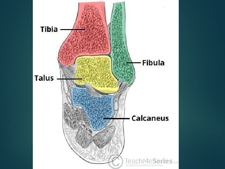
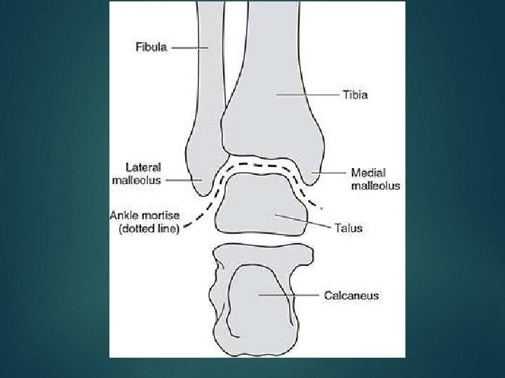
THE ANKLE JOINT • Hinge joint • Proximal articular outermost layer of the ankle joint is made by the articular facets of the: • – Lower end of tibia consisting of its medial malleolus. – Lateral malleolus. – Inferior transverse tibiofibular ligament. • These 3 together create a deep tibiofibular socket (also termed “tibiofibular mortise”). Distal articular outermost layer of the ankle joint is composed by: . – The articular facets on the upper, medial, and lateral aspects of the body of the talus
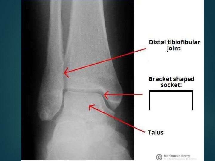
 Mortis formed by Tibia & Fibula")
Articulating Surfaces –Trochlea of Talus –Malleolar(lateral & medial) Mortis formed by Tibia & Fibula –Lateral & Medial Malleolus

Articulating Surfaces • The body of talus presents 3 articular surfaces: – Superior pulley-shaped articular surface (trochlear surface). • Articulate the inferior aspect of the lower end of tibia – Medial comma-shaped articular surface. • Articulates the lateral aspect of medial malleolus – Lateral triangular articular surface. • Articulates the medial aspect of lateral malleolus • The wedge shaped body of the talus fits into the socket above.

The socket –The socket is provided flexibility by • The tibiofibular ligaments, • The flexibility of the fibula, • Slight movements of the fibula at the superior tibiofibular joint

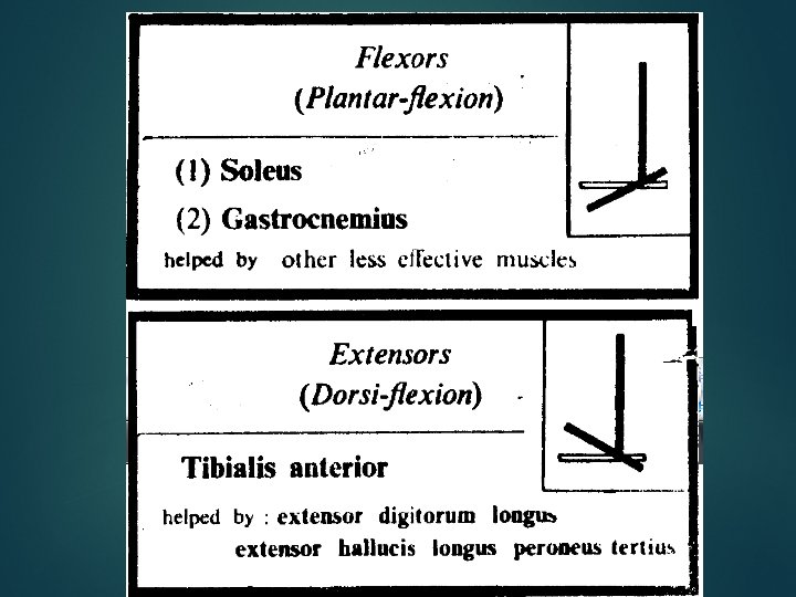
SOLIDITY OF THE ANKLE JOINT • The trochlear surface on the superior aspect of the body of talus is wider in front than behind. • During dorsiflexion, ankle joint of the anterior wider part of the trochlea moves posteriorly and fits correctly into the tibiofibular mortise, thus joint is stable. • During plantar flexion, the narrow posterior part of the trochlea doesn’t fit correctly in the tibiofibular mortise, thus the joint is unstable during plantar flexion.

VARIABLES KEEPING THE SOLIDITY OF ANKLE Close interlocking of articular surfaces. JOINT • • Strong, medial, and lateral collateral ligaments. • Deepening of tibiofibular socket posteriorly by the inferior transverse tibiofibular ligament. • Tendons (4 in front and 5 behind) crossing the ankle joint. • Other ligaments of the joint.

Stability of Ankle Joint • Ankle joint is a strong joint made stable by – Close interlocking of the articular surfaces – Strong collateral ligaments on the sides – The tendons that cross the joint (four in front, and five behind) • The depth of the superior articular socket is contributed to • The downward projection of the medial and lateral malleoli, • Transverse tibiofibular ligament.

Factors tend to displace the tibia and fibula forwards • Anatomically, two factors tend to displace the tibia and fibula forwards over the talus – The forward pull of tendons passes from the leg to the foot. – Pull of gravity. When standing line of gravity falls in front of ankle joint. • But following factors are responsible for prevention of displacement: – The talus is wedge shaped, being wider anteriorly. – The posterior border of the lower end of the tibia is prolonged downwards. – Ligamentous structures

LIGAMENTS • The essential ligaments of ankle joint are: 1. Capsular ligament. 2. Medial and lateral collateral ligaments.

JOINT CAPSULE • The articular capsule surrounds the joints, and is attached, • • – Above , to the borders of the articular surfaces of the tibia and malleoli; and – Below , to the talus around its upper articular surface. Anteriorly: – The joint capsule is a broad, thin, fibrous layer. Posteriorly: • – The fibres are thin and run mainly transversely blending with the transverse ligament. Laterally: – The capsule is thickened, and attaches to the hollow on the medial surface of the lateral malleolus. • The synovial membrane extends superiorly between Tibia & Fibula as far as the Interosseous Tibiofibular Ligament.

FIBROUS CAPSULE • The joint capsule is thin in front and behind to enable hinge movements and thick on either side where it combines with the collateral ligaments. • It encompasses the joint entirely. It is connected to the articular margins of the joint all around with two exceptions: – Posterosuperiorly it is connected to the inferior transverse tibiofibular ligament. – Anteroinferiorly it is connected to the dorsum of the neck of talus at some distance from the trochlear surface.

The synovial membrane • The synovial membrane lines the inner surface of the joint capsule, but ends at the periphery of the articular cartilages. –A small synovial process goes upward into the inferior tibiofibular syndesmosis.
 is attached to the medial")
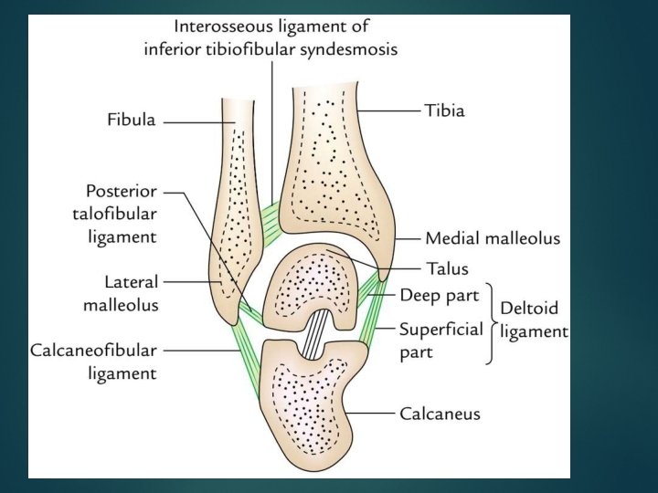
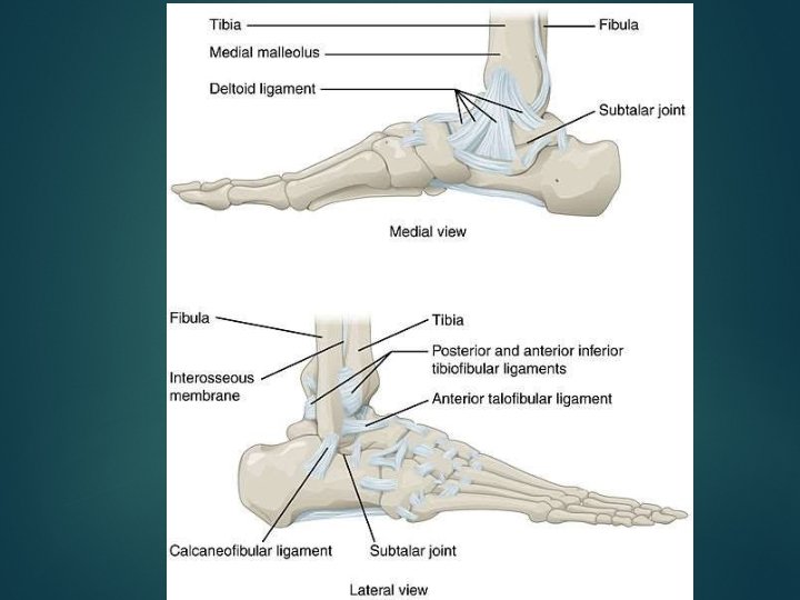
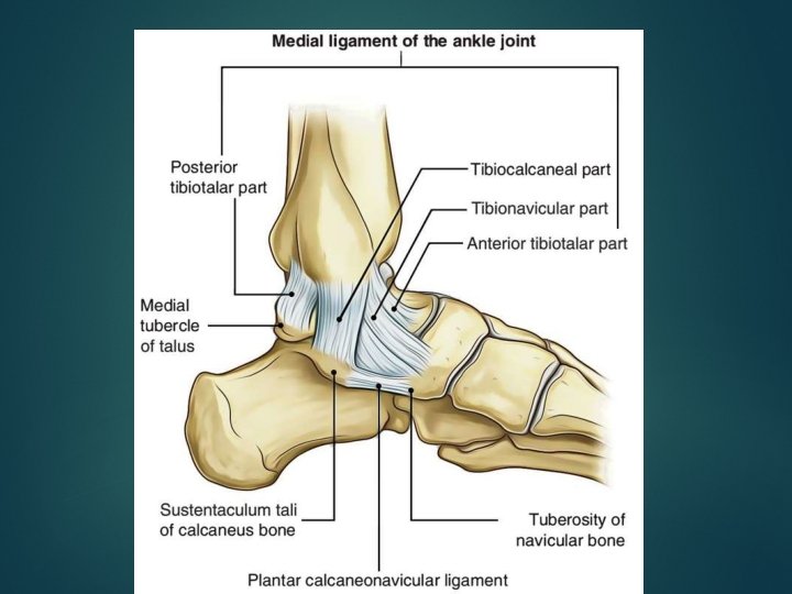
Medial Ligament • The medial ligament (or deltoid ligament) is attached to the medial malleolus (a bony prominence projecting from the medial aspect of the distal tibia). • It consists of an apex attached to tip and margins of medial malleolus and a base which fan out attaching to: (3 tarsal bones) – The talus, – The Calcaneus – The Navicular bones. • The primary action of the medial ligament is to resist over-eversion of the foot

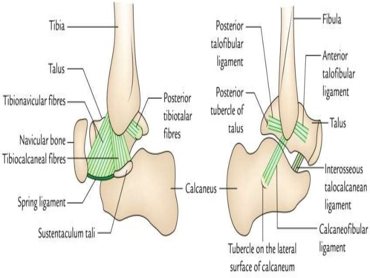
• The deltoid ligament is an extremely triangular ligament on the medial side of the ankle compensating the shortness of medial malleolus. • It splits into 2 parts: • Superficial and deep. – Above, both the parts have a common connection to the apex and margins of the medial malleolus. – Below, the connection of superficial and deep parts differs. • Superficial part: Its fibres are split into 3 parts: • Anterior : – Anterior fibres (tibionavicular) are connected to the tuberosity of navicular bone and the medial • Middle : – Middle fibres (tibiocalcanean) are connected to the entire length of sustentaculum tali. • Posterior: – Posterior fibres (posterior tibiotalar) to the medial tubercle and adjoining part of the medial surface of talu • Deep part (anterior tibiotalar) is connected to the s. anterior part of the medial surface of talus.

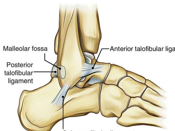
The lateral ligament • The lateral ligament is composed of 3 parts: • Anterior talofibular – (poor flat band) spans between the lateral malleolus and lateral aspect of the talus. • Posterior talofibular – (powerful band)spans between the lateral malleolus and the posterior aspect of the talus. • Calcaneofibular – (long rounded cord) spans between the lateral malleolus and the calcaneus • Superficially, the deltoid ligament is crossed by the tendons of tibialis posterior and flexor digitorum longus on the other hand lateral ligament is crossed superficially by the tendons of peroneus longus and brevis. • It resists over-inversion of the foot
 Flat Weak Band that extends Anteriomedially.")
LIGAMENT DESCRIPTION PROXIMAL ATTACHMENT Anterior Talofibular Ligament (ATFL) Flat Weak Band that extends Anteriomedially. Most commonly damaged ligament of the ankle. Lateral Malleolus DISTAL ATTACHMENT Neck of Talus ROLE • Restrain anterior displacement of the talus in respect to the fibula and tibia. • Resists Inversion in planterflexion. Posterior Talofibular Ligament (PTFL) Calcaneofibular Ligament (CFL) Thick, fairly strong band that runs horizontally medially. This ligament is under greater strain in full dorsiflexion of ankle. Rarely injured because bony stability protects ligaments when ankle in dorsiflexion. Malleolar Fossa of Fibula Round cord that passes posterioinferiorly Tip of Lateral Malleolus Lateral Tubercle of Talus Forms the back wall of the recipient socket for the talus' trochlea. Resists posterior displacement of the talus. Lateral Surface of Calcaneus • Aids Talofibular stability during Dorsiflexion. • Restrain inversion of the calcaneus with respect to the fibula. Prevent Talar tilt into Inversion.

LIGAMENTS DESCRIPTION Anterior Tibiotalar Ligament Posterior Tibiotalar Ligament PROXIMAL ATTACHMENT DISTAL ATTACHMENT ROLE Medial Malleolus Head of Talus Reinforces Ankle Joint. Control Plantarflexion & Eversion Talus Posteriorly Control Dorsiflexion Tibionavicular Ligament Forms most anterior part of the Deltoid Ligament Dorsomedial Aspect of Navicular Reinforces Ankle Joint Tibiocalcaneal Ligament Very thin ligament Sustentaculum Tali Reinforces Ankle Joint

Ankle‘Ring’ • The ankle joint and associated ligaments can be visualised as a ring in the coronal plane: • The upper part of the ring is formed by the articular surfaces of the tibia and fibula. • The lower part of the ring is formed by the subtalar joint (between the talus and the calcaneus). • The sides of the ring are formed by the medial and lateral ligaments. • A ring, when broken, usually breaks in two places (the best way of illustrating with is with a polo mint – it is very difficult to break one side without breaking the other).

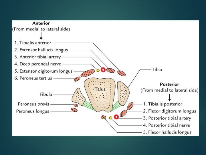
• RELATIONS OF ANKLE JOINT Anterior: Anteriorly from medial to lateral side the ankle joint is related to these structures: – – – Tibialis anterior. Extensor Hallucis longus. Anterior tibial Artery. Deep peroneal Nerve. Extensor Digitorum longus. Peroneus tertius. • Mnemonic: The Himalayas Are Not Dry Tablelands. • Posterior: Posteriorly from medial to lateral side the ankle joint is related to these structures: – – – Tibialis posterior. Flexion Digitorum longus. Posterio r tibial Artery. Posterio r tibial N erve. Flexor Hallucis longus. • Mnemonic: The Doctors Are Not Here.

POSTERIOR COMPARTMENT MUSCLE ACTION Gastrocnemius Plantarflexion when Knee Extended Flexion Knee Raises Heel during Walking PROXIMAL ATTACHMENT Lateral Head: Lateral Aspect of Lateral Femoral Condyle Medial Head: Popliteal Surface of Femur Superior to Medial Femoral Condyle Soleus Plantarflexion Steadies Leg on Foot Posterior Aspect of Head Fibula Superior ¼ Posterior Surface Tibia Soleal Line & Medial Border Tibia Plantaris Weakly Assists Gastrocnemius in Inferior end Lateral Supracondylar Line of Femur (superficial) DISTAL ATTACHMENT Posterior Surface Calcaneus via Calcaneal Tendon (Achilles Tendon) INNERVATION Tibial Nerve S 1 S 2
 MUSCLE ACTION PROXIMAL ATTACHMENT DISTAL ATTACHMENT INNERVATION Tibialis Posterior Plantarflexion Inversion")
POSTERIOR COMPARTMENT (DEEP) MUSCLE ACTION PROXIMAL ATTACHMENT DISTAL ATTACHMENT INNERVATION Tibialis Posterior Plantarflexion Inversion Supports Medial Longitudinal Arch Interosseous Membrane Posterior Surface Tibia inferior to Soleal Line Posterior Surface Fibula Navicular Tuberosity Cuneiform Cuboid Bases of Metatarsals 2 -4 Tibial Nerve L 4 -L 5 Flexor Digitorum Longus Plantarflexion Flexion Lateral Four Digits Supports Longitudinal Arch Medial Part Posterior Surface Tibia inferior to Soleal Line Broad Tendon to Fibula Base Distal Phalanges Digits 2 -4 Tibial Nerve Weak Plantarflexion Flexion Big Toe at all Joints Supports Medial Longitudinal Arch Inferior 2/3 Posterior Surface Fibula Inferior Part Interosseous Membrane Base Distal Phalanx of Big Toe Flexor Hallucis Longus S 2 -S 3

LATERAL COMPARTMENT MUSCLE ACTION Peroneus Brevis Weak Plantarflexion Eversion Peroneus Longus Weak Plantarflexion Eversion Supports Transverse Arch PROXIMAL ATTACHMENT DISTAL ATTACHMENT INNERVATION Inferior 2/3 of Lateral Surface Fibula Dorsal Surface Tuberosity of Base 5 th Metatarsal Superficial Peroneal Nerve Base 1 st Metatarsal L 5 - S 2 Head & Superior 2/3 of Lateral Surface Fibula Medial Cuniform (Superficial Fibular Nerve)

ANTERIOR COMPARTMENT MUSCLE ACTION PROXIMAL ATTACHMENT DISTAL ATTACHMENT INNERVATION Tibialis Anterior Dorsiflexion Inversion Supports Medial Longitudinal Arch Lateral Condyle Tibia Superior ½ Lateral Surface Tibia Interosseous Membrane Medial & Inferior Surfaces Medial Cuniform Base of 1 st Metatarsal Deep Peroneal Nerve (Deep Fibular Nerve) L 4 -L 5 Extensor Digitorum Longus Dorsiflexion Extends Lateral Four Digits Lateral Condyle Tibia Superior ¾ Anterior Surface Interosseous Membrane Middle & Distal Phalanges of Lateral Four Digits Deep Peroneal Nerve Extensor Hallucis Longus Dorsiflexion Extends Big Toe Middle Part Anterior Surface Fibula Interosseous Membrane Dorsal Aspect of Base Distal Phalanx of Big Toe Peroneus Tertius Dorsiflexion Aids Eversion Inferior 1/3 Anterior Surface Fibula Interosseous Membrane Dorsum Base 5 th Metatarsal (Deep Fibular Nerve) L 5 -S 1

ARTERIAL & NERVE SUPPLY • ARTERIAL SUPPLY • It is by the malleolar branches of anterior tibial, posterior tibial, and peroneal arteries. • NERVE SUPPLY • It is by the branches of deep peroneal and tibial nerves. (The segmental innervations is by L 4, L 5; S 1, S 2 spinal sections).

Ankle sprain • The excessive stretches and/or tearing of ligaments of the ankle joint is named the ankle sprain. The ankle sprains are normally caused by the falls from height or spins of ankle. • When the plantar-flexed foot is excessively inverted, the anterior and posterior talofibular and calcaneofibular ligaments are stretched and torn. The anterior talofibular ligament is most commonly torn. • When the plantar-flexed foot is excessively everted, the deltoid ligament isn’t torn; instead there’s an avulsion fracture of medial malleolus. • The inversion sprains are much more common than eversion sprains.

DISLOCATION OF THE ANKLE • The dislocations of ankle joint are uncommon because it is a quite stable joint because of tibiofibular mortise. • Nevertheless, whenever dislocation takes place it is constantly escorted by the fracture of one of the malleoli

POTT’S FRACTURE • It takes place when the foot is caught in the rabbit hole and everted forcibly. – Oblique fracture of the lateral malleolus because of internal rotation of the tibia. – Transverse fracture of the medial malleolus as a result of pull by powerful deltoid ligament. – Fracture of the posterior margin of the lower end of tibia (third malleolus) because it’s carried forward. • These phases are also referred to as first, 2 nd, and third degree of Pott’s fracture, respectively. The third degree of Potts fracture is also termed trimalleolar fracture.

• The optimum position of the ankle is one where ankle joint is in slight plantar flexion. • understanding of position is important for using plaster cast in the ankle region

THANKYOU
- Slides: 41