
The Anatomy of Running Mechanics Andrew Gaetano PT


- Slides: 52
The Anatomy of Running Mechanics Andrew Gaetano, PT, DPT, OCS, CSCS Evan Marsh, PT, DPT Capital Area Physical Therapy & Wellness, Malta NY
Andrew@Capital. Area. PT. com Parade Ground Village 7 Hemphill Place Suite 130 Malta NY 518 -289 -5242
Objectives � Understand basics of what is happening during normal running ◦ There is no “perfect”, running is individual � Understand basic anatomy in relation to normal running � Identify normal mechanics and potential compensatory patterns � Discuss some general techniques to correct compensatory patterns ◦ Physical Therapy examination would identify individual dysfunction and lead to individual treatment
Outline � Normal Running Phases � Foot/Ankle � Knee � Hip/Pelvis � Upper Extremity � Assessment of Functional Movement/Tips ◦ Next Week � Treadmill Analysis
Resources � Capital. Area. Healthytips. wordpress. com � Capital. Area. PT. com � http: //www. fleetfeetsports. com/resources � http: //www. fleetfeetsports. com/retail/injury- prevention
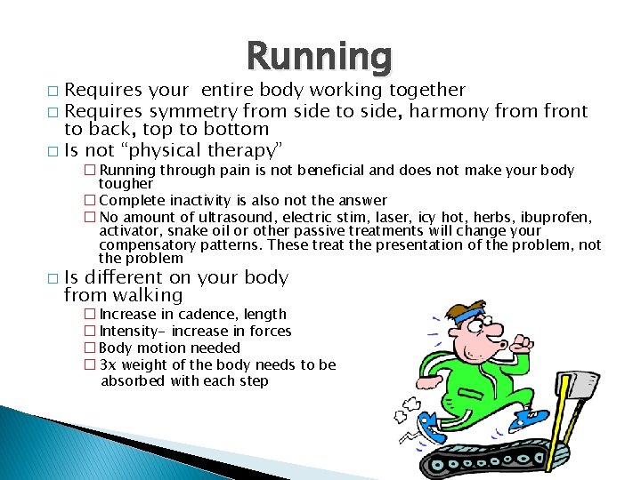
Running Requires your entire body working together � Requires symmetry from side to side, harmony from front to back, top to bottom � Is not “physical therapy” � � Running through pain is not beneficial and does not make your body tougher � Complete inactivity is also not the answer � No amount of ultrasound, electric stim, laser, icy hot, herbs, ibuprofen, activator, snake oil or other passive treatments will change your compensatory patterns. These treat the presentation of the problem, not the problem � Is different on your body from walking � Increase in cadence, length � Intensity- increase in forces � Body motion needed � 3 x weight of the body needs to be absorbed with each step
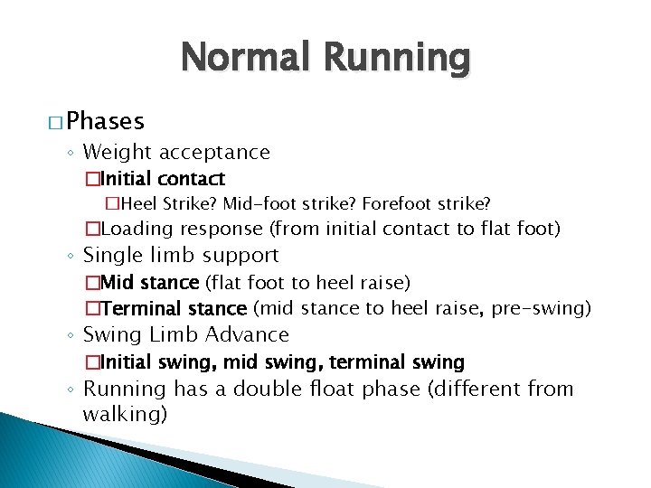
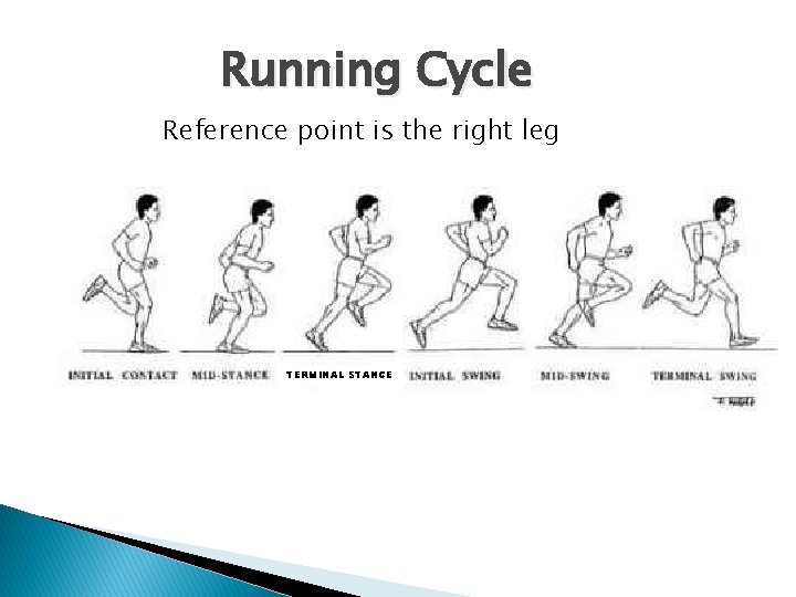
Normal Running � Phases ◦ Weight acceptance �Initial contact �Heel Strike? Mid-foot strike? Forefoot strike? �Loading response (from initial contact to flat foot) ◦ Single limb support �Mid stance (flat foot to heel raise) �Terminal stance (mid stance to heel raise, pre-swing) ◦ Swing Limb Advance �Initial swing, mid swing, terminal swing ◦ Running has a double float phase (different from walking)
Running Cycle Reference point is the right leg TERMINAL STANCE
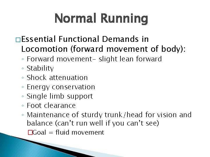
Normal Running � Essential Functional Demands in Locomotion (forward movement of body): ◦ ◦ ◦ ◦ Forward movement- slight lean forward Stability Shock attenuation Energy conservation Single limb support Foot clearance Maintenance of sturdy trunk/head for vision and balance (can’t run well if you can’t see) �Goal = fluid movement
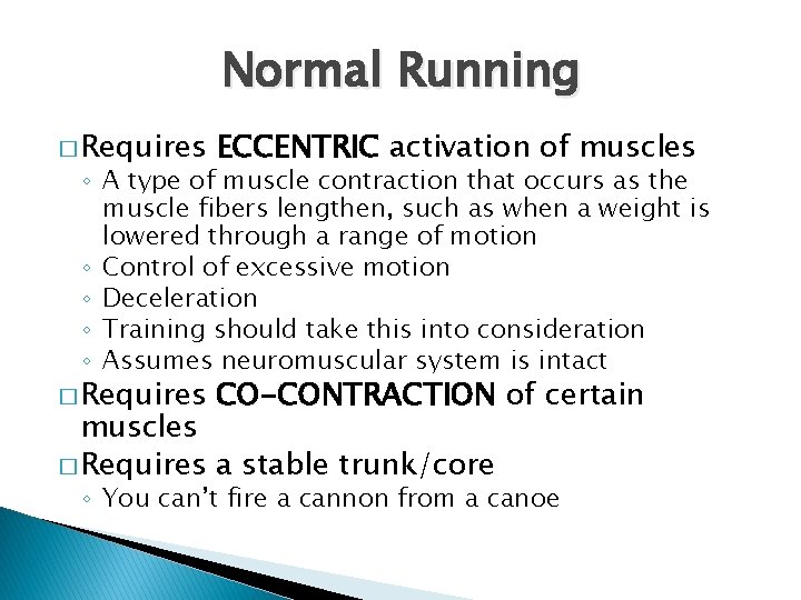
Normal Running � Requires ECCENTRIC activation of muscles � Requires CO-CONTRACTION of certain ◦ A type of muscle contraction that occurs as the muscle fibers lengthen, such as when a weight is lowered through a range of motion ◦ Control of excessive motion ◦ Deceleration ◦ Training should take this into consideration ◦ Assumes neuromuscular system is intact muscles � Requires a stable trunk/core ◦ You can’t fire a cannon from a canoe
Normal Running � It is just as abnormal to have too much motion as it is to have too little motion � Occurs in three planes of motion ◦ Sagittal ◦ Frontal ◦ Transverse � Goal: controlled mobility
What is happening during the phases of normal running? � Foot/Ankle � Knee � Pelvis/Lumbar Spine � Upper Extremities
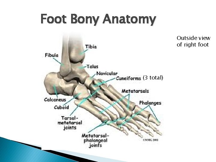
Foot Bony Anatomy Outside view of right foot (3 total)
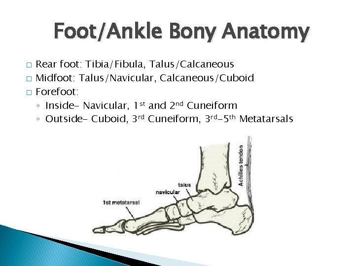
Foot/Ankle Bony Anatomy � � � Rear foot: Tibia/Fibula, Talus/Calcaneous Midfoot: Talus/Navicular, Calcaneous/Cuboid Forefoot: ◦ Inside- Navicular, 1 st and 2 nd Cuneiform ◦ Outside- Cuboid, 3 rd Cuneiform, 3 rd-5 th Metatarsals
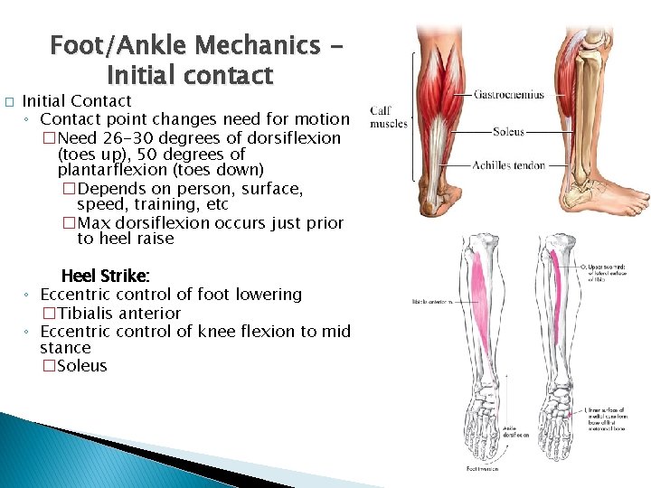
Foot/Ankle Mechanics Initial contact � Initial Contact ◦ Contact point changes need for motion �Need 26 -30 degrees of dorsiflexion (toes up), 50 degrees of plantarflexion (toes down) �Depends on person, surface, speed, training, etc �Max dorsiflexion occurs just prior to heel raise Heel Strike: ◦ Eccentric control of foot lowering �Tibialis anterior ◦ Eccentric control of knee flexion to mid stance �Soleus
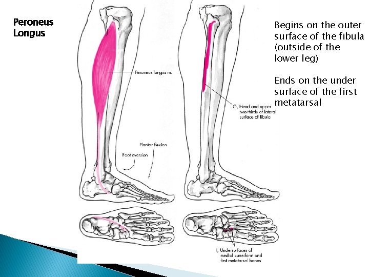
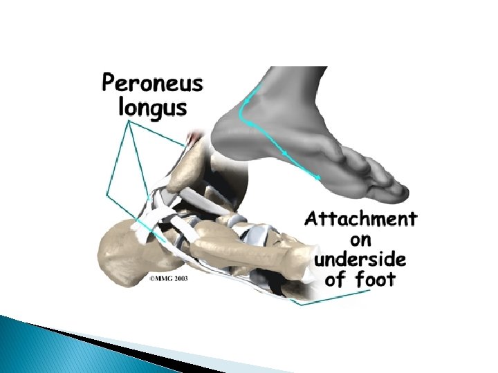
Peroneus Longus Begins on the outer surface of the fibula (outside of the lower leg) Ends on the under surface of the first metatarsal
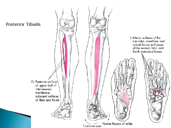
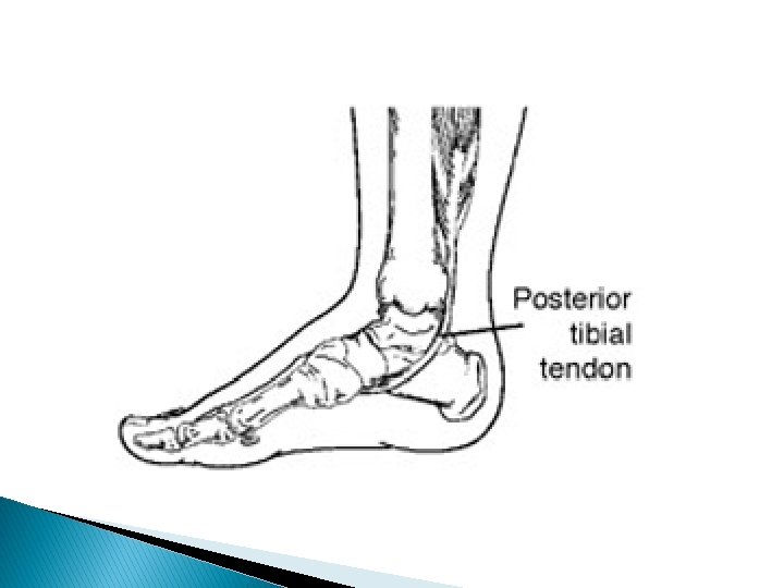
Posterior Tibialis
Foot/Ankle Mechanics � Weight Acceptance: ◦ As foot accepts weight it must go from a stable/locked position (initial contact), to a more flexible (midstance), back to a stable/locked position (terminal stance) �Supination -> pronation -> supination �“sling shot” ◦ The foot must have a balance between pronation and supination. Too much or too little of either motion at the wrong time of the gait cycle leads to inefficient foot function and potential dysfunction �Pronation is a normal part of the cycle and is necessary for shock absorption �
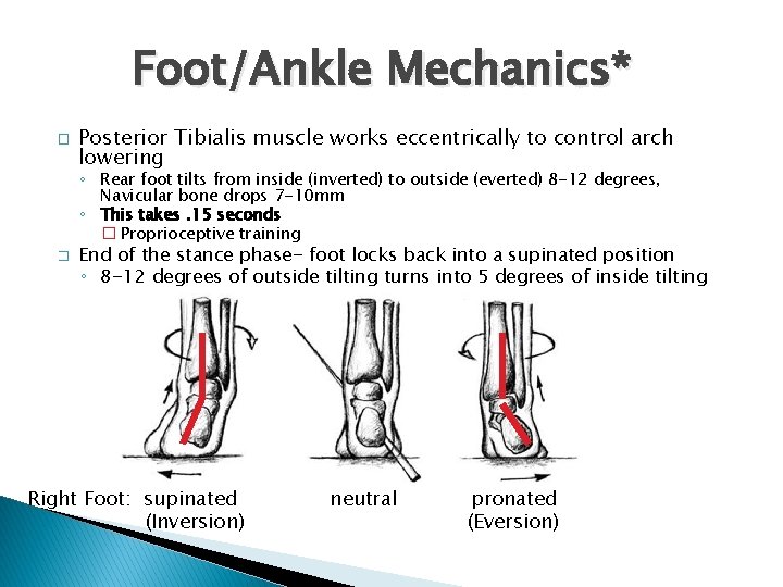
Foot/Ankle Mechanics* � Posterior Tibialis muscle works eccentrically to control arch lowering ◦ Rear foot tilts from inside (inverted) to outside (everted) 8 -12 degrees, Navicular bone drops 7 -10 mm ◦ This takes. 15 seconds � Proprioceptive training � End of the stance phase- foot locks back into a supinated position ◦ 8 -12 degrees of outside tilting turns into 5 degrees of inside tilting Right Foot: supinated (Inversion) neutral pronated (Eversion)
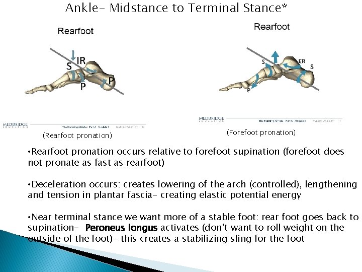
Ankle- Midstance to Terminal Stance* (Rearfoot pronation) (Forefoot pronation) • Rearfoot pronation occurs relative to forefoot supination (forefoot does not pronate as fast as rearfoot) • Deceleration occurs: creates lowering of the arch (controlled), lengthening and tension in plantar fascia- creating elastic potential energy • Near terminal stance we want more of a stable foot: rear foot goes back to supination- Peroneus longus activates (don’t want to roll weight on the outside of the foot)- this creates a stabilizing sling for the foot
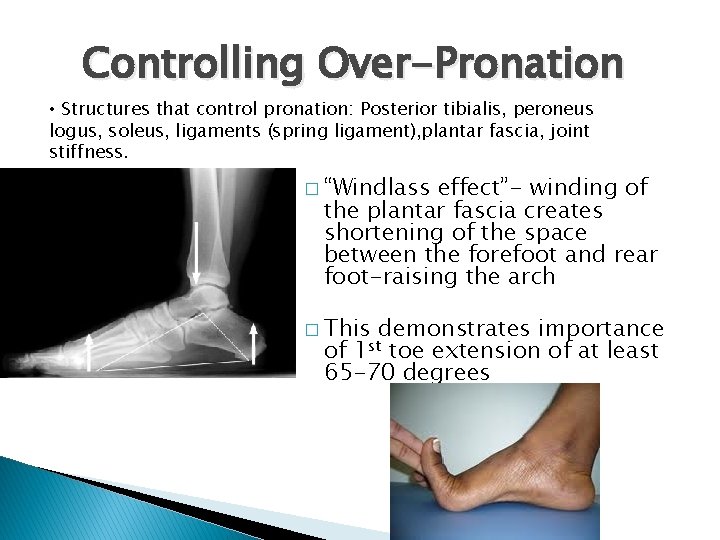
Controlling Over-Pronation • Structures that control pronation: Posterior tibialis, peroneus logus, soleus, ligaments (spring ligament), plantar fascia, joint stiffness. � “Windlass effect”- winding of the plantar fascia creates shortening of the space between the forefoot and rear foot-raising the arch � This demonstrates importance of 1 st toe extension of at least 65 -70 degrees
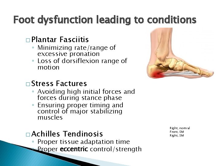
Foot dysfunction leading to conditions � Plantar Fasciitis ◦ Minimizing rate/range of excessive pronation ◦ Loss of dorsiflexion range of motion � Stress Factures ◦ Avoiding high initial forces and forces during stance phase ◦ Ensuring proper timing and control of major stabilizing muscles � Achilles Tendinosis ◦ Proper tissue adaptation time ◦ Proper eccentric control/strength Right, normal Front, SM Right, SM
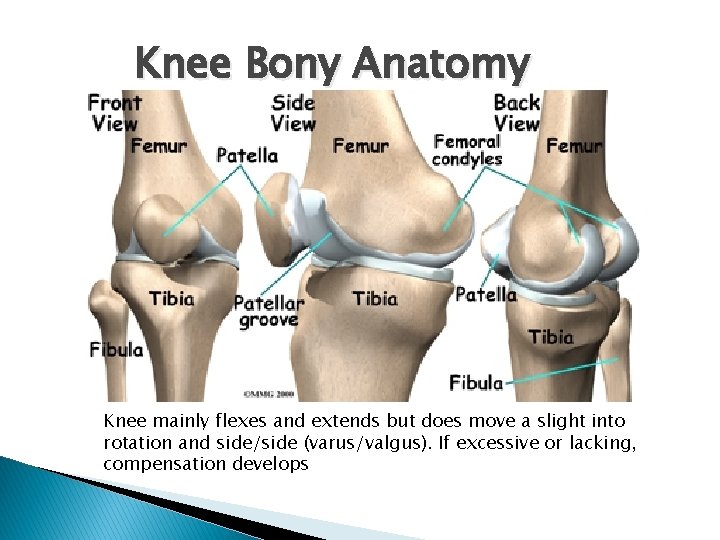
Knee Bony Anatomy Knee mainly flexes and extends but does move a slight into rotation and side/side (varus/valgus). If excessive or lacking, compensation develops
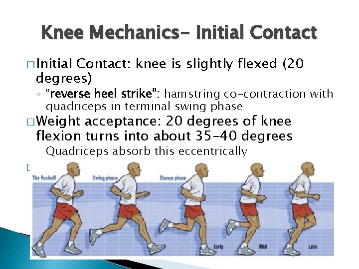
Knee Mechanics- Initial Contact � Initial Contact: knee is slightly flexed (20 degrees) ◦ “reverse heel strike”: hamstring co-contraction with quadriceps in terminal swing phase � Weight acceptance: 20 degrees of knee flexion turns into about 35 -40 degrees Quadriceps absorb this eccentrically �
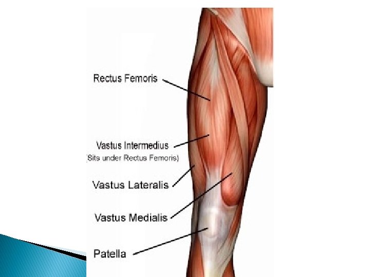
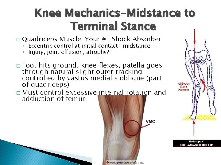
Knee Mechanics-Midstance to Terminal Stance � Quadriceps Muscle: Your #1 Shock Absorber ◦ Eccentric control at initial contact- midstance ◦ Injury, joint effusion, atrophy? Foot hits ground: knee flexes, patella goes through natural slight outer tracking controlled by vastus medialis oblique (part of quadriceps) � Must control excessive internal rotation and adduction of femur �
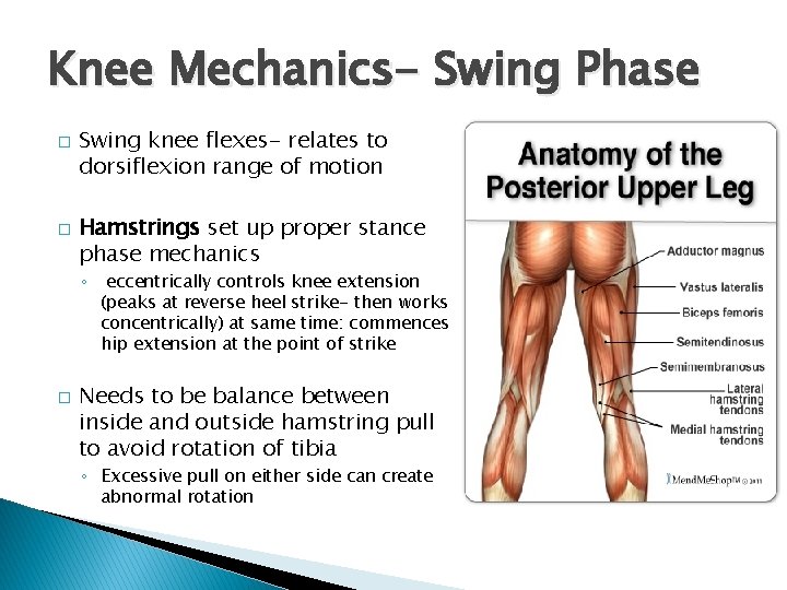
Knee Mechanics- Swing Phase � � Swing knee flexes- relates to dorsiflexion range of motion Hamstrings set up proper stance phase mechanics ◦ � eccentrically controls knee extension (peaks at reverse heel strike- then works concentrically) at same time: commences hip extension at the point of strike Needs to be balance between inside and outside hamstring pull to avoid rotation of tibia ◦ Excessive pull on either side can create abnormal rotation
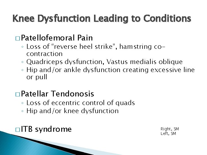
Knee Dysfunction Leading to Conditions � Patellofemoral Pain ◦ Loss of “reverse heel strike”, hamstring cocontraction ◦ Quadriceps dysfunction, Vastus medialis oblique ◦ Hip and/or ankle dysfunction creating excessive line or pull � Patellar Tendonosis ◦ Loss of eccentric control of quads ◦ Hip and/or knee dysfunction � ITB syndrome Right, SM Left, SM
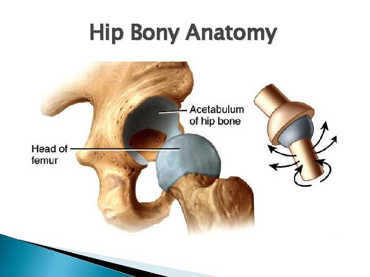
Hip Bony Anatomy
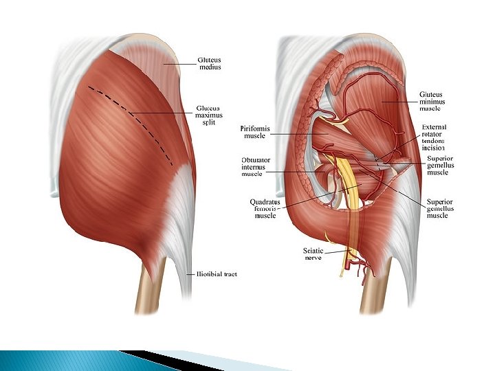
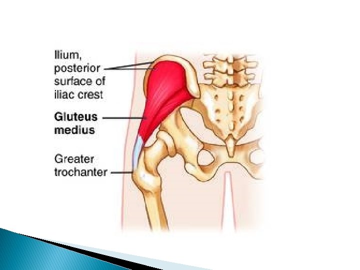
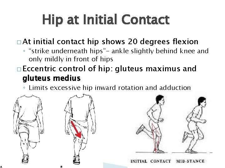
Hip at Initial Contact � At initial contact hip shows 20 degrees flexion ◦ “strike underneath hips”- ankle slightly behind knee and only mildly in front of hips � Eccentric control of hip: gluteus maximus and gluteus medius ◦ Limits excessive hip inward rotation and adduction
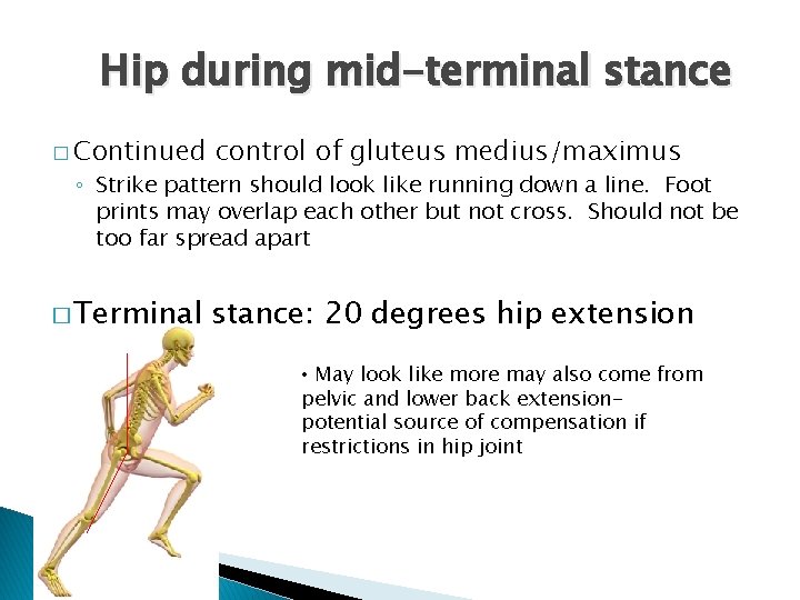
Hip during mid-terminal stance � Continued control of gluteus medius/maximus � Terminal stance: 20 degrees hip extension ◦ Strike pattern should look like running down a line. Foot prints may overlap each other but not cross. Should not be too far spread apart • May look like more may also come from pelvic and lower back extensionpotential source of compensation if restrictions in hip joint
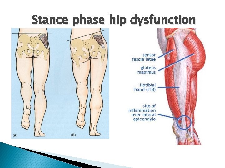
Stance phase hip dysfunction
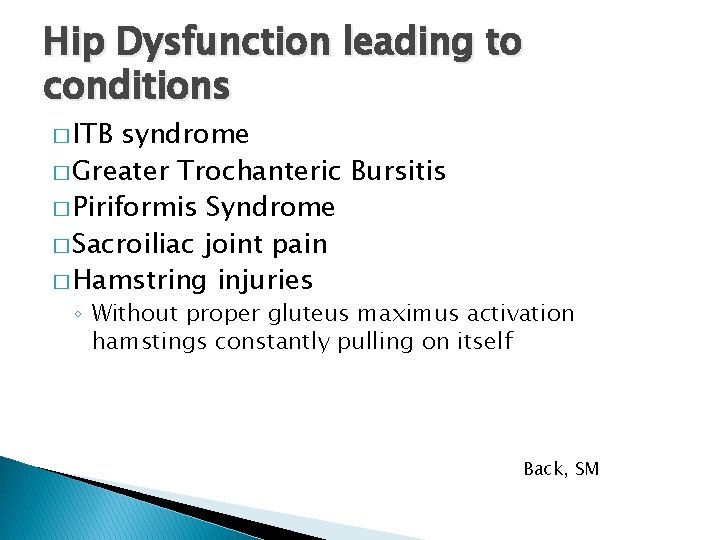
Hip Dysfunction leading to conditions � ITB syndrome � Greater Trochanteric Bursitis � Piriformis Syndrome � Sacroiliac joint pain � Hamstring injuries ◦ Without proper gluteus maximus activation hamstings constantly pulling on itself Back, SM
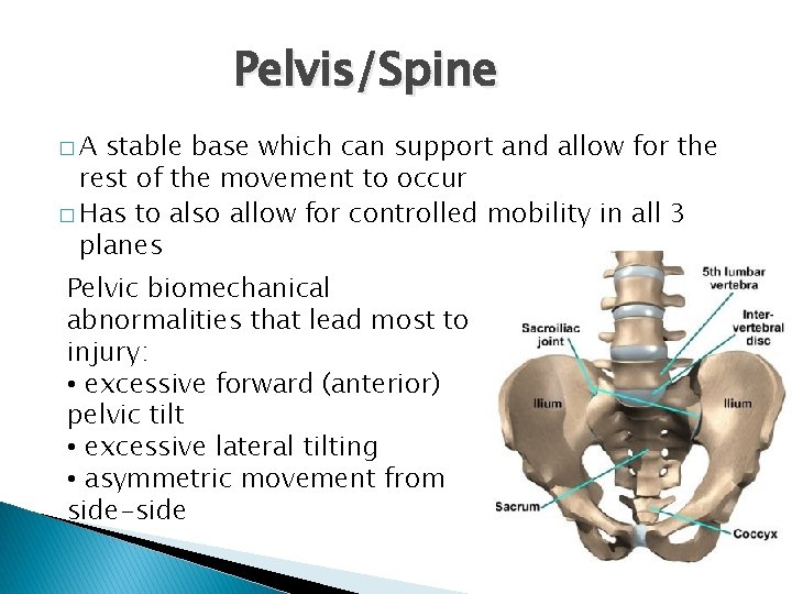
Pelvis/Spine �A stable base which can support and allow for the rest of the movement to occur � Has to also allow for controlled mobility in all 3 planes Pelvic biomechanical abnormalities that lead most to injury: • excessive forward (anterior) pelvic tilt • excessive lateral tilting • asymmetric movement from side-side
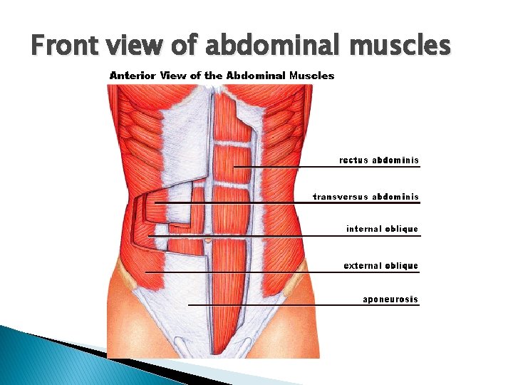
Front view of abdominal muscles
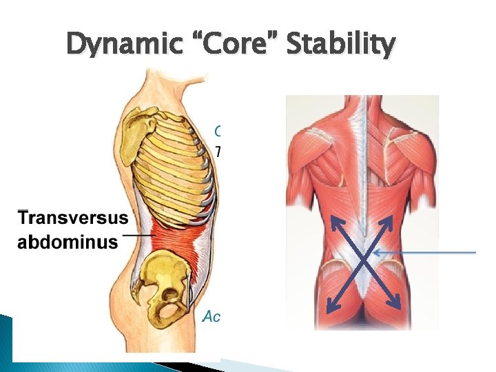
Dynamic “Core” Stability
Pelvis/Spine- Initial Contact � Pelvis slightly rotated backward on the same side at initial contact � Need: Pre-contraction of transversus abdominis and laying of core muscles with higher demand motion (i. e impact/loading phase) � Pelvis begins to rotate forward (anterior) with weight acceptance, peaking at toe off ◦ Excessive forward pelvic tilt may result from tight hips, tight hip flexor muscles
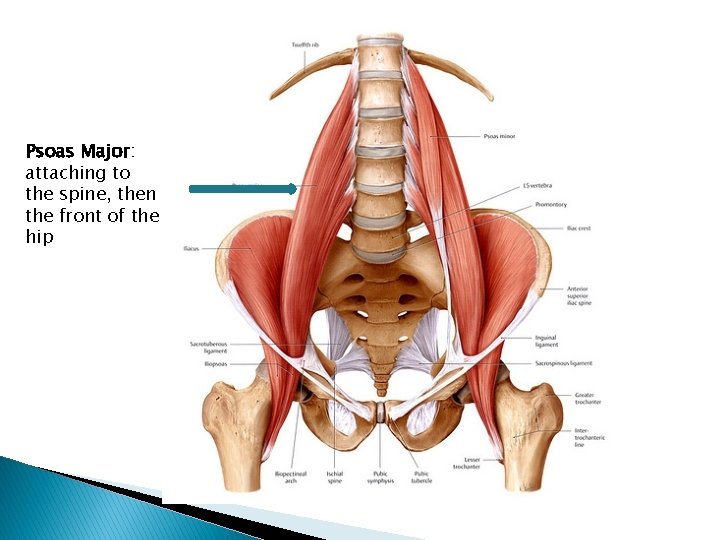
Psoas Major: attaching to the spine, then the front of the hip
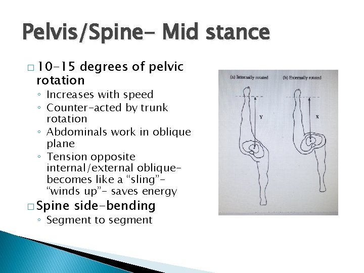
Pelvis/Spine- Mid stance � 10 -15 degrees of pelvic rotation ◦ Increases with speed ◦ Counter-acted by trunk rotation ◦ Abdominals work in oblique plane ◦ Tension opposite internal/external obliquebecomes like a “sling”“winds up”- saves energy � Spine side-bending ◦ Segment to segment
Pelvis/Spine � There are 29 “core” muscles that absorb and distribute impact forces, and allow body movements in a controlled efficient manner ◦ ◦ ◦ Abdominals Paraspinals Gluteal muscles Pelvic floor muscles Hip girdle Diaphragm � Endurance
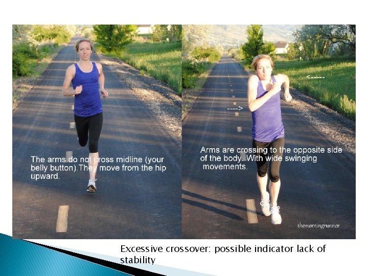
Upper Extremities � � � Provide balance and promote efficiency/stability Helps balance all planes of motion- if you don’t have this, creates excessive motion else where Each arm movement counter balances the opposite leg during swing phase ◦ Should match lower extremity motion � � Generates forward momentum All of this sequences with arms: as we flex one hip, flex opposite arm at same time ◦ Minimizes twisting of torso and pelvis- saves energy and allows for maximum stride
Excessive crossover: possible indicator lack of stability
Upper Extremities � If arm swing is not sufficient, hip flexion/adduction, knee flexion/adduction and ankle abduction have increase joint angles � Symmetry of motion important: if not sequenced appropriately, leads to compensation, dysfunction and creates breakdown/shear Back, SM Front, SM
Functional Movement Assessment � Identify potential areas of compensation, restriction, or loss of control � Patterns that require several areas working together � Movements that are required for day to day use, sport specific � Next week
Principles of good training � Consistency, rest days � Stretch dynamically, incorporate stability, sharpening/technical drills � Mileage- steady progressions (30% of total runs are the long runs) � Vary running surface � Intensity: 30% of total runs are the intense runs recovery techniques; training + rest= success (back off on a regular basis) � Monitoring techniques: listen to body for clueschange in heart rate, sleep patterns, stress levels, morning stiffness
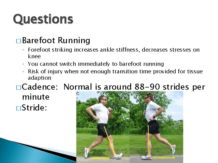
Questions � Barefoot Running ◦ Forefoot striking increases ankle stiffness, decreases stresses on knee ◦ You cannot switch immediately to barefoot running ◦ Risk of injury when not enough transition time provided for tissue adaption � Cadence: minute � Stride: Normal is around 88 -90 strides per
Questions Injury prevention � Training errors- what has changed? ◦ ◦ Mileage (sudden change, clustering) Intensity Surface: side of road, cushion, variety, shoes Consistency (erratic or unrelenting) � Ensuring adequate range, flexibility, and stability � Treadmill Analysis: part of our physical therapy, covered by insurance in those appropriate for PT http: //www. fleetfeetsports. com/training-programs
References � � � Cavanagh P (Ed. ) (1990), Biomechanics of Distance Running. Human Kinetics Nicola, Terry. The Anatomy and Biomechanics of Running. Clin Sports Med 31 (2012) 187 -201 Schache, Anthony. The coordinated movement of the lumbo-pelvic-hip complex during running: a literature review. Gate and Posture 10 (1999). 3046 Walsh, Matthew. The Running Athlete: Parts A, B, and C. http: //www. medbridgeeducation. com Http: //www. themanualtherapist. com