The Alimentary system or The digestive system Digestive


: I 2 C 1")

 Gum (gingiva)")


 Jejunum (2 m) Ileum (3")


 produce bile which collects and")


- Slides: 41
The Alimentary system or The digestive system
Digestive system • The digestive system is the collective name used to describe the alimentary canal (a long tube that runs through the body) along with accessory glands and variety of digestive processes that take place at different levels in the canal to prepare food eaten for absorption. • The digestive system is arranged as a series of organs along a tube called the gastrointestinal (GI) tract, into which various accessory glands release their secretions
The digestive system is divided into two major parts: • The digestive tract (alimentary canal) is a continuous tube with two openings: the mouth and the anus. It includes the mouth, pharynx, esophagus, stomach, small intestine, and large intestine. Food passing through the internal cavity, or lumen, of the digestive tract does not technically enter the body until it is absorbed through the walls of the digestive tract and passes into blood or lymphatic vessels. • Accessory organs include the teeth and tongue, salivary glands, liver, gallbladder, and pancreas.
Digestive system
Function of Digestive System § The function of the digestive system is digestion and absorption. § Digestion is the breakdown of food into small molecules, which are then absorbed into the body.
Treatment of food in the Digestive System Treatment involves seven processes: 1. Ingestion 2. Propulsion 3. Secretion of digestive enzymes and other substances (Liquefies, adjust p. H, chemically breakdown food) 4. Mechanical digestion (Chewing of food and continues with the muscular churning of the stomach) 5. Chemical digestion (Carried out by the enzymes in the stomach and small intestines) 6. Absorption (Entrance of digested food called nutrients into the body) 7. Defecation/Egestion (Eliminating undigested material through anus)
The Digestive System- layers of GIT Along most of its length, the wall of the digestive system has four basic layers: 1. Mucosa (Innermost layer) 2. Submucosa 3. Muscularis propria (Externa) 4. Serosa/Adventia (Outermost layer)
Lumen Blood vessels Lymphatic vessel Nerve The mucosa is a mucous membrane that lines the GI tract and secretes mucus that lubricates and protects the GI tract. The submucosa is a layer of connective tissue that contains blood vessels, lymph vessels, and nerves. The muscularis is made up of two layers of smooth muscle—one circular and one longitudinal. The serosa is a connective tissue covering that secretes a fluid to lubricate the outside of the GI tract.
The Digestive organs • Each organ helps to break down food into molecules small enough to be absorbed into the bloodstream and utilized by the body. • The digestive organs § § § Mouth Esophagus Stomach Small intestine Large intestine
• The digestive organs are aided by several accessory organs § Salivary glands Submandibular) § Pancreas § Gallbladder § Liver (Parotid, Sublingual, • Digestion begins in the mouth where food enters the digestive process. • The teeth bite, tear, and crush food into smaller pieces
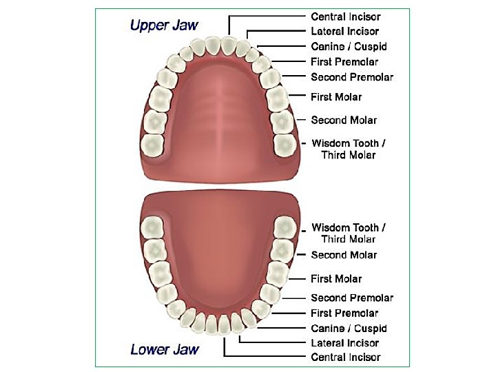
The teeth Formula of teeth (half of the upper jaw): I 2 C 1 P 2 M 3
Types of teeth and their functions • Each type of tooth has a slightly different shape and performs a different job. There are 4 types of teeth. 1. Incisors: Incisors are the eight teeth in the front and center of mouth (four on top and four on bottom). These are the teeth that used to take bites of food. 2. Canines: They are 4 in numbers. These are the sharpest teeth and are used for ripping and tearing food apart. 3. Premolars: Premolars, or bicuspids, are used for chewing and grinding food. We have four premolars on each side of our mouth, two on the upper and two on the lower jaw. 4. Molars: Primary molars are also used for chewing and grinding food. These appear between 12 and 15 months of age. These molars, also known as decidious molars.
The tooth Enamel Dentine Crown Pulp cavity (contains blood vessels and nerves) Gum (gingiva) Root canal Root Cementum Bone (b) The structure of the human tooth is suited for its function of breaking food into smaller pieces. Figure 15. 3 b
Different parts of Tooth • A tooth is divided into two basic parts. § The crown (Visible, white part of the tooth) § The root (Not visible, extent below the gum line and anchors the tooth) Teeth contain four kinds of tissue and each does a different job. 1. Enamel: Enamel is the visible substance that covers the tooth crown. It is harder than bone, and protects the tooth from decay. Enamel is made up of phosphorous and calcium. 2. Dentin: It is found underneath the enamel, which is calcified and looks similar to bone. Dentin is not quite as hard as enamel, so it is at greater risk for decay. 3. Cementum: This tissue covers the tooth root and helps anchor it (cement it) into the bone. It is softer than enamel and dentin. Cementum has a light yellow color and is usually covered by the gums. 4. Pulp: Pulp is found at the center of tooth and contains the blood vessels, nerves, and other soft tissues that deliver nutrients and signals to teeth.
Saliva is an important part of a healthy body. It is mostly made of water. But saliva also contains important substances that body needs to digest food and keep teeth strong. Saliva moves through the tube called salivary duct. Three pairs of major salivary glands release secretions, called saliva, into the mouth and hundreds of minor ones.
Salivary glands 1. Parotid gland § The two parotid glands are major salivary glands wrapped around the mandibular ramus in humans. § They are the largest of the salivary glands § They secrete saliva to facilitate swallowing, and amylase to begin the digestion of starches. § They produce 20% of the total salivary content in the oral cavity. 2. Submandibular gland § The submandibular glands are a pair of major salivary glands located beneath the lower jaws. § The secretion produced is a mixture of both serous fluid and mucus. § Approximately 65 -70% of saliva in the oral cavity is produced by the submandibular glands. § They are much smaller than the parotid glands.
3. Sublingual glands § The sublingual glands are a pair of major salivary glands located inferior to the tongue, anterior to the submandibular glands. § The secretion produced is mainly mucous in nature. § Approximately 5% of saliva entering the oral cavity comes from these glands.
Functions of Saliva 1. Chemical digestion: breaks down starch by the function of “salivary amylase” 2. Helps chewing and swallowing 3. Lubricating effect: moisturizes the inside of the mouth and creates smoother speech 4. Solvent effect: dissolves food and allows the tongue to taste food 5. Cleaning effect: washes away food debris and bacteria remaining in the mouth 6. Antibacterial effect: Lysozyme, peroxidase and lactoferrin fight against pathogenic microorganisms 7. p. H buffering effect: Prevents sudden changes in p. H 8. Supplies minerals, including calcium and phosphorus, to teeth
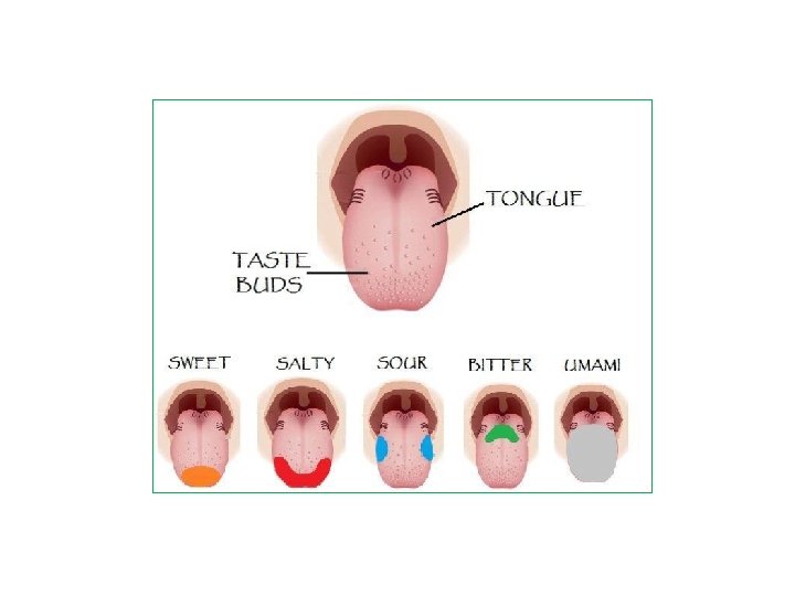
§ § § § § Tongue The tongue is a muscular organ in the mouth. It is covered with moist, pink tissue called mucosa. Tiny bumps called papillae give the tongue its rough texture. Thousands of taste buds cover the surfaces of the papillae. The tongue is anchored to the mouth by webs of tough tissue and mucosa. In the back of the mouth, the tongue is anchored into the hyoid bone. The four common tastes are sweet, sour, bitter, and salty. A fifth taste, called umami, results from tasting glutamate. Taste buds are collections of nerve-like cells that connect to nerves running into the brain. The tongue is vital for chewing and swallowing food, as well as for speech. The tongue has many nerves that help detect and transmit taste signals to the brain. Because of this, all parts of the tongue can detect these four common tastes.
The pharynx ü Commonly called the throat. ü Where the nasal and oral cavities join. ü During swallowing the epiglottis covers the trachea, or windpipe, to prevent choking The esophagus ü A tube that transports food from the mouth to the stomach ü No digestive processes occur here
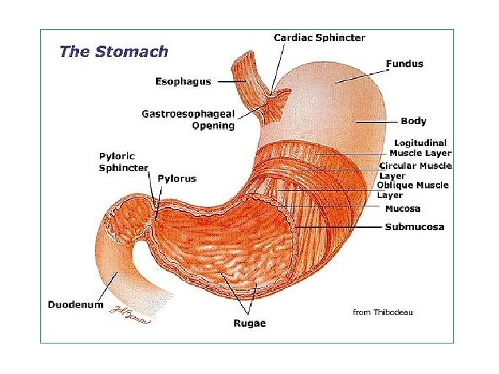
Stomach • The stomach is a muscular organ located on the left side of the upper abdomen. The stomach receives food from the esophagus. As food reaches the end of the esophagus, it enters the stomach through a muscular valve called the lower esophageal sphincter. • The stomach secretes acid and enzymes that digest food. The stomach muscles contract periodically (Peristalsis), churning food to enhance digestion. The pyloric sphincter is a muscular valve that opens to allow food to pass from the stomach to the small intestine.
Peristalsis • Food is pushed through our digestive system by a series of muscular contractions called peristalsis
Gastric acid/Gastric juice/Stomach acid • It is a digestive fluid, formed in the stomach. • Composed of hydrochloric acid (HCl) 0. 05– 0. 1 M, potassium chloride (KCl) and sodium chloride (Na. Cl). • The acid plays a key role in digestion of proteins, by activating digestive enzymes. • Gastric acid is produced by gastric parietal cells in the lining of the stomach. • Other cells in the stomach produce bicarbonate, a base, to buffer the fluid, ensuring that it does not become too acidic. • These cells also produce mucus, which forms a viscous physical barrier to prevent gastric acid from damaging the stomach.
Intestine • The intestines are a long, continuous tube running from the stomach to the anus. Most absorption of nutrients and water happen in the intestines. The intestines include the small intestine, large intestine, and rectum. • The small intestine (small bowel) is about 20 feet long and about an inch in diameter. Its job is to absorb most of the nutrients from food and liquid. The small intestine, which is divided into the duodenum, jejunum, and ileum. • The large intestine (colon or large bowel) is about 5 feet long and about 3 inches in diameter. The colon absorbs water from wastes, creating stool. As stool enters the rectum, nerves there create the urge to defecate.
Intestine • The small intestine : Duodenum (25 cm) Jejunum (2 m) Ileum (3 m)
Intestine • Large intestine: Undigested and indigestible materials that have not been absorbed by the small intestine move to the large intestine.
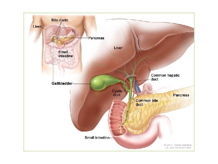
Liver § The liver is a large, meaty organ that sits on the right side of the belly. § Weighing about 3 pounds, § The liver is reddish-brown in color and feels rubbery to the touch. § It's protected by the rib cage. § The liver has two large sections, called the right and the left lobes. § The gallbladder sits under the liver, along with parts of the pancreas and intestines. § The liver and these organs work together to digest, absorb, and process food. § The liver's main job is to filter the blood coming from the digestive tract, before passing it to the rest of the body. § The liver also detoxifies chemicals and metabolizes drugs. § The liver also makes proteins important for blood clotting and other functions.
Bile is a digestive juice that is secreted by the liver and stored in the gallbladder. It has important functions: § It is a means for the body to excrete waste products from the blood. § Emulsify fats and break it down into small particles. This is a detergent-like action of bile. § Helps the body absorb the breakdown products of fat in the gut. § Bile salts bind with lipids to form micelles. This is then absorbed through the intestinal mucosa. § The other important function of bile is that it contains waste products from hemoglobin break down. This is known as bilirubin. § Bile also carries excess cholesterol out of the body and ‘dumps’ it into the gastrointestinal tract where it can be passed out with other waste matter.
Bile production Bile Production § The liver cells (hepatocytes) produce bile which collects and drains into the hepatic duct. § From here it enters into the small intestine to act on fats by traveling down the common bile duct § Or it can enter the gallbladder through the cystic duct, where it is stored. § The liver manufactures between 600 ml to 1 liter of bile in a day. § As bile travels down the ducts, the lining of these passages, secrete water, sodium and bicarbonate ions into the bile, thereby diluting it.
Bile salt Bile Salts Bile, whether from the liver or gallbladder, contains the following substances: ü Water ü Bile salts ü Bilirubin ü Cholesterol ü Fatty acids ü Lecithin ü Sodium ü Potassium ü Calcium ü Chlorine ü Bicarbonate ions
Gallbladder Ø The gallbladder is a small pouch that sits just under the liver. Ø The gallbladder stores bile produced by the liver. Ø After meals, the gallbladder is empty and flat, like a deflated balloon. Ø Before a meal, the gallbladder may be full of bile and about the size of a small pear. Ø In response to signals, the gallbladder squeezes stored bile into the small intestine through a series of tubes called ducts.
Gallstone § Gallstones are small stones, usually made of cholesterol, that form in the gallbladder. § These deposits may be as small as a grain of sand or as large as a golf ball; they may be hard or soft, smooth. § However, if a gallstone becomes trapped in an opening (duct) inside the gallbladder, it can trigger a sudden, intense abdominal pain that usually lasts between one and five hours. This type of abdominal pain is known as biliary colic. § Some people with gallstones can also develop complications, such as inflammation of the gallbladder (cholecystitis), which can cause: ü Persistent pain ü Jaundice ü Fever § When gallstones cause symptoms or complications, it's known as gallstone disease or cholelithiasis.
Gallstone
Pancreatic juice contains – Enzymes – Water – Ions
Summery………. . Digestive system and organs of digestive system (parts of GI tract with Diagram) Layers of GI Tract Glands in the Oral cavity /Salivary Gland Function of Saliva Description of – Teeth – Tongue – Esophagus – Pharynx – Stomach Parts of the intestines • Function of the intestines • Digestive glands in the body • Description of Liver, Pancreas , Gallbladder and bile Question: Write down the description of biliary tract/tree? • • •