
THE ABDOMINAL EXAM ACSASE Medical Student Simulationbased Surgical
THE ABDOMINAL EXAM ACS/ASE Medical Student Simulation-based Surgical Skills Curriculum
CREDITS n Author n n Contributors n n n Michael Hughes, MD , FACS (expert performance video) Richard Damewood, MD, FACS (modified score assessment tool) Duane Patterson, Ph. D (technical support) Paul Schreck (videographer ) Editors n n n Ebondo Mpinga, MD, FACS Keith Clancy, MD, FACS Amanda Beattie, MD , R 5 York Hospital Department of Surgery, York, PA
OBJECTIVES n After the completion of this module the student should be able to: 1. 2. 3. Perform a complete abdominal exam. Recognize the signs of peritonitis. Arrive at a differential diagnosis based upon the findings elicited during the exam.
ABDOMINAL EXAM n Although we will focus on the abdominal exam, it cannot be overemphasized that a thorough physical exam (head to toes) is important to help in arriving at a comprehensive differential diagnosis list. n Examples : n presence of jaundice may add consideration of a biliary /hepatic etiology n Irregularly irregular heart rate atrial fibrillation-> mesenteric ischemia n Crackle at lung bases pneumonia n Skin lesions (pyoderma gangrenosum) -> IBD
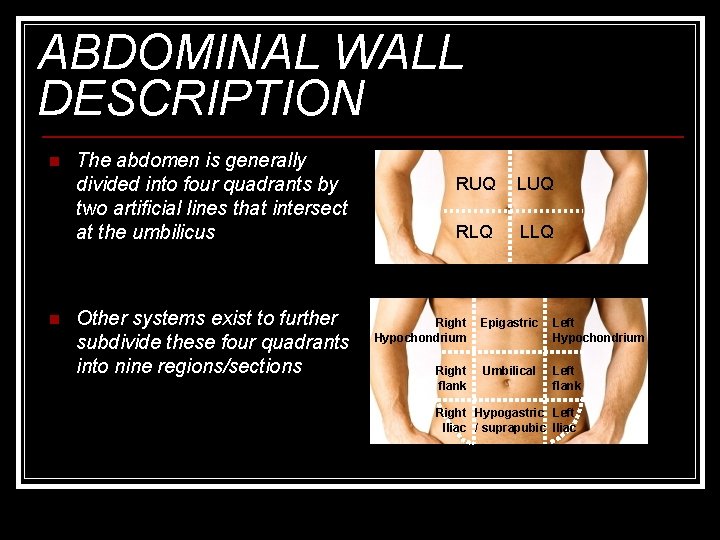
ABDOMINAL WALL DESCRIPTION n n The abdomen is generally divided into four quadrants by two artificial lines that intersect at the umbilicus Other systems exist to further subdivide these four quadrants into nine regions/sections RUQ LUQ RLQ LLQ Right Hypochondrium Epigastric Left Hypochondrium Right flank Umbilical Left flank Right Hypogastric Left Iliac / suprapubic Iliac
ABDOMINAL EXAM The exam should be performed in this specific order n General appearance n Vital signs n Inspection n Auscultation n Percussion n Palpation n It should include n An examination of the inguinal area n n including the external genitalia in males (testes) A rectal exam (discussed in a separate module) A pelvic exam in women (discussed in a separate module)

DESCRIPTION OF TECHNIQUES
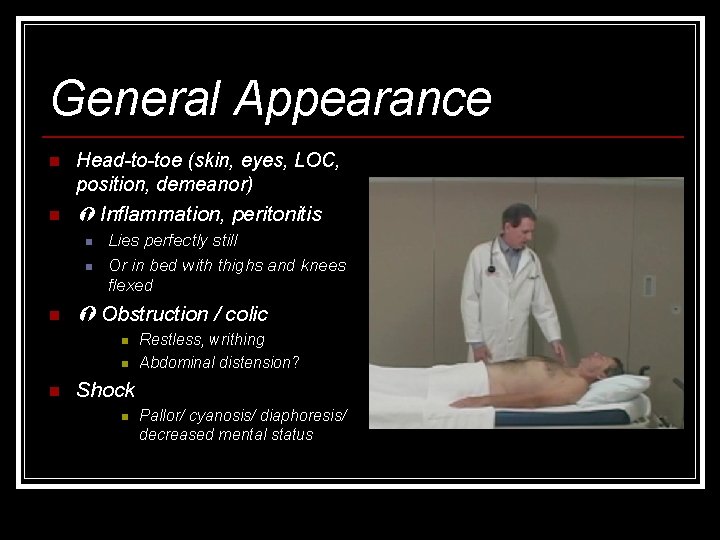
General Appearance n Head-to-toe (skin, eyes, LOC, position, demeanor) n Inflammation, peritonitis n n n Lies perfectly still Or in bed with thighs and knees flexed Obstruction / colic n n n Restless, writhing Abdominal distension? Shock n Pallor/ cyanosis/ diaphoresis/ decreased mental status
Vital Signs n Tachycardia n n n Rapid shallow breathing (splinting) n n ? Early shock (may present prior to hypotension) May be absent if on Beta blockers Peritonitis Hypotension n May be late finding depending on pre-existing state of health Fever Infectious etiology or perforation
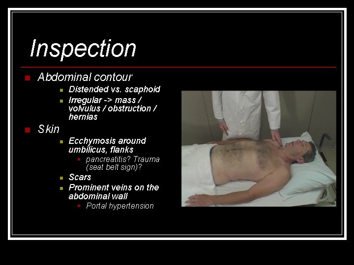
Inspection n Abdominal contour n n n Distended vs. scaphoid Irregular -> mass / volvulus / obstruction / hernias Skin n Ecchymosis around umbilicus, flanks § pancreatitis? Trauma (seat belt sign)? n n Scars Prominent veins on the abdominal wall § Portal hypertension

Auscultation n Bowel Sounds n n Auscultate all regions Listen in each region Listen before feeling Absent bowel sounds § ileus, peritonitis, shock n Hyperactive § Enteritis / obstruction (high pitched or distant) n Bruits § AAA / Reno-vascular diseases § Iliac and Femoral arteries Aorta Renal Iliac arteries Femoral arteries
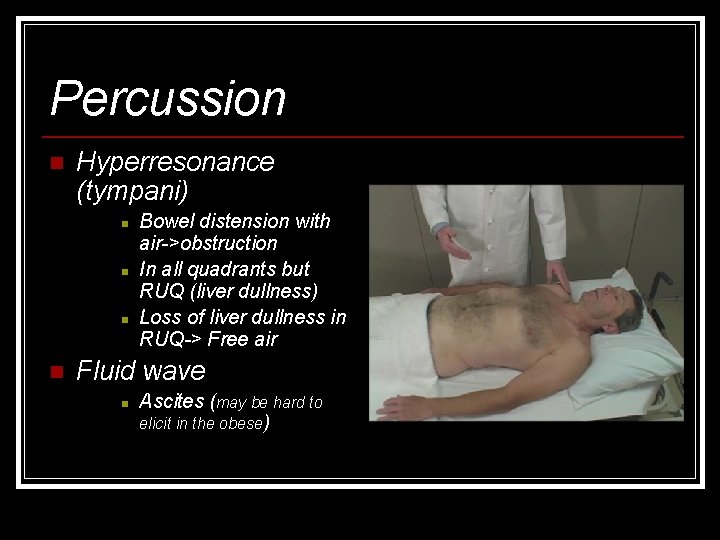
Percussion n Hyperresonance (tympani) n n Bowel distension with air->obstruction In all quadrants but RUQ (liver dullness) Loss of liver dullness in RUQ-> Free air Fluid wave n Ascites (may be hard to elicit in the obese)
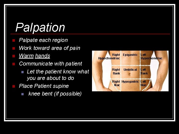
Palpation n n Palpate each region Work toward area of pain Warm hands Communicate with patient n Let the patient know what you are about to do Place Patient supine n knee bent (if possible) Right Hypochondriac Epigastric Left Hypochondriac Right flank Umbilical Left flank Right Hypogastric Left Iliac
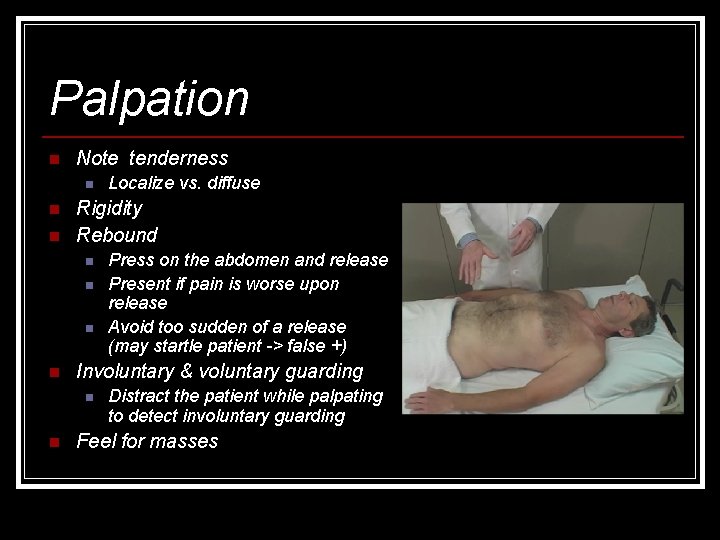
Palpation n Note tenderness n n n Rigidity Rebound n n Press on the abdomen and release Present if pain is worse upon release Avoid too sudden of a release (may startle patient -> false +) Involuntary & voluntary guarding n n Localize vs. diffuse Distract the patient while palpating to detect involuntary guarding Feel for masses
Signs highly suggestive of peritonitis n Tenderness to percussion n Tenderness elicited when the examiner firmly taps on the Iliac crest n Tenderness elicited when the examiner firmly taps on the heel of the patient’s extended leg n Tenderness when the bed is gently shaken or the patient coughs n Rebound tenderness
Abdominal exam: findings that suggest specific etiology n Biliary / hepatic etiology n Courvoisier' sign § Palpable gallbladder in the presence of painless jaundice § periampullary tumor n Caput medusa (Cruveilhier sign) § Varicose veins at umbilicus cirrhosis with portal HTN n Murphy’s sign § Pain caused during inspiration while palpating the RUQ-> acute cholecystitis n Ransohoff sign § Periumbilical yellow discoloration -> ruptured CBD
Abdominal exam: findings that suggest specific etiology n Appendicitis n Rovsing’s sign n n Ten Horn test n n Palpation on the LLQ produces tenderness at Mc. Burney’s point Pain caused by gentle traction of the right testicle Aaron sign n Persistent pressure applied at Mc. Burney ‘s point causes pressure in the epigatrium and upper chest wall
Abdominal exam: findings that suggest specific etiology n Pelvic inflammation/abscess n Iliopsoas sign n n Obturator sign n Allow patient to lie on the opposite side of the pain Extend the thigh on the affected side This should cause pain if there is irritation of the iliopsoas muscle (seen with appendicitis as well) Flexion and internal rotation of the right thigh while supine elicits hypogastric pain Indicates irritation of obturator internus muscle (seen with appendicitis as well) Chandelier sign n Extreme lower abdominal/pelvic pain with movement of the cervix
Abdominal exam: findings that suggest specific etiology n Hemoperitoneum n Hemorrhagic pancreatitis § Cullen’s sign § periumbilical bruising-> hemoperitoneum § Grey Turner’s sign § Local area of discoloration around the flanks-> acute hemorrhagic pancreatitis n Danforth sign § shoulder pain on inspiration-> hemoperitoneum n Kehr’s sign § Left shoulder pain when supine or pressure applied to LUQ-> splenic rupture
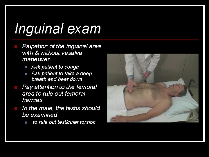
Inguinal exam n Palpation of the inguinal area with & without vasalva maneuver n n Ask patient to cough Ask patient to take a deep breath and bear down Pay attention to the femoral area to rule out femoral hernias In the male, the testis should be examined n to rule out testicular torsion
COMMON ERRORS n n n n Focus only on the abdomen Begin with palpation prior to inspection, auscultation and percussion Not asking the patient to localize the pain and therefore beginning palpation of the affected area first, exacerbating the pain and thus precluding complete examination of the abdomen Skipping the rectal, pelvic and groin exam Putting too much weight on the absence of rebound tenderness to r/o peritonitis Putting to much weight on the physical exam in an immunosuppressed patient who may not exhibit normal signs of peritonitis Forgetting to consider mesenteric ischemia when there is pain out of proportion to clinical exam
GROUPING OF SIGNS AND SYMPTOMS DIFFERENTIAL DIAGNOSIS
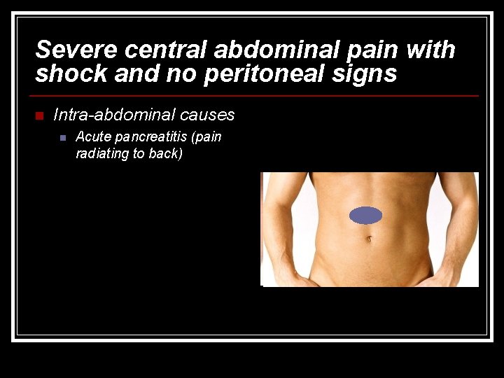
Severe central abdominal pain with shock and no peritoneal signs n Intra-abdominal causes n Acute pancreatitis (pain radiating to back)
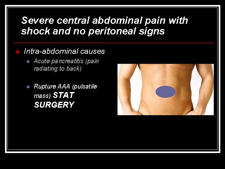
Severe central abdominal pain with shock and no peritoneal signs n Intra-abdominal causes n Acute pancreatitis (pain radiating to back) n Rupture AAA (pulsatile mass) STAT SURGERY
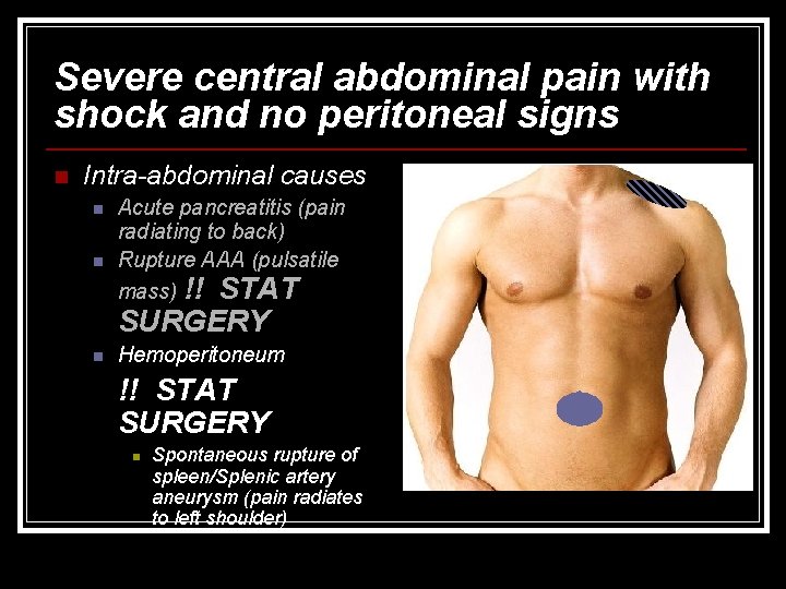
Severe central abdominal pain with shock and no peritoneal signs n Intra-abdominal causes n n Acute pancreatitis (pain radiating to back) Rupture AAA (pulsatile mass) !! STAT SURGERY n Hemoperitoneum !! STAT SURGERY n Spontaneous rupture of spleen/Splenic artery aneurysm (pain radiates to left shoulder)
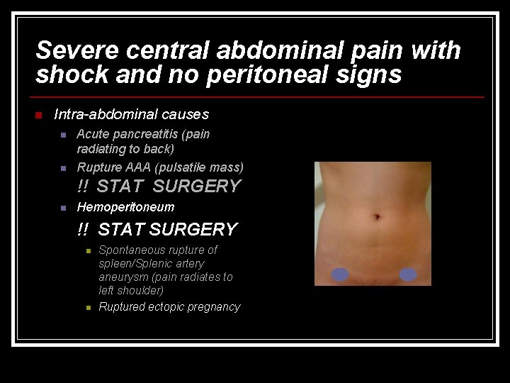
Severe central abdominal pain with shock and no peritoneal signs n Intra-abdominal causes n n Acute pancreatitis (pain radiating to back) Rupture AAA (pulsatile mass) !! STAT SURGERY n Hemoperitoneum !! STAT SURGERY n n Spontaneous rupture of spleen/Splenic artery aneurysm (pain radiates to left shoulder) Ruptured ectopic pregnancy
Severe central abdominal pain with shock and no peritoneal signs n Intra-abdominal causes n Acute pancreatitis (pain radiating to back) Rupture AAA (pulsatile mass) n Hemoperitoneum n !! STAT SURGERY n n Spontaneous rupture of spleen/Splenic artery aneurysm Rupture ectopic pregnancy Late mesenteric ischemia Extra- abdominal causes n Acute MI with cardiogenic shock
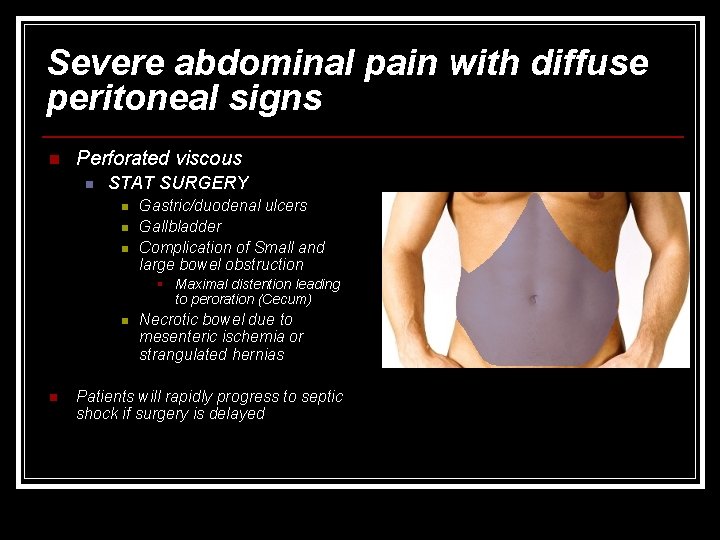
Severe abdominal pain with diffuse peritoneal signs n Perforated viscous n STAT SURGERY n n n Gastric/duodenal ulcers Gallbladder Complication of Small and large bowel obstruction § Maximal distention leading to peroration (Cecum) n n Necrotic bowel due to mesenteric ischemia or strangulated hernias Patients will rapidly progress to septic shock if surgery is delayed
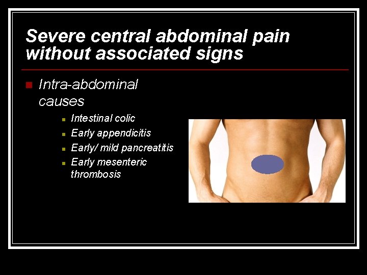
Severe central abdominal pain without associated signs n Intra-abdominal causes n n Intestinal colic Early appendicitis Early/ mild pancreatitis Early mesenteric thrombosis
Severe central abdominal pain without associated signs n Intra-abdominal causes n n n Intestinal colic Early appendicitis Early/ mild pancreatitis Early mesenteric thrombosis Extra- abdominal causes n n Herpes Zoster (rash in dermatome distribution) CAD (ECG/Enzymes) Glaucoma Tabes dorsalis (rare)
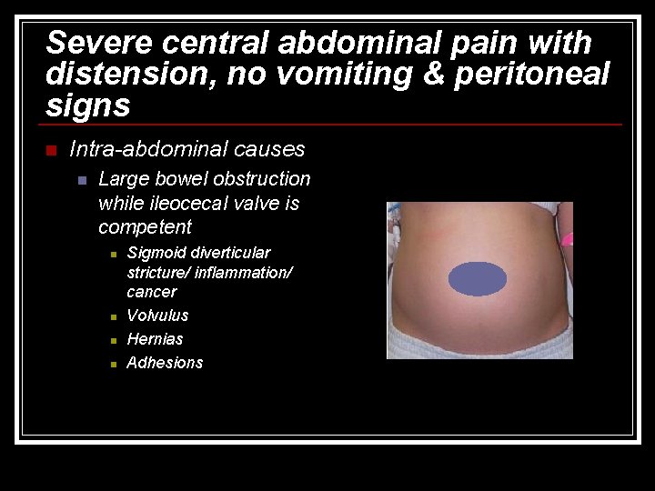
Severe central abdominal pain with distension, no vomiting & peritoneal signs n Intra-abdominal causes n Large bowel obstruction while ileocecal valve is competent n n Sigmoid diverticular stricture/ inflammation/ cancer Volvulus Hernias Adhesions
Severe central abdominal pain with distension, no vomiting & peritoneal signs n Intra-abdominal causes n Large bowel obstruction while ileocecal valve is competent n n n Sigmoid diverticular stricture/ inflammation/ cancer Volvulus Hernias Adhesions Extra- abdominal causes n Uremia
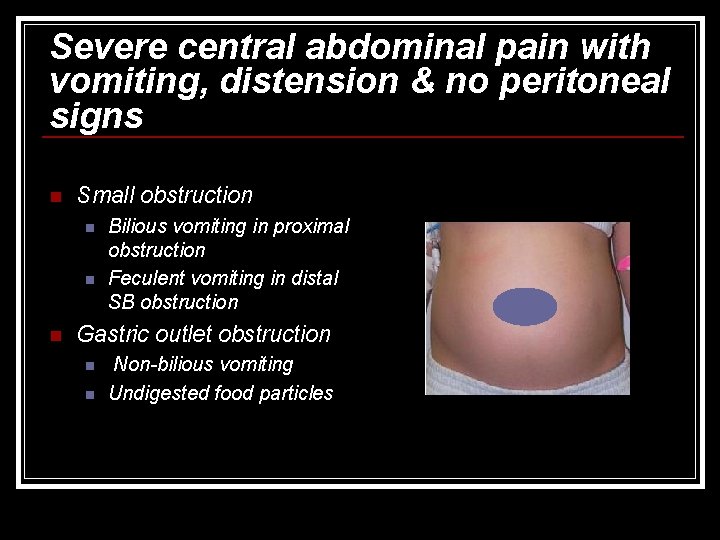
Severe central abdominal pain with vomiting, distension & no peritoneal signs n Small obstruction n Bilious vomiting in proximal obstruction Feculent vomiting in distal SB obstruction Gastric outlet obstruction n n Non-bilious vomiting Undigested food particles
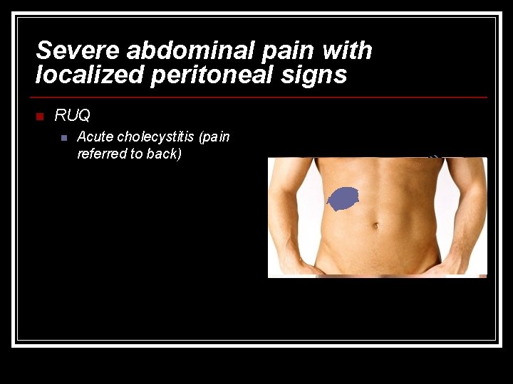
Severe abdominal pain with localized peritoneal signs n RUQ n Acute cholecystitis (pain referred to back)
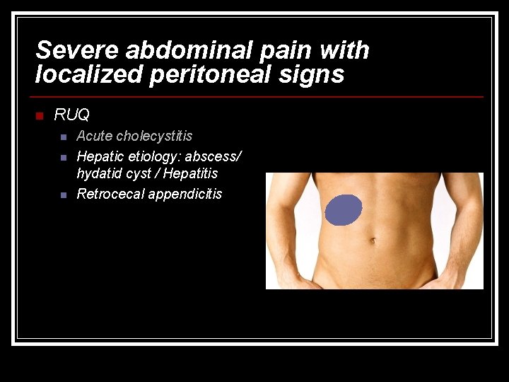
Severe abdominal pain with localized peritoneal signs n RUQ n n n Acute cholecystitis Hepatic etiology: abscess/ hydatid cyst / Hepatitis Retrocecal appendicitis
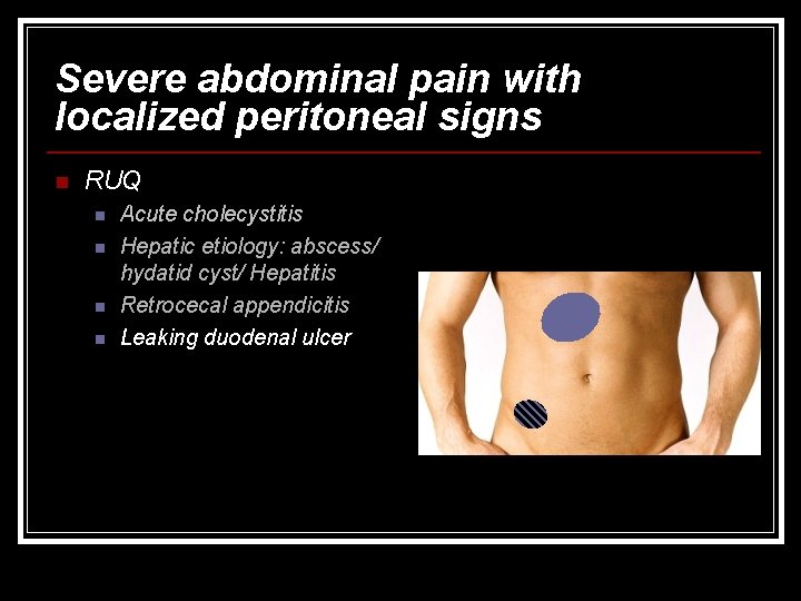
Severe abdominal pain with localized peritoneal signs n RUQ n n Acute cholecystitis Hepatic etiology: abscess/ hydatid cyst/ Hepatitis Retrocecal appendicitis Leaking duodenal ulcer
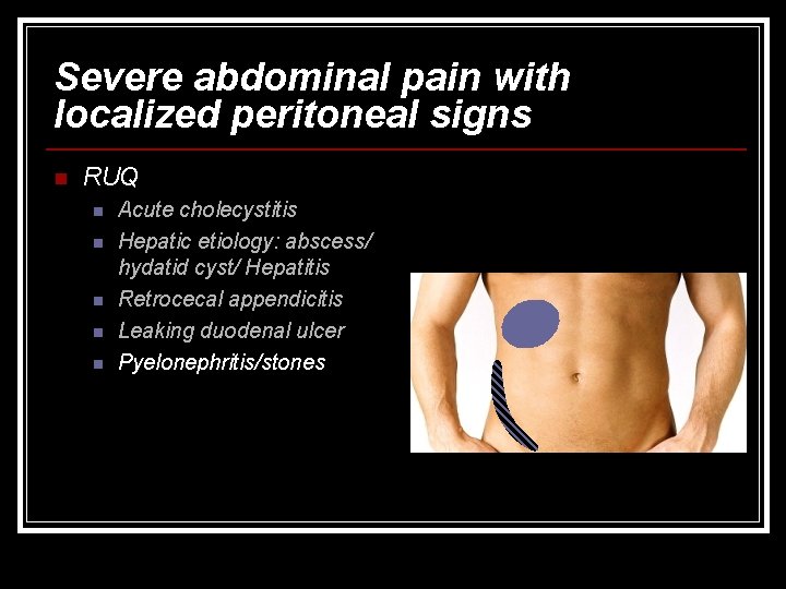
Severe abdominal pain with localized peritoneal signs n RUQ n n n Acute cholecystitis Hepatic etiology: abscess/ hydatid cyst/ Hepatitis Retrocecal appendicitis Leaking duodenal ulcer Pyelonephritis/stones
Severe abdominal pain with localized peritoneal signs n RUQ n n n Acute cholecystitis Leaking duodenal ulcer Hepatic etiology: abscess/ hydatid cyst/ Hepatitis Retrocecal appendicitis Pyelonephritis/stones Extra- abdominal causes n Lobar pneumonia
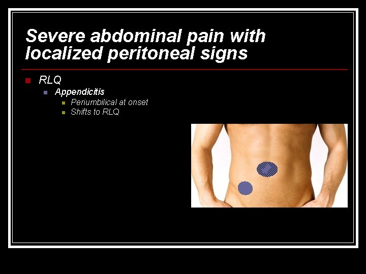
Severe abdominal pain with localized peritoneal signs n RLQ n Appendicitis n n Periumbilical at onset Shifts to RLQ
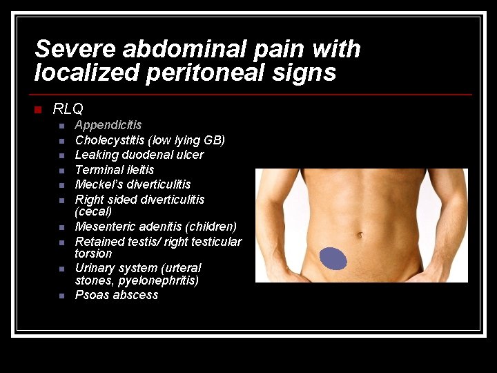
Severe abdominal pain with localized peritoneal signs n RLQ n n n n n Appendicitis Cholecystitis (low lying GB) Leaking duodenal ulcer Terminal ileitis Meckel’s diverticulitis Right sided diverticulitis (cecal) Mesenteric adenitis (children) Retained testis/ right testicular torsion Urinary system (urteral stones, pyelonephritis) Psoas abscess
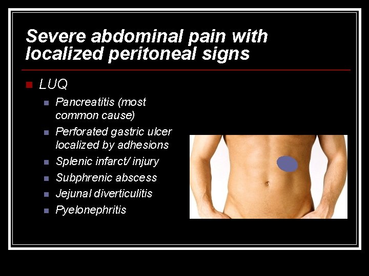
Severe abdominal pain with localized peritoneal signs n LUQ n n n Pancreatitis (most common cause) Perforated gastric ulcer localized by adhesions Splenic infarct/ injury Subphrenic abscess Jejunal diverticulitis Pyelonephritis
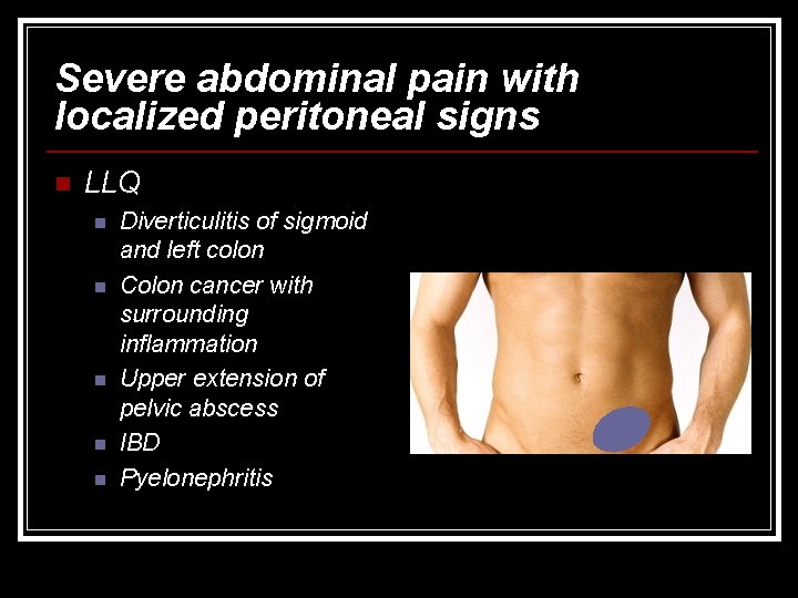
Severe abdominal pain with localized peritoneal signs n LLQ n n n Diverticulitis of sigmoid and left colon Colon cancer with surrounding inflammation Upper extension of pelvic abscess IBD Pyelonephritis
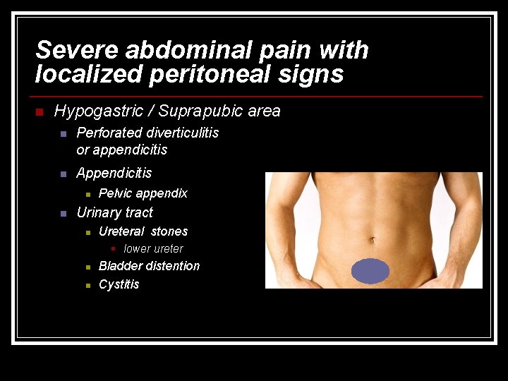
Severe abdominal pain with localized peritoneal signs n Hypogastric / Suprapubic area n Perforated diverticulitis or appendicitis n Appendicitis n n Pelvic appendix Urinary tract n Ureteral stones § lower ureter n n Bladder distention Cystitis
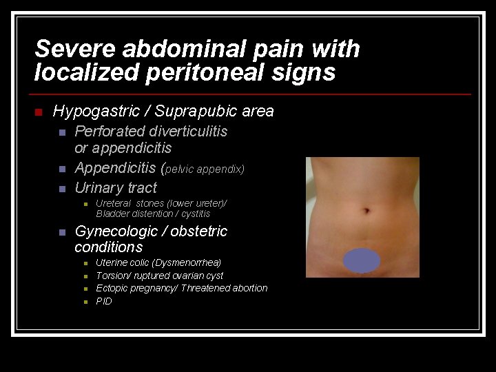
Severe abdominal pain with localized peritoneal signs n Hypogastric / Suprapubic area n n n Perforated diverticulitis or appendicitis Appendicitis (pelvic appendix) Urinary tract n n Ureteral stones (lower ureter)/ Bladder distention / cystitis Gynecologic / obstetric conditions n n Uterine colic (Dysmenorrhea) Torsion/ ruptured ovarian cyst Ectopic pregnancy/ Threatened abortion PID
- Slides: 44