The abdomen is the region of trunk that

The abdomen is the region of trunk that lies between the diaphragm above and the inlet of pelvis below.

Structure of the abdominal walls The diaphragm separates the abdomen from the chest superiorly. n Inferiorly the abdomen is continuous with the pelvis. n Anteriorly the anterior abdominal wall. n Posteriorly the abdominal wall is made by the lumbar vertebrae, twelfth rib, muscles, and upper part of bony pelvis. Note/ these walls are lined by fascia and parietal peritoneum. n

Structure of the anterior abdominal wall ANTERIOR ABDOMINAL WALL skin superficial fascia deep fascia muscles Extraperitoneal fascia parietal peritoneum

Anterior abdominal wall SKIN The skin is loosely attached to the underlying structures except at the umbilicus, where it is tethered to the scar tissue. n The natural lines of cleavage in the skin are constant and run downward and forward almost horizontally around the trunk. n

SURGICAL INCISIONS l l If possible, all surgical incisions should be made in the lines of cleavage where the bundles of collagen fibers in the dermis run in parallel rows. An incision along a cleavage line will heal as a narrow scar, whereas one that crosses the lines will heal as wide or heaped-up scars.

SURGICAL INCISIONS
 Deep membranous layer")
ANTERIOR ABDOMINAL WALL Superficial Fascia Superficial fatty layer (fascia of Camper) Deep membranous layer (Scarpa’s fascia).

ANTERIOR ABDOMINAL WALL Superficial Fascia n The fatty layer is continuous with the superficial fat over the rest of the body and may be extremely thick (3 in. ) or more in obese patients.

ANTERIOR ABDOMINAL WALL Superficial Fascia n The membranous layer is thin and fades out laterally and above, where it becomes continuous with the superficial fascia of the back and the thorax, respectively.

ANTERIOR ABDOMINAL WALL Superficial Fascia n lnferiorly, the membranous layer passes onto the front of the thigh, where it fuses with the deep fascia one fingerbreadth below the inguinal ligament.

ANTERIOR ABDOMINAL WALL Superficial Fascia n In the midline inferiorly, the membranous layer of fascia is not attached to the pubis but forms a tubular sheath for the penis (or clitoris).

ANTERIOR ABDOMINAL WALL Superficial Fascia n n Below in the perineum, it enters the wall of the scrotum (or labia majora). From there it passes to be attached on each side to the margins of the pubic arch; it is here referred to as Colles’ fascia.

ANTERIOR ABDOMINAL WALL Superficial Fascia n Posteriorly, it fuses with the perineal body and the posterior margin of the perineal membrane.

ANTERIOR ABDOMINAL WALL Superficial Fascia n n In the scrotum, the fatty layer of the superficial fascia is represented as a thin layer of smooth muscle, the dartos muscle. The membranous layer of the superficial fascia persists as a separate layer.

GENERAL APPEARANCES OF THE ABDOMINAL WALL v v v The normal abdominal wall is soft and pliable and undergoes inward and outward excursion with respiration. The contour is subject to considerable variation and depends on the tone of its muscles and the amount of fat in the subcutaneous tissue. Well-developed muscles or an abundance of fat can prove to be a severe obstacle to the palpation of the abdominal viscera.

MEMBRANOUS LAYER OF SUPERFICIAL FASCIA AND THE EXTRAVASATION OF URINE l l The membranous layer of the superficial fascia is important clinically because beneath it is a potential closed space that does not open into the thigh but is continuous with the superficial perineal pouch via the penis and scrotum. Rupture of the penile urethra may be followed by extravasation of urine into the scrotum perineum and penis and then up into the lower part of the anterior abdominal wall deep to the membranous layer of fascia.

MEMBRANOUS LAYER OF SUPERFICIAL FASCIA AND THE EXTRAVASATION OF URINE

MEMBRANOUS LAYER OF SUPERFICIAL FASCIA AND THE EXTRAVASATION OF URINE l The urine is excluded from the thigh because of the attachment of the fascia to the fascia Iata.

MEMBRANOUS LAYER OF SUPERFICIAL FASCIA AND THE EXTRAVASATION OF URINE l l When closing abdominal wounds it is usual for a surgeon to put in a continuous suture uniting the divided membranous layer of superficial fascia This strengthens the healing wound, prevents stretching of the skin scar, and makes for a more cosmetically acceptable result.

ANTERIOR ABDOMINAL WALL Deep Fascia n The deep fascia in the anterior abdominal wall is merely a thin layer of connective tissue covering the muscles; it lies immediately deep to the membranous layer of superficial fascia.

ANTERIOR ABDOMINAL WALL Muscles of ant. abd. wall external oblique internal oblique transversus abdominis In addition to the rectus abdominis in the midline & a small muscle called pyramidalis

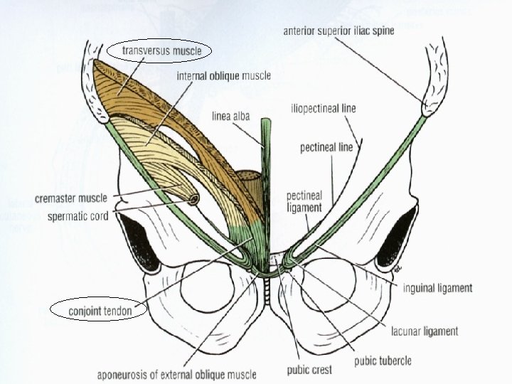
ANTERIOR ABDOMINAL WALL External Oblique n n n Is a broad, thin, muscular sheet. Arises from the outer surfaces of the lower eight ribs Inserted by a broad aponeurosis into the: - xiphoid process, - linea alba, - pubic crest, - pubic tubercle, - anterior half of the iliac crest.

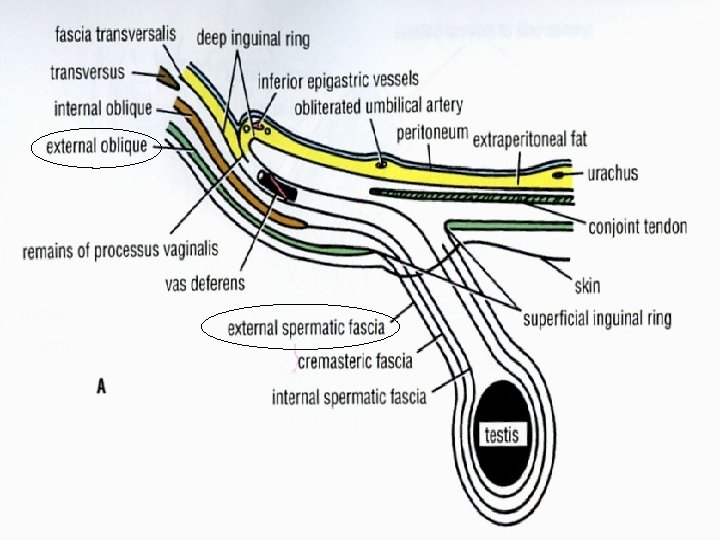
ANTERIOR ABDOMINAL WALL External Oblique v The superficial inguinal ring is a shaped defect in the external oblique aponeurosis lies immediately above and medial to the pubic tubercle.

ANTERIOR ABDOMINAL WALL External Oblique n It transmits the spermatic cord (or round ligament of the uterus) which carries the external spermatic fascia (or the external covering of the round ligament of the uterus) from the margins of the ring.

ANTERIOR ABDOMINAL WALL External Oblique n n Between the anterior superior iliac spine and the pubic tubercle the lower border of the aponeurosis is folded backward on itself, forming the inguinal ligament. From the medial end of the ligament, the lacunar ligament extends backward and upward to the pectineal line on the superior ramus of the pubis.
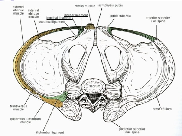

ANTERIOR ABDOMINAL WALL External Oblique n n The sharp, free crescentic edge of the lacunar ligament forms the medial margin of the femoral ring. On reaching the pectineal line, the lacunar ligament becomes continuous with a thickening of the periosteum called the pectineal ligament.

ANTERIOR ABDOMINAL WALL External Oblique v To the inferior rounded border of the inguinal ligament is attached the deep fascia of the thigh, the fascia lata.

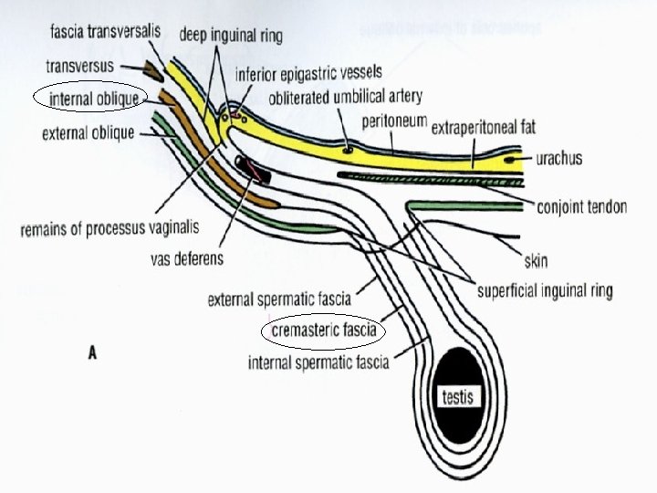
ANTERIOR ABDOMINAL WALL Internal Oblique n n A broad, thin, muscular sheet that lies deep to the external oblique; most of its fibers run at right angle s to those of the external oblique (upward and forward). It arises from: -lumbar fascia, -the anterior two thirds of the iliac crest, -the lateral two thirds of the inguinal ligament.

ANTERIOR ABDOMINAL WALL Internal Oblique n The muscle is inserted into: -the lower border of the lower three ribs, -the xiphoid process, -the linea alba, -the symphysis pubis.

ANTERIOR ABDOMINAL WALL Internal Oblique n It has a lower free border that arches over the spermatic cord (or round ligament of the uterus) and then descends behind it to be attached to the pubic crest and the pectineal line.

ANTERIOR ABDOMINAL WALL Internal Oblique n Near their insertion, n the lowest tendinous fibers are joined by similar fibers from the transversus abdominis to form the conjoint tendon. The conjoint tendon is attached medially to the linea alba, but it has a lateral free border.

ANTERIOR ABDOMINAL WALL Internal Oblique n n n The spermatic cord carries with it some of the muscle fibers that are called the cremaster muscle. The cremasteric fascia is the term used to describe the cremaster muscle and its fascia. Is supplied by the genital branch of the genitofemoral nerve.

ANTERIOR ABDOMINAL WALL Transversus abdominis n n A thin sheet of muscle that lies deep to the internal oblique, and its fibers run horizontally forward. It arises from: -the deep surface of the lower six costal cartilages (interdigitating with the diaphragm), -the lumbar fascia, -the anterior two thirds of the iliac crest, -the lateral third of the inguinal ligament.

ANTERIOR ABDOMINAL WALL Transversus abdominis n It is inserted into the: -xiphoid process, -the linea alba, -the smphysis pubis.

ANTERIOR ABDOMINAL WALL Transversus abdominis n The lowest tendinous fibers join similar fibers from the internal oblique to form the conjoint tendon, which is fixed to the pubic crest and the pectineal line.

The posterior border of the external oblique muscle is free, whereas the posterior borders of the internal oblique and transversus muscles are attached to the lumbar vertebrae by the lumbar fascia.

ANTERIOR ABDOMINAL WALL Rectus abdominis n A long strap muscle that extends along the whole length of the anterior abdominal wall, it is broader above and lies close to the midline, being separated from its fellow by the linea alba.

ANTERIOR ABDOMINAL WALL Rectus abdominis n It arises by two heads, from the front of n the symphysis pubis and from the pubic crest. It is inserted into the fifth, sixth, and seventh costal cartilages and the xiphoid process.

ANTERIOR ABDOMINAL WALL Rectus abdominis n When it contracts, its lateral margin forms a cowed ridge that can be palpated and often seen and is termed the linea semilunaris. This extends from the tip of the ninth costal cartilage to the pubic tubercle.

ANTERIOR ABDOMINAL WALL Rectus abdominis n The rectus abdominis muscle is divided into distinct segments by three transverse tendinous intersections: -at the xiphoid process, -at the umbilicus, -one halfway between these two.

ANTERIOR ABDOMINAL WALL Pyramidalis The pyramidalis muscle is often absent, it arises by its base from the anterior surface of the pubis and is inserted into the linea alba. n It lies in front of the lower part of the rectus abdominis. n

ANTERIOR ABDOMINAL WALL Rectus sheath n n n Is a long fibrous sheath that encloses the rectus abdominis muscle and pyramidalis muscle (if present). contains the: anterior rami of the lower six thoracic nerves, superior and inferior epigastric, vessels lymph vessels. It is formed mainly by the aponeuroses of the three lateral abdominal muscles.

ANTERIOR ABDOMINAL WALL Rectus sheath n Above the costal margin: - the anterior wall is formed by the aponeurosis of the external oblique. - the posterior wall is formed by the thoracic wall (the fifth, sixth, and seventh costal cartilages and the intercostal spaces).

ANTERIOR ABDOMINAL WALL Rectus sheath n Between the costal margin and the level of the anterior superior iliac spine: the aponeurosis of the internal oblique splits to enclose the rectus muscle; the external oblique aponeurosis is directed in front of the muscle, and the transversus aponeurosis is directed behind the muscle.

ANTERIOR ABDOMINAL WALL Rectus sheath Between the level of the anterosuperior iliac spine and the pubis: - the anterior wall by the aponeuroses of all three muscles. - the posterior wall is absent, and the rectus muscle lies in contact with the fascia transversalis. n

ANTERIOR ABDOMINAL WALL Rectus sheath n n It should be noted that where the aponeuroses forming the posterior wall pass in front of the rectus at the level of the anterior superior iliac spine, the posterior wall has a free, curved lower border called the arcuate line. At this site, the inferior epigastric vessels enter the rectus sheath and pass upward to anastomose with the superior epigastric vessels.
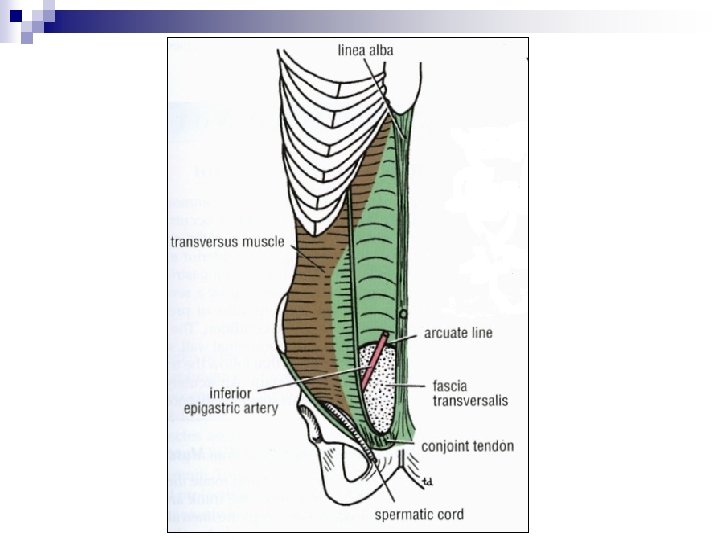

ANTERIOR ABDOMINAL WALL Rectus sheath n The rectus sheath is separated n n from its fellow on the opposite side by a fibrous band called the linea alba. This extends from the xiphoid process down to the symphysis pubis and is formed by the fusion of the aponeuroses of the lateral muscles of the two sides. It is wider above the umbilicus and narrows down below it to be attached to the symphysis pubis.

ANTERIOR ABDOMINAL WALL Rectus sheath n The posterior wall of the rectus sheath is not attached to the rectus abdominis muscle. The anterior wall is firmly attached to it by the muscle’s tendinous intersections.

HEMATOMA OF THE RECTUS SHEATH l l l Hematoma of the rectus sheath is uncommon but Important. since it is often overlooked. It occurs most often on the right side below the level of the umbilicus. The source of the bleeding is the inferior epigastric vein or, more rarely, the inferior epigastric artery.

HEMATOMA OF THE RECTUS SHEATH

HEMATOMA OF THE RECTUS SHEATH l l l These vessels may be stretched during a severe bout of coughing or in the later months of pregnancy, which may predispose to the condition. The cause is usual blunt trauma to the abdominal wall, such as a fall or a kick. The symptoms that follow the trauma include midline abdominal pain. An acutely tender mass confined to one rectus sheath is diagnostic.

Function of the Anterior Abdominal Wall Muscles n n The oblique muscles laterally flex and rotate the trunk. The rectus abdominis flexes the trunk and stabilizes the pelvis, and the pyramidalis keeps the linea alba taut during the process.

Function of the Anterior Abdominal Wall Muscles The muscles of the anterior and lateral abdominal walls assist the diaphragm during inspiration by relaxing as the diaphragm descends so that the abdominal viscera can be accommodated. The muscles assist in the act of forced expiration that occurs during coughing and sneezing by pulling down the ribs and sternum. Their tone plays an important part in sup porting and protecting the abdominal viscera. By contracting simultaneously with the diaphragm, with the glottis of the larynx closed, they increase the intra-abdominal pressure and help in micturition, defecation , vomiting, and parturition.

Nerve Supply of Anterior Abdominal Wall Muscles n The oblique and transversus abdominis muscles the lower six thoracic nerves and the iliohypogastric and ilioinguinal nerves (L 1). n The rectus muscle n The pyramidalis lower six thoracic nerves). the twelfth thoracic nerve.

ABDOMINAL MSCLES, ABDOMINO THORACIC RHYTHM AND VISCEROPTOSIS • The shape of the anterior abdominal wall depends on the tone of its muscles. • A middle-aged woman with poor abdominal muscles who has had multiple pregnancies is often incapable of supporting her abdominal viscera. • The lower part of the anterior abdominal wall protrudes forward, a condition known as visceroptosis. This should not be confused with an abdominal tumor such as an ovarian cyst or with the excessive accumulation of fat in the fatty layer of the superficial fascia.

ANTERIOR ABDOMINAL WALL Fascia Transversalis n n Is a thin layer of fascia that lines the transversus abdominis muscle and is continuous with a similar layer lining the diaphragm and the iliacus muscle. It is important to understand that the fascia transversalis, the diaphragmatic fascia, the iliacus fascia, and the pelvic fascia form one continuous lining to the abdominal and pelvic cavities.

ANTERIOR ABDOMINAL WALL Fascia Transversalis n The femoral sheath for the femoral vessels in the lower limbs is formed from the fascia transversalis and the fascia iliaca that covers the iliacus muscle.

ANTERIOR ABDOMINAL WALL Extraperitoneal Fat n The extraperitoneal fat is a thin layer of connective tissue that contains a variable amount of fat and lies between the fascia transversalis and the parietal peritoneum.

ANTERIOR ABDOMINAL WALL Parietal Peritoneum n The walls of the abdomen are lined with parietal peritoneum. This is a thin serous membrane and is continuous below with the parietal peritoneum lining the pelvis.

Nerves of the Anterior Abdominal Wall the lower six thoracic nerves the first lumbar nerve

Nerves of the Anterior Abdominal Wall n n The anterior rami of the lower six thoracic nerves. The first lumbar nerve (the iliohypogastric and ilioinguinal nerves). They supply the skin of the anterior abdominal wall, the muscles, and the parietal peritoneum. They pass forward in the interval between the internal oblique and the transversus muscles.
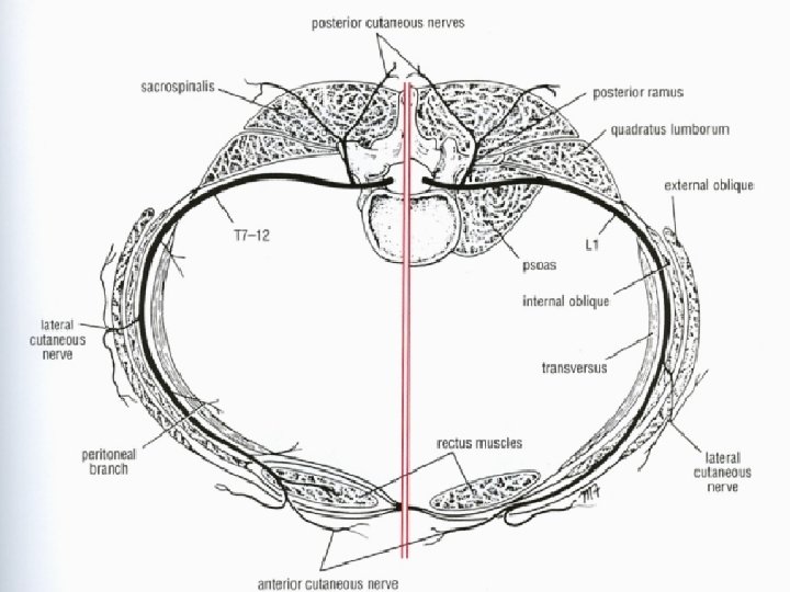

Nerves of the Anterior Abdominal Wall n The lower six thoracic nerves pierce the posterior wall of the rectus sheath to supply the rectus muscle and the pyramidalis (T 12 only). They terminate by piercing the anterior wall of the sheath and supplying the skin.

Nerves of the Anterior Abdominal Wall n n n The first lumbar nerve has a similar course, but it does not enter the rectus sheath. It is represented by the iliohypogastric nerve, which pierces the external oblique aponeurosis above the superficial inguinal ring, and by the ilioinguinal nerve, which emerges through the ring. They end by supplying the skin just above the inguinal ligament and symphysis pubis.

ABDOMINAL PAIN Muscle Rigidity and Referred Pain l l Sometimes it is difficult for a physician to decide whether the muscles of the anterior abdominal wall of a patient are rigid because of underlying inflammation of the parietal peritoneum or whether the patient is voluntarily contracting the muscles because he or she resents being examined or because the physician’s hand is cold. This problem is usually easily solved by asking the patient, who is lying supine on the examination table, to rest the arms by the sides and draw up the knees to flex the hip joints.

ABDOMINAL PAIN Muscle Rigidity and Referred Pain l l It is practically impossible for a patient to keep the abdominal musculature tensed when the thighs are flexed. Needless to say, the examiner’s hand should be warm. A pleurisy involving the lower costal parietal pleura causes pain in the overlying skin that may radiate down into the abdomen. Although it is unlikely to cause rigidity of the abdominal muscles, it may cause confusion in making a diagnosis unless these anatomic facts are remembered.

ANTERIOR ABDOMINAL NERVE BLOCK Area of Anesthesia l l The nerves of the anterior and lateral abdominal walls are the anterior rami of the seventh through the twelfth thoracic nerves and the first lumbar nerve (ilioinguinal and iliohypogastric nerves) Indications : Repair of lacerations of the anterior abdominal wall.

ANTERIOR ABDOMINAL NERVE BLOCK Area of Anesthesia l l l Procedure : The anterior ends of intercostal nerves. T 7 through T 1 inter the abdominal wall by passing posterior to the costal cartilages. An abdominal field block is most easily carried out along the lower border of the costal margin and then infiltrating the nerves as they emerge between the xiphoid process and the tenth or eleventh rib along the costal margin.

ANTERIOR ABDOMINAL NERVE BLOCK Area of Anesthesia l l l The ilioinguinal nerve passes forward in the inguinal canal and emerges through the superficial inguinal ring. The iliohypogastric nerve passes forward around the abdominal wall and pierces the external oblique aponeurosis above the superficial inguinal ring. The two nerves are easily blocked by inserting the anesthetic needle 1 in. (2. 5 cm) above the anterior superior iliac spine on the spinoumbilical line.

Arteries of the Anterior Abdominal Wall superior epigastric artery inferior epigastric artery deep circumflex iliac artery lower two posterior intercostal arteries four lumbar arteries

Arteries of the Anterior Abdominal Wall n The superior epigastric artery, n one of the terminal branches of the internal thoracic artery, enters the upper part of the rectus sheath between the sternal and costal origins of the diaphragm. It descends behind the rectus muscle, supplying the upper central part of the anterior abdominal wall, and anastomoses with the inferior epigastric artery.

Arteries of the Anterior Abdominal Wall n n n The inferior epigastric artery is a branch of the external iliac artery just above the inguinal ligament. It runs upward and medially along the medial side of the deep inguinal ring. It pierces the fascia transversalis to enter the rectus sheath anterior to the arcuate line. It ascends behind the rectus muscle, supplying the lower central part of the anterior abdominal wall, and anastomoses with the superior epigastric artery.

Arteries of the Anterior Abdominal Wall n The deep circumflex iliac artery is a branch of the external iliac artery just above the inguinal ligament. It runs upward and laterally toward the anterosuperior iliac spine and then continues along the iliac crest, It supplies the lower lateral part of the abdominal wall.

Arteries of the Anterior Abdominal Wall n The lower two posterior intercostal arteries, branches of the descending thoracic aorta, and the four lumbar arteries, branches of the abdominal aorta, pass forward between the muscle layers and supply the lateral part of the abdominal wall.

Veins of the Anterior Abdominal Wall Superficial Veins Deep Veins

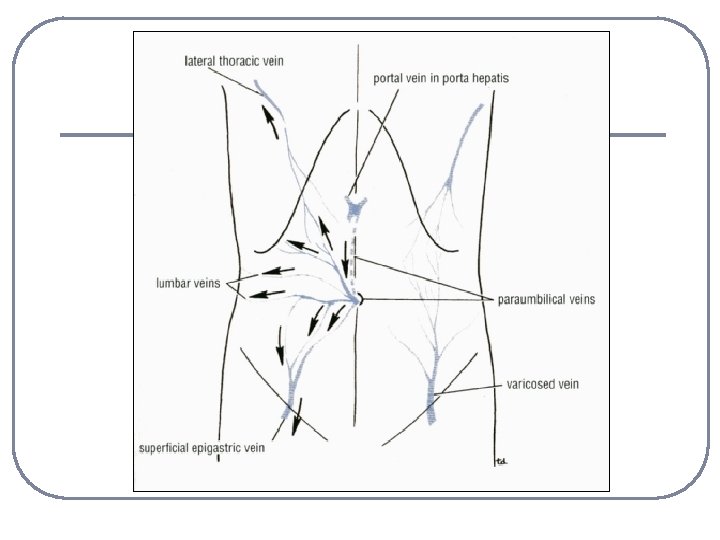
Veins of the Anterior Abdominal Wall Superficial Veins n n The superficial veins form a network that radiates out from the umbilicus. Above, the network is drained into the axillary vein via the lateral thoracic vein Below, into the femoral vein via the superficial epigastric and great saphenous veins. A few small veins, the paraumbilical veins, connect the network through the umbilicus and along the ligamentum teres to the portal vein. This forms an important porto- systemic venous anastomosis.

Veins of the Anterior Abdominal Wall Deep Veins n n The deep veins of the abdominal wall, the superior epigastric, inferior epigastric, and deep circumflex iliac veins, follow the arteries of the same name and drain into the internal thoracic and external iliac veins. The posterior intercostal veins drain into the azygos veins, and the lumbar veins drain into the inferior vena cava.

PORTAL VEIN OBSTRUCTION l l In cases of portal vein obstruction, the superficial veins around the umbilicus and the paraumbilical veins become grossly distended. The distended subcutaneous veins radiate out from the umbilicus, producing in severe cases the clinical picture referred to as caput medusae.

PORTAL VEIN OBSTRUCTION

CAVAL OBSTRUCTION l l If the superior or inferior vena cava is obstructed, the venous blood causes distension of the veins running from the anterior chest wall to the thigh. The lateral thoracic vein anastomoses with the superficial epigastric vein, a tributary of the great saphenous vein of the leg. In these circumstances, a tortuous varicose vein may extend from the axilla to the lower abdomen

Lymph Drainage of the Anterior Abdominal Wall Superficial lymph vessels Deep lymph vessels

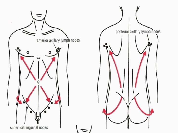
Lymph Drainage of the Anterior Abdominal Wall Superficial Lymph Vessels n n The lymph drainage of the skin of the anterior abdominal wall above the level of the umbilicus is upward to the anterior axillary nodes. Below the level of the umbilicus, the lymph drains downward and laterally to the superficial inguinal nodes.

Lymph Drainage of the Anterior Abdominal Wall Superficial Lymph Vessels n n The lymph of the skin of the back above the level of the iliac crests is drained upward to the posterior axillary nodes Below the level of the iliac crests, it drains downward to the superficial inguinal nodes.

Lymph Drainage of the Anterior Abdominal Wall Deep Lymph Vessels n The deep lymph vessels follow the arteries and drain into the internal thoracic, external iliac, posterior mediastinal, and para-aortic (lumbar) nodes.

SKIN AND ITS REGIONAL LYMPH NODES l l A knowledge of the areas of the skin that drain into a particular group of lymph nodes is clinically important. For example. it is possible to find a swelling in the groin (enlarged superficial inguinal node) caused by an infection or malignant tumor of the skin of the lower part of the anterior abdominal wall or that of the buttock.

Umbilicus n The umbilicus is a consolidated scar representing the site of attachment of the umbilical cord in the fetus; it is situated in the linea alba.

INFECTION OF THE UMBILICUS l In the adult, the umbilicus often receives scant attention in the shower and is consequently a common site of infection.

- Slides: 97