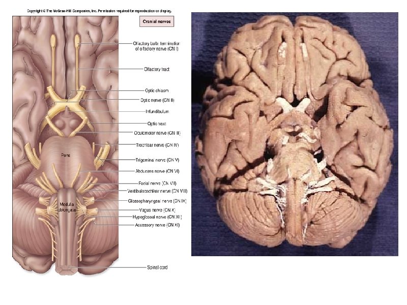
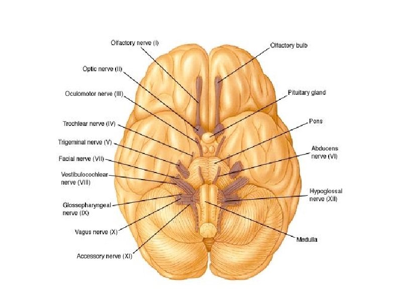
The 12 Pairs of Cranial Nerves Figure 14


.")

")

 - the only cranial nerve that travels")


 12 pairs")


 • Parasympathetic (resting)")


- Slides: 35
The 12 Pairs of Cranial Nerves Figure 14. 8
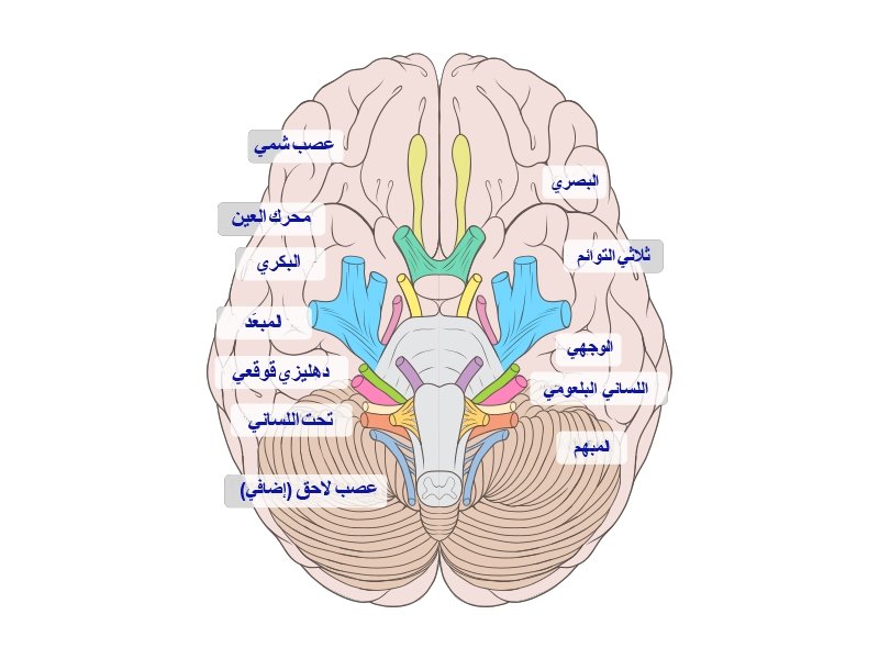
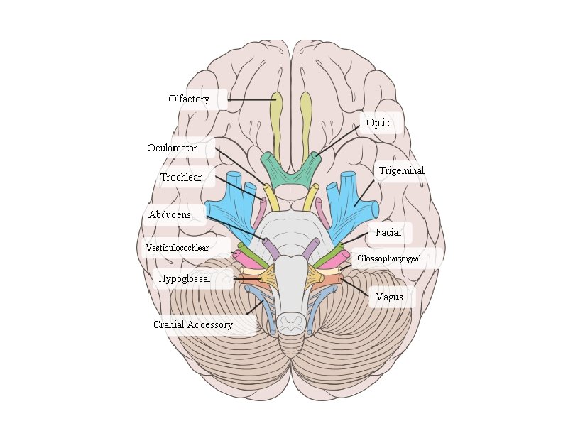
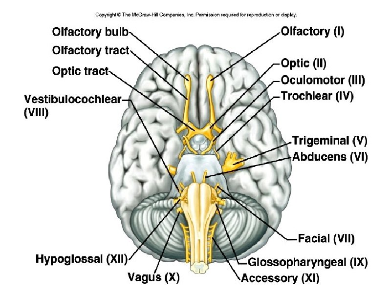
I. OLFACTORY • Transmit the sense of smell. Outside of the CNS they are called olfactory nerves, and inside of the CNS they are called the olfactory tract.
II. OPTIC NERVE • Transmits information from the eye’s retina.
III Occulomotor Nerve • This controls most of the extrinsic muscles of the eye (that move the eyeball).
IV. Trochlear Nerve • Innervates an extrinsic eye muscle
V. Trigeminal Nerve • This is the main sensory nerve of the face. It has a large branch that passes through the foramen ovale of the skull. It has three parts.
VI: Abducens Controls one of the eye muscles (lateral rectus).
VII Facial Nerve • This innervates the muscles of facial expression. • A person who cannot blink or smile may have damage to this nerve. • BELL’S PALSY is damage of the facial nerve causing paralysis on one side.
VIII. VESTIBULOCOCHLEAR • Transmits hearing and balance. (also called Auditory nerve)
IX: GLOSSOPHARYNGEAL • carries information from the head and neck to the brainstem. • Information about blood pressure (baroreceptors)
X Vagus Nerve • (vagrant = “wanders”) - the only cranial nerve that travels into the abdomen. • This is the most important cranial nerve because it innervates all of the organs in the thoracic and abdominal cavities
XI: ACCESSORY NERVE • Enters the skull through foramen magnum and leaves through the jugular foramen. • It just supplies the shoulder muscles.
XII. HYPOGLOSSAL NERVE • Supplies the tongue. • Damage causes impairment of speech.
Need to know all of the cranial nerves ? On Old Olympus Towering Top A Fin And German Viewed A Hop
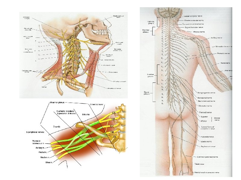
Spinal Nerves 8 pairs of cervical nerves (C 1 - C 8) 12 pairs of thoracic nerves (T 1 -T 12) 5 pairs of lumbar nerves (L 1 -L 5) 5 pairs of sacral nerves (S 1 -S 5) 1 pair of coccygeal nerves (Co) 31 Total
*Spinal cords ends at the level between the 1 st and 2 nd lumbar vertebrae *The lumbar, sacral, coccygeal nerves descend from the end of the cord – CAUDA EQUINA (horse’s tail)
ROOTS
Each nerve emerges from the spinal cord at points called ROOTS Dorsal Root Ganglion Ventral root ganglion
PLEXUSES • Main portions of the spinal nerves combine to form complex networks called PLEXUSES
1. Cervical Plexuses - neck 2. Brachial Plexus - shoulders, arms, hands ulnar, median, radial, axillary nerves 3. Lumbrosacral Plexuses - pelvic area sciatic nerve, femoral nerve
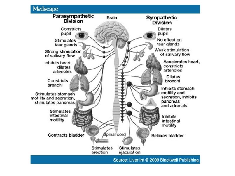
AUTONOMIC NERVOUS SYSTEM • Sympathetic (fight or flight) • Parasympathetic (resting)
9. 15 Autonomic Nervous System Sympathetic - energy, high stress, emergency Fight or Flight Parasympathetic resting, normal Divisions act antagonistically - one is exhitatory, other inhibits
More Images of The Cranial Nerves
The sheep brain also has the 12 cranial nerves, but they can be difficult to find
Assignment: Coloring of the Cranial Nerves