Thalamus and its connections Introduction Anatomically the thalamus


- Slides: 18
Thalamus and its connections
Introduction ● ● Anatomically the thalamus is a large ovoid mass of grey matter lying above the midbrain. two thalami situated one on each side of a slitlike cavity, the third ventricle The thalami and third ventricle as seen from above
● ● The long axes of the thalami are set obliquely running backwards and laterally The pointed anterior ends are nearer to the median plane whereas the wider posterior ends are separated from each other by pineal body, superior colliculi and habenular triangles. The thalami and third ventricle as seen from above
● ● The thalami are usually attached across the median plane by a narrow interthalamic connexus of grey matter (also called interthalamic adhesion ). Each thalamus forms most of the lateral wall of the third ventricle and floor of the central part of the lateral ventricle.
● ● Functionally, the thalamus is generally considered as the great sensory gateway to the cerebral cortex. It receives impulses from the opposite half of the body and transmits most of them to the sensory area of the cerebral cortex (Brodmann areas 3, 2, and 1).
External features Each thalamus has two ends and four surfaces. Ends • The anterior end is narrow and constitutes the tubercle of thalamus. It forms the posterior boundary of the interventricular foramen. • The posterior end is expanded and is known as pulvinar. It overhangs the medial and lateral geniculate bodies, and superior colliculi with their brachia.
Superior surface. Its lateral part forms the floor of the central part of the lateral ventricle and its medial part is covered by the tela choroidea of the third ventricle. Inferior surface. Hypothalamus anteriorly and subthalamus posteriorly. Medial surface. It forms the greater part of the lateral wall of the third ventricle. Lateral surface. It forms the medial boundary of the posterior limb of internal capsule.
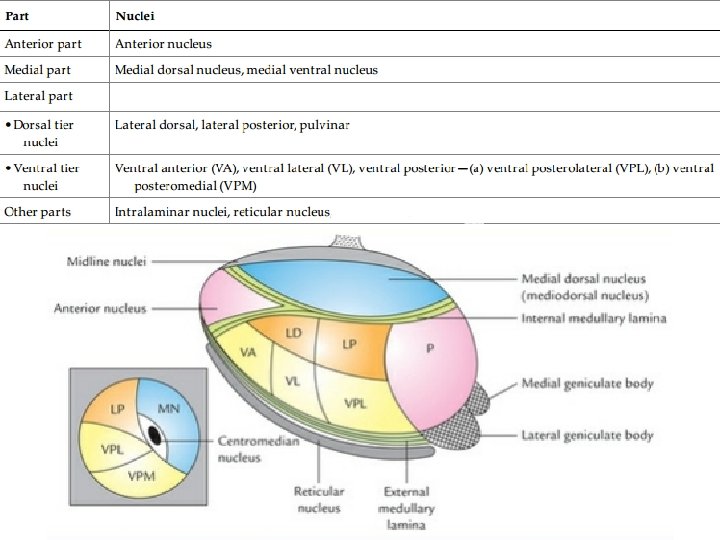
internal structure The thalamus consists mainly of grey matter and only a small amount of white matter. Horizontal section of the thalamus (schematic) to show the location of various thalamic nuclei. The diagram in the inset is the coronal section of thalamus passing in front of pulvinar showing ventral posteromedial (VPM), ventral posterolateral (VPL) nuclei, and centromedian nucleus. (P = pulvinar, LD = lateral dorsal nucleus, LP= lateral posterior nucleus, VA= ventral anterior nucleus, VL = ventral lateral nucleus, VPL = ventral posterolateral nucleus, MN = Medio- dorsal nucleus. )
White matter ● ● The lateral surface of the thalamus is covered by a thin layer of white matter called external medullary lamina and its superior surface by a similar layer of white matter called stratum zonale. A vertical Y-shaped sheet of white matter within the thalamus is called internal medullary lamina.
grey matter ● ● ● Y-shaped internal medullary lamina divides the thalamus into three main parts: anterior, medial, and lateral. The anterior part includes the anterior tubercle and lies between the ‘limbs’ of the Y, the medial and lateral parts lie on either side of the ‘stem’ of the Y. Each of these parts consists of number of nuclei.
The thalamic nuclei are classified into three main functional groups: specific, nonspecific and reticular. Connections of the specific nuclei These nuclei receive input from certain ascending tracts and project it to the specific (primary) cortical areas. Nuclei of this group comprise ventral tier nuclei ,
Connections of the nonspecific nuclei ● ● ● Nonspecific nuclei do not receive afferents from ascending tracts, but have abundant connections with other diencephalic nuclei. They mostly project to the cortical ‘association areas’ in the frontal and parietal lobes. Nuclei of this group comprise anterior nucleus, dorsal medial nucleus and dorsal tier nuclei of thalamus.
Connections of the reticular nuclei The reticular nuclei of thalamus include reticular nucleus, intralaminar nuclei and median nuclei. These nuclei are connected with the reticular formation.
Functions ● It is a sensory relay station of all the sensory pathways except for the olfactory pathway, which is projected directly to the cerebral cortex without being relayed in the thalamus. ● Modify the cortical activities: It influences voluntary movements by receiving impulses from basal ganglia and cerebellum and relaying them to the motor cortex, which in turn influences lower motor neurons through corticonuclear and corticospinal pathways. ● Higher center for autonomic system and viscera(Dorsomedial nucleus) ● Appreciation of pain.
applied Thalamic syndrome It usually occurs subsequent to a vascular lesion of the thalamus (viz. thrombosis of thalamogeniculate artery). Characteristic features The threshold for pain, touch and temperature is decreased on the opposite side of the body (thalamic overreaction) the sensations are exaggerated, perverted and disagreeable. For example, the prick of a pin may be felt as a severe burning sensation, and even music that is ordinarily pleasing may be disagreeable. Sometimes even light touch may produce excruciating pain. fail to respond to powerful analgesics (pain relieving) drugs.
Thalamic hand ● ● ● It is sometimes seen in thalamic lesions. The opposite hand is held in an abnormal posture. The forearm is pronated, wrist flexed, metacarpophalangeal joints flexed and interphalangeal joints extended. The fingers can be moved but slowly, due to altered muscle tone in the different muscle groups. The integrity of anterior nucleus and its connections is necessary for attention and recent memory, therefore a lesion involving them can lead to loss of recent memory.
thank you