Th 17 AntiCytokine Autoantibodies IL17 A IL 17
 and abnormal Th 17")
“Th 17” Anti-Cytokine Autoantibodies (IL-17 A, IL 17 F, IL-22) and abnormal Th 17 T cell function Associated with Mucocutaneous Candidiasis of APS-1 Kisand…Meager et al Chronic mucocutaneous candidiasis in APECED or thymoma patients correlates with autoimmunity to Th 17 -associated cytokines. J Exp Med 2010 207: 299 -308 Puel…. Casanova et al Autoantibodies against IL-17 A, IL-17 F, and IL-22 in patients with chronic mucocutaneous candidiasis and autoimmune polyendocrine syndrome type I. J Exp Med 2010, 207: 264 -265. Ahlgren…Lobell et al Increased IL-17 A secretion in response to Candida albicans in autoimmune polyendocrine syndrome type 1 and its animal model. Eur J Immunol 2011, 41: 235 -245 Anti-Cytokine Autoantibodies

Baker et al. Haplotype Analysis discriminates Genetic Risk for DR 3 Associated Endocrine Autoimmunity and Helps Define Extreme Risk for Addison’s Disease. JCEM 2010 95: E 263 -E 270. Multiplex Addison’s Families Greater DR 3/4 -B 8 80% Multiplex Simplex Control Less Complete DR 3 -B 8 -A 1 Extended Haplotype

General Paradigm • Identify Genetic Susceptibility • Detect Initial Autoantibodies • Monitor Metabolic Decompensation • Treat Overt Disease Prior to Morbidity/Mortality • Basic/Clinical Research to Allow Prevention

Associated Autoimmune Illnesses

Premature Mortality in Patients with Addison’s Disease: A Population-Based Study J clin endocrinol Metab 91: 4859, 2006 Percent Dying 6. 7 yr follow-up; mean start age 52. 8 N=507 deaths of 1675 patients N=199 deaths
 APS-I (AIRE mutation) XPID: (Scurfy Mutation)")
Autoimmune Polyendocrine Syndromes • • APS-II (Autoimm Polyendocrine) APS-I (AIRE mutation) XPID: (Scurfy Mutation) Anti-insulin Receptor Abs + “Lupus” Hirata (Anti-insulin Autoantibodies) POEMS (Plasmacytoma, . . ) Thymic Tumors + Autoimmunity Congenital Rubella + DM +Thyroid

Polyendocrine non-Autoimmune Syndromes • Wolfram’s Syndrome – DIDMOAD Diabetes Insipidus, Diabetes Mellitus, Optic Atrophy, and Deafness (WFS 1 gene mutation on Chromosome 4) • Kearns-Sayre Syndrome External Ophthalmoplegia, Retinal Degeneration, Heart Block- Diabetes, Hypoparathyroidism, Thyroiditis reported (Mitochondrial deletions, rearrangments)

APS-Syndromes Betterle et al. Endocrine Reviews 2002 Neufeld and Blizzard: 1980, Pinchera, in Symposium Autoimmune Endocrine Aspects of Endocrine Disorders • APS-I: >=2 of Candidiasis, Hypopara, Addison’s • APS-II: Addison’s + Autoimmune Thyroid and/or Type 1 Diabetes (Addison’s must be present) • APS-III: Thyroid Autoimmune + other autoimmune [not Ad, hypopara, candidiasis] • APS-IV: Two or more organ-specific autoimmune, not I, II, or III.

Comparison APS-I and APS-II • Onset Infancy • Siblings AIRE gene mutated • Not HLA Associated • Immunodeficiency Asplenism • • Mucocutaneous Candidiasis • 18% Type 1 DM BDC Older Onset Multiple Generations DR 3/4 Associated No Defined Immunodeficiency • 20% Type 1 DM
")
APS-I • Autoimmune Polyendocrine Syndrome Type 1 • Autosomal Recessive mutations AIRE (Autoimmune Regulator) gene • Mucocutaneous Candidiasis/Addison’s Disease/Hypoparathyroidism • 18% Type 1 Diabetes • “Transcription Factor” in Thymus BDC

Diagnosis • Classic criterion – At least two: • • Chronic recurrent mucocutaneous candidiasis Hypoparathyroidism Addison’s disease Prevalence of these criterion by 30 years is only 94% – High index of suspicion with individuals presenting with multiple autoimmune disease – In siblings one autoimmune disease is required for diagnosis • Mutation analysis – Three most common mutations may miss 5%

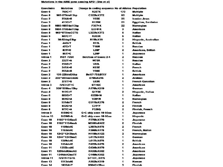
200 C 322 fs. X 372 300 Plextrin Homolo gy 1 Homolo gy 2 LXLL 100 SAND Domain NLS LXLL 0 LXLL Homogeneously Staining Domain 400 LXLL AIRE (Autoimmune Regulator) and Percentage Mutations APS-I: Halonen JCEM 87: 2568, 2002 500

MODEL AIRE Role in Preventing Autoimmunity Autoreactive thymocyte TCR Tolerization of autoreactive thymocyte MHC + Peptide Thymic Medullary Epithelial Cells AIRE Self-peptides from "peripheral" antigens Mathis/Benoist

Highly variable expression of tissue-restricted self-antigens in human thymus: Implications for self-tolerance and autoimmunity Richard Taubert, Jochen Schwendemann and Bruno Kyewski Division of Developmental Immunology, Tumor Immunology Program, German Cancer Research Center, Heidelberg, Germany

Insulin Message but not GAD 67 thymic meduallary epithelial expression is tremendously variable and correlates with AIRE message Log scale 100 -fold differences Continuous not step-wise variation Taubert et al, 2007 EJI

Gene Dosage-limiting Role of Aire in Thymic Expression, Clonal Deletion, and Organ-Specific Autoimmunity Liston et al. J. Exp Med 200: 1015, 2004 Rip-HEL Antigen+CD 4 T Cell Receptor anti-HEL Model X 107 CD 4+Cd 8 -1 G 12 -CD 69 -

Halonen JCEM 87: 2568, 2002 104 APS-I International Series Patients Greater % Addison’s and Candidiasis with R 257 X Nonsense (X) Mutation

APS-I Patients Protected from Diabetes by DQB 1*0602 Diabetes DQB 1*0602+ 0 Not Diabetic 25 P=. 03 DQB 1*0602 - 13 66 16. 4% Halonen et al JCEM 87: 2574, 2002

NALP 5: Hypoparathroidism NACHT leucine-rich-protein 5 • • • >>Expression Parathyroid and Ovary 41% Hypopara+APS-1 Positive 0% Not APS-1 Animal Model “Have Abs” 68% Hypogonad+; 29% not Hypogonad Day 3 Thymectomy model + Kampe et al NEJM 358: 1018, 2008
 • Check oral Candida, Autontibodies, Ca, Pi, Na,")
A. 6 Month Evaluation APS-I (Perheentupa) • Check oral Candida, Autontibodies, Ca, Pi, Na, K, Mg, Alkaline phosphatase, ACTH, TSH, HCG, renin, Hb. A 1 c, Howell-Jolly smear, platelets • Autoantibodies: 21 -hydroxylase (Addison’s), GAD 65 (Diabetes), 17 OH, CYP 450 scc (hypogonadism/Addison’s); Tryptophane hydroxylase (intestine chromaffin cell loss), H/K ATPase and Intrinsic factor (Pernicious anemia), Thyroid peroxidase (hypothyroidism) • If hypoparathyorid: every 6 to 8 weeks check Ca • Intense control oral candida (e. g. amphotericin lozenge, fluconazole or ketoconazole if needed) with prompt biopsy suspicious lesion. Careful mouth hygiene with elimination of sharp points of teeth and plastic materials from mouth. • No live virus immunization • Patient web site: http: //www. empower. org. nz
 • Carry written warning of disease symptoms/complications •")
B. 6 Month Evaluation APS-I (Perheentupa) • Carry written warning of disease symptoms/complications • If Howell-Jolly bodies on smear, ultrasound spleen • Asplenic patients need meningococcal and hemophilus influenza type b immunization and pneumococcal vaccine with measured response. If no response to pneumococcal vaccine, prophylactic daily antibiotics • Keratoconjunctivitis: Topical steroid and vitamin A • Potential immunosuppression for hepatitits, refractory diarrhea and other refractory disorders

Check List APS-I Visit New Symptoms History New Signs Physical Oral Candidiasis New Antibodies (21 -OH, GAD, IA-2) Ca, Pi, Mg Na, K ALT ACTH, TSH, (LH, FSH) Hb. A 1 c Blood Smear (Howell-Jolly) Platelet Count Other

Oral Cancer Prevention APS-I • Aggressive Therapy Oral Candidiasis Amphoteracin Lozenges for early infection Fluconazole/Keotoconazole(2 -3 weeks) Itaconazole (4 -6 months) for nail candida • Prompt biopsy of suspicious oral lesion BDC

Immunodeficiency APS-I • Live virus vaccination avoided • If splenic atrophy present (Howell-Jolly bodies of blood smear, ultrasound) -Pneumococcal vaccine with Antibody response monitoring(6 -8 weeks) -If no antibody response daily antibiotic prophylaxis BDC

Gastrointestinal disease • Pernicious anemia • Autoimmune hepatitis • Diarrhea – – Hypocalcemia from hypoparathyroidism Celiac disease Intestinal infection (candida) Autoimmune destruction of endocrine cells of duodenal mucosa • Severe constipation

Table 8. 5 Unusual manifestations of disease – APS-I • Pituitary hormone deficiency (diabetes insipidus, growth hormone, gonoadotropic, ACTH deficiency) • Autoimmune disease (hyperthyroidism, rheumatoid arthritis, Sjogren’s syndrome, periodic fever with rash, antisperm autoimmunity, hemolytic anemia) • Hemetologic manifestations (pure red cell aplasia, autoimmune hemolytic anemia, splenomegaly and pancytopenia, Ig A deficiency) • Ocular disease (iridocyclitis, optic nerve atrophy, retinal degeneration) • Other organ system involvement (nephritis, cholelithiasis, Bronchiolitis obliterans organizing pneumonia, Lymphocytic myocarditis) • Hypokalemia with or without hypertension • Metaphyseal dysostosis

XPID: X-linked polyendocrinopathy, immune dysfunction and diarrhea • Other Names IPEX: Immunodysregulation, Polyendocrinopathy, Enteropathy, X-linked XLAAD: X-Linked Autoimmunity Allergic Dysregulation • Foxp 3 Gene Mutation • Loss of Regulatory T Lymphocytes Bone Marrow Transplant with Chimera “Cures” Scurfy Mouse and Man BDC

Mutations for XPID Syndrome Scurfy/Foxp 3/JM 2 Gene Zn Fork Head Homology Zip ORF X D XLAAD-200 X Zn = Zinc-finger domain, Zip = Zip Motif ORF = Predicted Open Reading Frame Modified from Review by Patel, JCI, 2000 XLAAD-100 Scurfy

Type II Syndrome Diseases BDC

Addison’s: DR 3/4 DQ 8 DRB 1*0404 U. S. Odds Ratio: 3/4 DQ 8= 32; 3/4 DQ 8 DRB 1*0404 = 98 U. S. Risk= 1/200 Addison’s with 3/4 DQ 8 DR 0404 (1/500 Norway) Information from Yu et al JCEM, 84: 328 -335, Myhre et al JCEM, 87: 618 -623, 2002

PTPN 22 Lymphoid Tyrosine Phosphatase R 620 W Allele in Graves’ and Addison’s Disease Odds ratio T allele Graves=1. 88 Odds ratio T Addison’s=1. 69 Velaga et al The codon 620 Tryptophan Allele of Lymphoid Tyrosine Phosphase (LYP) Gene is a major determinant of Graves’ Disease JCEM 89: 5862, 2004

MIC-A MHC Class I Chain-related Genes • • • Near HLA B No Classical Binding Groove Predominantly expressed in intestine NK cell Receptor; gamma delta cells Addison’s Association Sanjeevi et al. Triplet repeat within gene, and allele 5. 1 has 1 extra nucleotide=frameshift, no transmembrane BDC

JO 30 G: Odds ratio 1. 5 for combined

Percent 21 -OH Autoantibody Positive/ Patients with type 1 DM N=208 53 Yu et al, JCEM, 1999 57 55 307 BDC

Yu et al, JCEM, 1999

Stages Adrenal Function 21 -hydroxylase Positive Patients: Modified from Betterle Endocrine Reviews 23: 327 -364, 2002 Stage ACTH Cort 0 Cort 60 Renin Aldos Sign 0’ O’ Addis 0’ 60’ 0’ 0(Potential) normal normal No 1 (Subclin) normal Incr +/- No 2 (Subclin) normal Decr Incr +/- No 3(Subclin) N/Incr Decr No 4(Clinical) Incr Decr yes Decr

Serositis Tucker WS, et al. Serositis with Autoimmune Endocrinopathy: Clinical and Immunogenetic Features. Medicine. 1987. – Retrospective review of 20 pts presenting with serositis and autoimmune endocrinopathies between 1967 and 1984 at Vanderbilt University – Could include: Thyroiditis, Grave’s, Addison’s, 1 o hypogonadism, Type 1 DM, 1 o Hypoparathyroidism – Serositis = idiopathic pleuritis, pleural effusion, pericardial effusion, peritonitis or ascites – Checked Abd: microsomal, thyroglobulin, TSH receptor, islet cells, adrenal cortical cells and ovarian follicular cells – Extensive Rheumatologic tests – Immunogenetic tests (HLA antigens) Tucker WS. Medicine. 1987. Adochio slide

Results: Serositis – – – 7 pts with APS-II 4 pts with SLE (? ) No pt with hypopara or candidiasis 45 total episodes of serositis 25 episodes in the hospital = 10% of all inpatient cases of idiopathic/rheumatologic serositis – 4 episodes of pericardial tamponade – Fevers, pleuritis, dyspnea, pericarditis – Some episodes occurred simultaneously with onset of endocrinopathy Tucker WS. Medicine. 1987. Adochio slide
 • 73% HLA-DR")
Serositis 15 unrelated Caucasian pts: • 80% HLA-B 8 (17% controls) • 73% HLA-DR 3 (22% controls) 17 pts phenotyped for C 4: • 52% C 4 AQ 0 phenotype (all B 8 & DR 3) Tucker WS. Medicine. 1987. Adochio slide

A family of diseases occurring in families Type 1 A Diabetes Celiac Disease Addison’s Disease BDC

WHICH HLA LOCI ARE INVOLVED APS-II? DP DQ DR B C A ? +++ ? ? + MIC-A Modified from Noble

Major DR/DQ Associations • Type 1 Diabetes DR 3: DRB 1*0301/DQA 1*0501/DQB 1*0201 DR 4: DRB 1*0401/DQA 1*0301/DQb 1*0302 • Celiac Disease The same as Type 1 DM plus DR 5/DR 7 = DQA 1*0501/DQB 1*0201 in trans • Addison’s Disease The same as Type 1 DM but DRB 1*0404 preference (Yu, JCEM 84: 328, 1999) BDC

Known Initiators

IL-21 drives secondary autoimmunity in patients with multiple sclerosis, following therapeutic lymphocyte depletion with alemtuzumab (Campath-1 H) Joanne L. Jones, et al JCI 119: 2052 -2061, 2009
")
Mediator/Autoantigen(s)
 • 1/200 General Population U.")
Celiac Disease • Intestinal Autoimmune Disorder • Anti-Transglutaminase (EMA) • 1/200 General Population U. S. /Europe 1/20 Patients with Type 1 DM 1/6 Patients Type 1 DM who are DR 3/DR 3 • Gliadin Induction • Hypothesis: transglutaminase+gliadin

Celiac disease introduction • Also known as “gluten sensitive enteropathy” • Celiac disease is considered an autoimmune disease, mediated by T cells • Associated with other autoimmune diseases – Type 1 diabetes, autoimmune thyroid • Autoantibodies to tissue transglutaminase are one of the hallmark features of celiac disease Liu

Celiac disease introduction • Gluten is the environmental trigger – Comes from a group of plant storage proteins called prolamins • Found in wheat (gliadin), rye (secalin), and barley (hordien) – Treatment is lifelong dietary avoidance of gluten (gluten-free diet, GFD) • Found in pastas, bread, most marinated meats, salad dressings, beer Liu

A brief historical perspective Early 19 th century Dr. Mathew Baillie described a chronic diarrheal disorder causing malnutrition characterized by a gas-distended abdomen. “Some patients have appeared to derive considerable advantage from living almost entirely upon rice. ” 75 years later Samuel Gee sensed that “if the patient can be cured at all, it must be by means of diet. ” Described a child “who was fed upon a quart of the best Dutch mussels daily, throve wonderfully, but relapsed when the seasons for mussels was over. ” 1918 Sir Frederick Still, Royal College of Physicians "Unfortunately one form of starch which seems particularly liable to aggravate the symptoms is bread. I know of no adequate substitute. “ 1924 Haas Cornerstone of therapy: the high-banana diet. Specifically excluded bread, crackers and all cereals. Decades of success. Professor Dicke 1950 Bread shortages in Netherlands coincided with improvements in children with celiac disease. When Allied plans dropped bread into the Netherlands, they quickly deteriorated. Doctoral thesis reported that celiac children benefited dramatically when wheat, rye and oats flour were excluded from the diet 1950’s Charlotte Anderson extracted wheat starch and determined that the resulting “gluten mass” was the harmful component of wheat. Formed the basis of today’s “gluten-free diet” Liu

Celiac disease in London, 1938 • • • diarrhea distention vomiting abdominal pain weight loss malnutrition Liu

Clinical Presentations • Intestinal – diarrhea, distention, vomiting, abdominal pain, weight loss • Extra-intestinal – rash, pubertal or growth delay, anemia, osteopenia • Asymptomatic – Type 1 diabetes, relative with CD or diabetes Liu

The Celiac Iceberg: Clinical symptoms 1: 5000 Liu

Antibodies and Celiac Disease • Anti-Gliadin antibodies – Less Specific/Less Sensitive ? Utility • Calreticulin antibodies – calcium binding protein – Not disease specific – No studies to correlate with degree of intestinal injury • Anti-actin antibodies - against cytoskeletal structure – Correlation with degree of intestinal injury – Needs further study • EMA – Endomysial antibody – Immunofluorescent test human umbilical cord – Probably = high TG autoantibodies (highly specific/ less sensitive) • Transglutaminase autoantibodies (TG) Liu
 include loss of")
Diagnosis of celiac disease Endoscopic findings suggestive of celiac disease (CD) include loss of duodenal folds, scalloped folds Normal Celiac

Histologic Features of CD Normal IELs Villous atrophy Liu
 is required for: – Deamidation")
Role of transglutaminase in celiac disease • Transglutaminase (TG) is required for: – Deamidation of Glutamine (Q) to Glutamic Acid (E) on gliadin peptides • Enhances the immunogenicity of gliadin – Crosslinks proteins (ie TG-gliadin complexes) • Similar to deimination of arginine to citrulline by peptidylarginine deiminase (PAD) to create citrullinated antibodies in RA and MS
 to Glutamic Acid (E) QXP")
Ovalbumin vs wheat gliadin Selective deamidation of Glutamine (Q) to Glutamic Acid (E) QXP into EXP and other algorithms 1 61 121 181 241 301 361 MGSIGAASME FCFDVFKELK VHHANENIFY CPIAIMSALA MVYLGAKDST RTQINKVVRF DKLPGFGDSI EAQCGTSVNV HSSLRDILNQ ITKPNDVYSF SLASRLYAEE RYPILPEYLQ CVKELYRGGL EPINFQTAAD QARELINSWV ESQTNGIIRN VLQPSSVDSQ TAMVLVNAIV FKGLWEKAFK DEDTQAMPFR VTEQESKPVQ MMYQIGLFRV ASMASEKMKI LELPFASGTM SMLVLLPDEV SGLEQLESII NFEKLTEWTS SNVMEERKIK VYLPRMKMEE KYNLTSVLMA MGITDVFSSS ANLSGISSAE SLKISQAVHA AHAEINEAGR EVVGSAEAGV DAASVSEEFR ADHPFLFCIK HIATNAVLFF GRCVSP 1 61 121 181 241 MKTFLILALL AIVATTATTA VRVPVPQPQP QNPSQPQPQR QVPLVQQQQF PGQQQQFPPQ QPYPQPQPFP SQQPYLQLQP FPQPQPFPPQ LPYPQPPPFS PQQPYPQPQP QYPQPQQPIS QQQAQQQQQQ QQILPQILQQ QLIPCRDVVL QQHNIAHARS QVLQQSTYQP LQQLCCQQLW QIPEQSRCQA IHNVVHAIIL HQQQQQQQPS SQVSLQQPQQ QYPSGQGFFQ PSQQNPQAQG SVQPQQLPQF EEIRNLALQT LPRMCNVYIP PYCSTTTAPF GIFGTN Proline content Glutamine/Glutamic acid content ~ 14% of gliadin ~ 46% of gliadin

Significance of TG autoantibodies • Data controversial 2 suggest inhibition of enzymatic activity, 2 suggest insufficient inhibition – Latest study by Schuppan suggests that patient’s TG autoantibody is insufficient to block TG enzymatic activity • Pathogenic role? – Celiac disease common in selective Ig. A deficiency – No evidence to suggest pathogenic role in enteropathy

Proposed formation of TG autoantibodies TG Gliadin TG-reactive B cell DQ 2 T cell help Gliadin peptides 1. TG crosslinks to gliadin 2. Gliadin-TG complexes taken up by B cells Gliadin-reactive T cell – Function as a hapten 3. Prossessed and presented 4. DQ 2 -gliadin recognized by gliadin -reactive T cell 5. T cell help to B cells to make TG autoantibodies Adapted from Sollid L, Gut 1997

828 Deaths Intest malignancy 21 Non-Hodg Lymph 33 Peters, Arch Int Med 163: 1566 -1572

Prevalence of TGA by HLA-DR amongst patients with type 1 DM, relatives of DM patients and general population Prevalence HLA-DR BDC

0. 05 0. 1 0. 25 0. 75 Higher TG levels are more 0. 76 0. 80 0. 89 0. 96 1 predictive 1 of villous 1 0. 75 atrophy 0. 65 0. 39 TG Index PPV NPV 1. 6 TG Index 1. 4 1. 2 1. 0 0. 8 0. 6 0. 4 0. 2 0. 0 0 1 2 3 (Marsh Score) Increasing villous atrophy Liu E et al. Clin Gastroenterol Hepatol 2003
")
DGP antibodies resolved sooner than TG on GFD (mean follow-up was 2 years)

Clinical Features of Children With Screening. Identified Evidence of Celiac Disease Hoffenberg et al, Pediatrics 113: 1254, 2004 • 13/18 (2. 3 -7. 3 years old) of Transglutaminase autoantibody+ abnormal small bowel biopsy • Decreased Z-score weight for height (-0. 3) • Decreased BMI Z-score (-0. 3) • Zinc concentration inversely correlated with intestinal biopsy • Post antibody increased symptoms (irritability/lethargy; distention/gas; poor weight gain)

Bone Mass Subclinical Celiac Disease Corazza Bone 18: 525, 1996 Z-Scores Before Median age 28. 5, 7/11 relatives CD patients Gluten Free Diet
 The role of HLA-DQ 8 57 polymorphism")
Nature 456, 534 -538 (27 November 2008) The role of HLA-DQ 8 57 polymorphism in the anti-gluten T-cell response in coeliac disease Zaruhi Hovhannisyan, Angela Weiss, Alexandra Martin, Martina Wiesner, Stig Tollefsen, Kenji Yoshida, Cezary Ciszewski, Shane A. Curran, Joseph A. Murray, Chella S. David, Ludvig M. Sollid, Frits Koning, Luc Teyton & Bana Jabri Department of Medicine, Pathology, Pediatrics and Committee of Immunology, University of Chicago, Illinois 60637, USA Department of Molecular Biology, Princeton University, Princeton, New Jersey 08544, USA Department of Immunohematology and Blood Transfusion, Leiden University, 2300 RC, Leiden, The Netherlands Centre for Immune Regulation, Institute of Immunology, Rikshospitalet University Hospital, 0027 Oslo, Norway The Scripps Research Institute, La Jolla, California 92037, USA Department of Immunology, Mayo Clinic College of Medicine, Rochester, Minnesota 55905, USA Centre for Immune Regulation, Institute of Immunology, University of Oslo, 0027 Oslo, Norway J COHEN

Celiac Disease Antigen is a gliadin, a proline/glutamine rich protein in wheat, barley and rye. There are several gliadins, which combine with glutenins to form gluten, the crosslinked elastic protein which allows bread to rise. All gliadin-specific CD 4 T cells from the intestines of adult patients see an immunodominant gluten peptide on HLA-DQ 2 or HLA-DQ 8. MHC → 40% of risk. The immunogen studied here is α 2 gliadin 219 -242: QQPQQQYPSGQGSFQPSQQNPQAQ From which the epitope (DQ 8 -α-I) recognized by many HLA-DQ 8 restricted CD 4 cells is: QGSFQPSQQ “Q” while most see a deamidated version, EGSFQPSQE “E” [Gln 229 and 237 are targets of tissue transglutaminase. ] J COHEN
 Glutamic acid (Glu, E) Tissue Transglutaminase (TG 2) is activated during")
Glutamine (Gln, Q) Glutamic acid (Glu, E) Tissue Transglutaminase (TG 2) is activated during gut inflammation, and converts many gliadin Q residues to E. J COHEN
 From Fig 4. of: A structural and immunological")
The basic P 9 pocket (blue) From Fig 4. of: A structural and immunological basis for the role of human leukocyte antigen DQ 8 in celiac disease. Henderson KN, Tye-Din JA, Reid HH, Chen Z, Borg NA, Beissbarth T, Tatham A, Mannering SI, Purcell AW, Dudek NL, van Heel DA, Mc. Cluskey J, Rossjohn J, Anderson RP. Immunity. 2007 Jul; 27(1): 23 -34. J COHEN

Previous/Supplemental: Can get strong responses to native peptides that cannot be demonstrated to bind to HLA-DQ 8! They should have a negative charge to bind to the strongly positive P 9 pocket in DQ 8. But they don’t. Can these peptides can be stabilized in the MHC Class II cleft if the TCR has a negative amino acid at CDRβ 3 position 3? J COHEN
 and proline (P) content, gluten")
Conclusions and speculation: 1. Because of high glutamine (Q) and proline (P) content, gluten peptides are difficult to digest fully, so immunogenic peptides may linger. 2. If absorbed, they associate poorly with HLA-DQ 8 because its positive P 9 pocket interacts weakly with their uncharged Q. 3. However, the structure of the peptide – DQ 8 complex can be stabilized by a TCR with a negative amino acid in CDR 3β position 3. 4. Most responsive clones respond as well or better on deamidated peptides where Q → E. 5. TTG is activated in inflammation, causing more Q → E. 6. T cell clones responding to deamidated peptides have no special restrictions on CDR amino acids, so many more clones are recruited. 7. So things go from bad to worse. J COHEN

Barbara Davis Center • New Onset Patients Anti-Islet Autoantibodies ½ Hispanic/African American Children not 1 A • All type 1 A patients periodic TSH, transglutaminase and 21 -OH Abs 21 -OH autoantibody positive: Annual ACTH, cortrosyn Tg+: Biopsy when level >0. 5: Diet Rx if + Biopsy

Demyelinating Neuropathy in Diabetes Mellitus Sharma et al. Arch Neurol 2002: 758 -765 CIDP: Chronic Inflammatory Demyelinating Polyneuropathy • Sensory symptoms, limb weakness, pain, poor balance (Type 1 and Type 2 DM) • Conduction block, prolonged distal motor latency, slowed conduction, delayed or absent F waves • Odds ratio 11 fold re diabetes present with CIDP than other neurologic disorders • Treatment response to IV immunoglobulin

Disruption of Intestinal Motility by a Calcium Channel-Stimulating Autoantibody in Type 1 Diabetes Jackson, Gordon, Waterman Gastroenterology 2004: 126: 819 • “Functional” autoantibody bioassays in vitro and in vivo (note also Narcolepsycholinergic: Lancet 2004: 364) • Type 1 DM: 8/16 patients: Antibodies (Protein A Purified) mouse colon and vas deferens) • L-type channel Voltage Gated Calcium Channels apparent target (block DHPdihydropyridine antagonist) • Clinical GI Correlates: Unknown
- Slides: 75