Text Only in Females slide Only in Males


 Only in Females’ Slides 7")


 Only in Males’ Slides Only in Females’ Slides Regulation")


 � 350 ml of food is adaptable, which means the")


")


 either by:")


 Cephalic phase (30%): So")


- Slides: 31
§ § § § Text Only in Females’ slide Only in Males’ slides Important Numbers Doctor notes Notes and explanation 1 Lecture No. 3 " " ﻭﺃﻦ ﺛﺎﺑﺮ ﻓﻲ ﻃﻠﺐ ﺍﻟﻌﻠﻢ
Physiology of the stomach and regulation of gastric secretions Objective: 1. Functions of stomach. 2. Gastric secretion. 3. Mechanism of HCl formation. 4. Gastric digestive enzymes. 5. Neural & hormonal control of gastric secretion. 6. Phases of gastric secretion. 7. Motor functions of the stomach. 8. Stomach Emptying. 2
Only in Females’ Slides Overview 3 Just read it
Anatomy and Physiology of the Stomach Anatomicall y Fundus � � Physiologically The orad portion (Fundus and upper two thirds of the body) Reservoir part (tonic contraction)*** The main functions of reservoir part: 1. To maintain a continuous compression. * 2. To accommodate the received food without significant gastric wall distention or pressure (Storage of food). ** Caudad Body (Lower third of the body plus antrum) Antral pump (phasic contraction). **** Rugae = large folds. Muscular wall (only in Females’ slides) Longitudinal (Outer layer) Circular (Middle layer) - (Very thick alkaline Mucus = protects lining of stomach Oblique (inner layer) Antrum mucus layer formed by surface mucus cells to protect the mucus membrane by neutralizing the acidity). ***Receptive dilatation to receive food coming. ****For strong contractions 4 The reservoir part: for storage and always contracted. The antrum: contraction and relaxation Only in Females’ Slides *on the food. **Up to a certain limit: 350 ml> fullness 500 ml> discomfert/pain
Motor functions of the stomach ( ﻟﻬﻢ ﺗﻜﻤﻠﺔ 6 & 4 & 2 ﺧﻄﻮﺓ ، ﺍﻧﺘﻬﻮﺍ ﻣﻦ ﻛﻞ ﺧﻄﻮﺓ ﺍﺫﺍ ﻛﺎﻥ ﻣﻜﺘﻮﺏ ﺃﻦ ﻟﻬﺎ ﺗﻜﻤﻠﺔ ﻭﺑﻌﺪﻳﻦ ﺭﻭﺣﻮﺍ ﻟﻠﻲ ﺑﻌﺪﻫﺎ ، ) ﺍﺩﺭﺳﻮﻫﺎ ﺑﺎﻟﺘﺮﺗﻴﺐ 1. Storage & emptying are most important functions. - Storage of f large quantities of food. - The stomach can store (accommodate up to) 0. 8 -1. 5 L of food. - Gastric contents may remain unmixed for 1 hour in the corpus (body). - When the stomach is stretched by food, a vagovagal reflex is initiated from the stomach to the brain stem and back to the muscular wall of the stomach resulting in reduction in muscular wall tone in the body of the stomach which allows storage (stomach wall bulges progressively). - The pressure in the stomach remains low until the volume reaches ~1. 5 L of food. - To accommodate the food without pressure and expose it to HCl we need neural response (enteric nervous system). ﻷﻦ ﺍﻟﻤﻌﺪﺓ ، ﻋﺸﺎﻥ ﻛﺬﺍ ﺑﻤﺴﺎﺑﻘﺎﺕ ﺍﻷﻜﻞ آﺨﺮﻫﺎ ﻳﺮﻭﺣﻮﻥ ﺍﻹﺳﻌﺎﻑ ﺍﻣﺘﻠﺖ ﻭﻣﺎﻫﻲ ﻗﺎﺩﺭﺓ ﺗﺘﺤﻤﻞ ﺯﻳﺎﺩﺓ ﻋﻠﻰ ﻗﺪﺭﺗﻬﺎ ﻓﻴﺤﺼﻞ vomiting or rupture. ! ﻭﺑﻜﺬﺍ ﺧﻠﺼﻨﺎ ﺃﻮﻝ ﻭﻇﻴﻔﺔ 2. Mixing and propulsion 3. Absorption Mixing and propulsion of food in the stomach with gastric secretions to produce chime & preparing the chyme for digestion in the small intestine. Absorption of water and lipidsoluble substances (alcohol and drugs). Stomach is a poor absorptive area of GIT. It lacks the villous type of absorptive membrane and It has tight junctions between epithelial cells. Only a few highlylipid soluble substances can be absorbed such as: Alcohol & Aspirin ﺗﻜﻤﻠﺔ ﻫﺬﻩ ﺍﻟﻮﻇﻴﻔﺔ ، ﻓﻲ ﺍﻟﺴﻼﻳﺪ ﺍﻟﻘﺎﺩﻡ ﻛﻤﻠﻮﻫﺎ ﻗﺒﻞ ﺍﻹﻧﺘﻘﺎﻝ . ﻟﻠﻮﻇﻴﻔﺔ ﺍﻟﺜﺎﻟﺜﺔ 4. Emptying 5 ( 12 )ﻣﺸﺮﻭﺣﺔ ﻓﻲ ﺳﻼﻳﺪ (Just read it) The most impotant function Emptying of the chyme from the stomach into the small intestine at a suitable rate for proper digestion & absorption. ﺗﻜﻤﻠﺔ ﻫﺬﻩ ﺍﻟﻮﻇﻴﻔﺔ ﻓﻲ ،9 ﻭ 8 ﺍﻟﺴﻼﻳﺪ ﺭﻗﻢ ﻛﻤﻠﻮﻫﺎ ﻗﺒﻞ ﺍﻹﻧﺘﻘﺎﻝ . ﻟﻠﻮﻇﻴﻔﺔ ﺍﻟﺨﺎﻣﺴﺔ ! ﻭﺑﻜﺬﺍ ﺧﻠﺼﻨﺎ ﺛﺎﻟﺚ ﻭﻇﻴﻔﺔ This function is regulated by receptive relaxation reflex (vagovagal ): triggered by swallowing reflex. 5. Hunger contractions • • • Hunger contractions occur when the stomach has been empty for several hours. They are rhythmical peristaltic contractions in the body of the stomach that can become very strong and fuse into a continuing tetanic contraction lasting 2 -3 min. Hunger contractions are intense in young healthy people and increase by low blood glucose levels. Sometimes they cause mild pain (hunger pangs) begin 12 -24 hrs after last meal In starvation they reach greatest intensity in 3 -4 days. Leptin and Ghrelin hormones are important in regulating hunger. ! ﻭﺑﻜﺬﺍ ﺧﻠﺼﻨﺎ ﺧﺎﻣﺲ ﻭﻇﻴﻔﺔ 6. Digestion (Only in females’ slides) 1. Mechanical: Mix – churn (Mixed with anzymes) 2. Chemical: Protein digestion The real digestion happens in small intestine. ﺗﻜﻤﻠﺔ ﻫﺬﻩ ﺍﻟﻮﻇﻴﻔﺔ ﻓﻲ . 11 ﺍﻟﺴﻼﻳﺪ ﺭﻗﻢ Chyme: ( ) ﻣﺜﻞ ﻋﺠﻴﻨﺔ ﺍﻟﻜﻴﻚ ü Chyme: is a murky semi-fluid or paste composed of food that is thoroughly mixed with gastric secretions. ü Gastric juice: converts meal to acidic chyme. • HCl: ph 2, kills bacteria, denatures proteins.
* • So it causes 3 slow waves/sec. • And this is the only place in the GIT where slow waves cause contractions. • It works even without food its always contacting. Cont. Mixing and Propulsion � Major mixing activities take place in the antrum (antral pump region, phasic contraction). � The digestive gastric juices are secreted by gastric glands and these secretions come in contact with the food lying against the mucosal surface of the stomach. � The weak peristaltic constriction is produced by slow wave without action potential. This part is the only part of git that is contracted by the slow wave. � The Basic Electrical Rhythm of the Stomach Wall: � The presence of food (As long as food is in the stomach) causes weak peristaltic constrictor* waves called mixing waves begin in the mid to upper portions of the stomach wall and move toward the antrum once every 15 -20 sec. � These waves are initiated by the gut wall basic electrical rhythm (slow spontaneous electrical waves). � These waves progress from the body to → antrum. � and become intense forcing the chyme to mix and move under high pressure from the antrum toward → pylorus. � Each time a peristaltic wave passes from to the antrum to the pylorus, few millimeters of antral content move into the duodenum through the pyloric sphincter. � 6 Some become extremely intense providing peristaltic action potential-driven constrictor rings that force antral contents under higher pressure toward the pylorus. � Constrictor rings play an important role in mixing the stomach contents: � Each time it digs deeply into the food contents in the antrum � The opening of the pylorus allows only a few millimeters of antral contents to be expelled into the duodenum with each wave. � As each wave approaches the pylorus the pyloric muscle contracts. � Most of the antral content are squeezed upstream through the peristaltic ring toward the body. � The moving peristaltic ring + upstream squeezing action called Retropulsion is an important mixing mechanism. Retropulsion Phenomena: � As the trailing contraction approaches the closed pylorus, the gastric contents are forced into an antral compartment of everdecreasing volume and progressively increasing pressure. � This results in jet-like retropulsion through the orifice formed by the trailing contraction. � Repetition at 3 cycles/min reduces particle size to the 1 -mm to 7 mm range that is necessary before a particle can be emptied into the duodenum. � These intense peristaltic contractions that cause emptying increase the pressure in the stomach to 50 -70 cm* of H 2 O (compared to a pressure of ~10 cm of H 2 O during the mixing peristaltic contractions). * The pressures mentioned here is important.
Summary of Mixing & Propulsion (from slides) Only in Females’ Slides 7
Cont. Emptying � Stomach Emptying is the result of: intense peristaltic antral contractions against resistance to passage of chyme at the pylorus. � Role of the Pylorus in Controlling Stomach Emptying: � � Pylorus is the distal opening of the stomach. The pyloric sphincter is characterized by strong circular muscle (as compared to the antrum) (Thickness of circular muscles is 50 -100% greater than in the antrum) and remains tonically contracted most of the time. � It is named the pyloric sphincter. � However, during pyloric constriction, watery chyme can still pass through the pylorus into the duodenum, but not food particles (It is usually open enough to allow water & fluids). � Pyloric constriction is determined by nervous and humoral reflex signals from the stomach and the duodenum. Pyloric pump: � Most of the time contractions are weak and cause mixing of food with gastric secretions. � 20% of the time contractions in the form of tight ringlike constrictions cause stomach emptying. � ﻣﻨﻬﺎ ﺑﺲ ﻫﻲ ﺍﻟﻠﻲ ﺭﺍﺡ ﺗﺴﻤﺢ ﺑﻤﺮﻭﺭ ﺍﻟﻜﺎﻳﻢ 20 ﻛﻮﻧﺘﺮﺍﻛﺸﻦ 100 ﻳﻌﻨﻲ ﻟﻮ ﻋﻨﺪﻱ � They are 6 times as powerful as mixing waves. 8
Regulation of the emptying of the stomach The rate of stomach emptying is controlled by signals from the duodenum and stomach. The signals from the duodenum are far stronger and control emptying of chyme at a rate that allows the proper digestion and absorption in the small intestines Duodenum (more potent) More potent because it’s the receiver part Stomach (gastric factors) Powerful duodenal factors that inhibit stomach emptying Gastric factors that promote stomach emptying 1. Inhibitory effect of enterogastric nervous reflexes from the duodenum 2. Hormonal feedback When food enters the duodenum, multiple nervous reflexes are initiated from the duodenal wall and pass back to the stomach to regulate stomach emptying depending on the volume of chyme in the duodenum. These duodenal reflexes are mediated by three routes: 1. Directly from the duodenum to stomach through the enteric nervous system in the gut wall. 2. Through extrinsic nerves that go to the prevertebral sympathetic ganglia and then back through inhibitory sympathetic nerve fibers to the stomach. 3. Through the vagus nerves reflex to the brain stem to inhibit the normal excitatory signals that are transmitted to the stomach through the vagus nerves. These reflexes inhibit the pyloric pump and increase the tone of the pyloric sphincter thus > decreasing stomach emptying. The duodenal factors that can initiate these include: (1) duodenal distention. (2) duodenal irritation. (3) Acidity of the duodenum activates S cells to release Secretin which constricts the antrum. (4) osmolality of the chyme in the duodenum (Hyperosmotic or hyposmotic solutions delay gastric emptying). (5) protein (and maybe fat, monoglycerides) content of the chyme in the duodenum activates different cells to produce CCK and GIP that delay gastric emptying. (6) Amino acids 9 elicit inhibitory enterogastric reflexes; by slowing the rate of stomach emptying. Fat entering the duodenum(is the main stimulus for releasing these inhibitory hormones) or acidity of chyme or excess quantities of chyme causes (probably a receptor mediated mechanism through receptors on epithelial cells) the release of: • Cholecystokinin (CCK), the most potent hormone. Released from jejunum by fat. • Other inhibitory hormones such as secretin, released from duodenal mucosa in response to acid. • And gastric inhibitory peptide (gip) from the epithelium of the duodenum and jejunum. From upper small intestine mainly by fat in chyme and carbohydrates. - When released, cck (and probably secretin and gip) circulates and inhibit the pyloric pump and increase the tone (contraction) of the pyloric sphincter thus> decreasing stomach emptying. - Cck also acts as an inhibitor to block 1. Effect of gastric food volume on rate of stomach emptying. 2. Effect of the hormone gastrin on stomach emptying. An increase in gastric food volume results in: Increased stretch in the stomach wall which elicits local myenteric reflexes that: 1. Increase the activity of the pyloric pump. 2. Inhibit the pylorus (the tonic contraction of the pyloric sphincter) leading to increased stomach emptying. - Gastrin is released from the antral mucosa in response to the presence of digestive products of meat. - In turn, gastrin promotes the secretion of acidic gastric juices (ex. Hcl) by the stomach gastric glands (or oxyntic glands) located on the inside surface of the body and fundus of the stomach; (i. E. Proximal 80% of the stomach). - Gastrin also increases the activity of the pyloric pump and motor stomach function (moderate effect) and probably Gastrin hormone comes from g cells in the antrum, when there is distention in this part g cells release gastrin which goes to the parietal cells and promotes acid release in the body of the stomach thus increasing the motility in the antrum part so it pushes food down to the duodenum.
Summary of Emptying (from slides) Only in Males’ Slides Only in Females’ Slides Regulation of stomach emptying Gastric factors that promote emptying Food volume Gastrin hormone Powerful duodenal factors that inhibit stomach emptying At the presence of food in the duodenum, multiple nervous reflexes are initiated from the duodenal wall that pass back to the stomach to slow or even stop stomach emptying via one of the following routes: Increased Enhances Directly Through extrinsic Through food volume the activity through nerves that go to the in the of the pyloric ENS. prevertebral vagus stomach pump. Thus, sympathetic ganglia nerves. promotes it, too, and then back through emptying probably inhibitory sympathetic from the promotes nerve fibers to the stomach. (inhibits the emptying. Constriction of Pyloric Sphincter pylorus). Hormones promote constriction Sympathetic innervation 1. Cholecystokinin (CCK) 2. Secretin 3. Glucose-dependent insulinotropic peptide (GIP) 10 -
Only in Females’ Slides Cont. Digestion of carbohydrate in mouth & stomach Digestion of proteins in the stomach - Pepsin Hydrochloric acid - Food mixed with saliva that contain ptyalin (an - secreted by chief (peptic) cells. α amylase) secreted by parotid gland. - It is active at p. H 2 -3 and inactive - It hydrolysis starch to maltose. at p. H 5. - It continues in stomach for 1 horse - Initiate protein digestion (10 -20% - Gastric acid deactivate it. of protein digestion). - Can digest collagen. ﻭﺑﻜﺬﺍ ﻧﻜﻮﻥ ﺧﻠﺼﻨﺎ ﻣﻦ Motor Functions of the Stomach ﻭﺃﺨﻴﺮ 11 secreted by parital (oxyntic) cells
Relaxation Reflexes in Gastric Reservoir Part Three Kinds of Relaxation Occur in the Gastric Reservoir Receptive Relaxation Reflex • • • Only in Males’ Slides Adaptive relaxation Triggered by swallowing reflex. When the esophageal peristaltic waves reach the stomach, the stomach relaxes through inhibition of myenteric neurons which prepares the stomach to receive the food that is propelled into the esophagus during swallowing. • • It relieves the tonic contractions. Pushes the food to the stomach by: relaxing the sphincter and relaxing the reservoir part. the receptive relaxation reflex is not enough to store food, that’s why there is other reflex witch is adaptive reflex. • • • Triggered by stretch receptors (vago-vagal reflex). Normally, when food stretches the stomach, a “vagovagal reflex” from the stomach to the brain stem and then back to the stomach reduces the tone in the muscular wall of the body of the stomach so that the wall bulges progressively outward. Adaptive relaxation is lost in patients who have undergone a vagotomy. Following a vagotomy, increased tone in the musculature of the reservoir decreases the wall compliance, which in turn affects the responses of gastric stretch receptors to distention of the reservoir. Pressure–volume curves obtained before and after vagotomy reflect the decrease in compliance of the gastric wall. The loss of adaptive relaxation after a vagotomy is associated with a lowered threshold for sensations of fullness and pain. Feedback Relaxation • The presence of nutrients in the small intestine triggers feedback relaxation. • It can involve both local reflex connections between receptors in the small intestine and the gastric ENS or hormones that are released from endocrine cells in the small intestinal mucosa and transported by the blood to signal the gastric ENS and stimulate firing in vagal afferent terminals in the stomach. The main goal of this is to inhibit the emptying but by doing so it causes it relax so it can adapt the amount of food and keep it. Enterogastric reflex is example of Feedback Relaxation. This mechanism is more important for adaptation and accommodation. ﺷﺮﺡ ﻫﺬﺍ ﺍﻟﻨﻮﻉ ﻭﺍﻟﺮﺳﻤﺔ ﻓﻲ ﺍﻟﺴﻼﻳﺪ ﺍﻟﺠﺎﻱ • • if someone eat too much food, stomach wall distention will happen, then he will feel pain and discomfort. 12 The reservoir part undergo tonic contraction similar to lower esophageal sphincter but differ in the severity •
Doctor explanation (Adaptive relaxation) � 350 ml of food is adaptable, which means the stomach is able to accommodate certain amount/size of food without generating pressure. How does the adaptation reflex do that? Via certain threshold. � 350 ml> normal>threshold of fullness> activates the adaptive relaxation reflex>sends info to the medulla “I’m full stop eating”. � That’s why its important. � 500 ml> threshold of discomfort/pain> vomits the extra volume or the all of it. ﺍﻷﺸﺨﺎﺹ ﺍﻟﻤﺼﺎﺑﻴﻦ ﺑﺎﻟﺴﻤﻨﺔ ﻳﻘﺪﺭﻭﻥ ﻳﺎﻛﻠﻮﻥ ﻛﻤﻴﺔ ﻛﺒﻴﺮﺓ ﻣﻦ ﺍﻷﻜﻞ ﻣﻦ ﻭ ﻫﺬﺍ ﺑﺴﺒﺐ ، ﻏﻴﺮ ﻣﺎ ﻳﺤﺴﻮﻥ ﺑﻌﺪﻡ ﺍﻟﺮﺍﺣﺔ ﻋﻜﺲ ﺍﻷﺸﺨﺎﺹ ﺍﻟﻨﺤﻴﻔﻴﻦ ﺃﻦ ﺍﻟﻤﺼﺎﺑﻴﻦ ﺑﺎﻟﺴﻤﻨﺔ ﺗﻌﻮﺩﻭﺍ ﻟﻤﺪﺓ ﻃﻮﻳﻠﺔ ﺟﺪﺍ ﺃﻨﻬﻢ ﻳﺎﻛﻠﻮﻥ ﻛﻤﻴﺔ ﻛﺒﻴﺮﺓ ﻣﺮﺗﻔﻊ ﻣﻘﺎﺭﻧﺔ ﺑﺎﻟﻨﺎﺱ Adaptive reflex ﻣﻦ ﺍﻷﻜﻞ ﻓﺒﺎﻟﺘﺎﻟﻲ ﻣﺪﻯ ﺍﻟـ . ﺍﻟﻨﺤﻴﻔﻴﻦ ﺗﻠﻌﺐ ﺩﻭﺭ ﻣﻬﻢ ﻓﻲ ﺗﻌﻮﺩ ﺍﻟﺸﺨﺺ ﻟﻜﻤﻴﺔ adaptive reflex ﺇﺫ ﺍﻟـ . ﺃﻜﻠﻪ 13 • Threshold of fullness = 350 ml • Threshold of discomfort = 500 ml
Only in Males’ Slides Gastric Action Potential � Motor Behavior of the Antral Pump Is Initiated by a Dominant Pacemaker (Motility in the Antrum). � Gastric action potentials determine the duration and strength of the phasic contractions of the antral pump. � They are initiated by a dominant pacemaker ICC (interstitial cells of cajal). � Each Action Potential gives 2 contraction The action potentials propagate rapidly around the gastric circumference and trigger a ring-like contraction. � The action potentials and associated ring-like contraction then travel more slowly toward the gastroduodenal junction. � Electrical syncytial properties of the gastric musculature account for propagation of the action potentials from the pacemaker site to the gastroduodenal junction. � The pacemaker region in humans generates action potentials and associated antral contractions at a frequency of 3/min. � The gastric action potential lasts about 5 seconds and has a rising phase (depolarization), a plateau phase, and a falling phase (repolarization). Electrical action potentials in gastrointestinal muscles occur in four phases, determined by specific ionic mechanisms Phase 0 - Resting membrane potential. Phase 1 - - outward potassium current 14 Rising phase (upstroke depolarization). activation of voltagegated calcium channels and voltage-gated potassium channels. Phase 3 Plateau phase. balance of inward calcium current and outward potassium current Phase 4 - Falling phase (repolarization). inactivation of voltage -gated calcium channels and activation of calciumgated potassium channels.
Doctor Explanation � In the antrum pump region: � In addition to the weak peristatic wave of contraction that’s usually generated by a slow wave, we have very severe contraction. � Slow waves are 3/sec they all will succeed, each will give 10 spikes and each will last 1— 20 ms, so it’s a very long action potential, 40 times the duration in large nerves- normal nerve is 0. 1 ms. � So 1 slow wave > 1 -10 spikes >1 -10 action potentials > each lasts 10 -20 ms � The doctor: I don’t like asking about numbers but I just want you to know its long. � Because we have a long action potential and two phases of Ca influx, we will get two types of contractions: leading and trailing (more severe ). � All contractions will succeed why? 1. the region is well stretched. 2. well supplied by parasympathetic acetylcholine so the firing is excellent. � These contractions will keep going through the stomach till they reach the pyloric sphincter, if the food particle is still larger than 7 mm 2 it will go back for further digestion, if it does go back we call it retropulsion. � How will it happen? The intestines has somatosensory receptors and proprioceptors which will sense the size of the chyme so it will close the sphincter more and the leading contraction will help it to close even more. � The trailing contraction will squeeze the region and return the food for digestion. So the contraction that responses to retropulsion is the trailing contraction. 15
Only in Males’ Slides Cont. The gastric action potential triggers two kinds of contractions: The gastric action potential is responsible for two components of the propulsive contractile behavior in the antral pump. A trailing contraction Is associated with the plateau phase. Of variable amplitude The trailing contraction follows the leading contraction by a few seconds. 16 A leading contraction Is associated with the rising phase of the action potential. Which has relatively constant amplitude. Have negligible amplitude as they propagate to the pylorus. As the rising phase reaches the terminal antrum and spreads into the pylorus, contraction of the pyloric muscle closes the orifice between the stomach and duodenum.
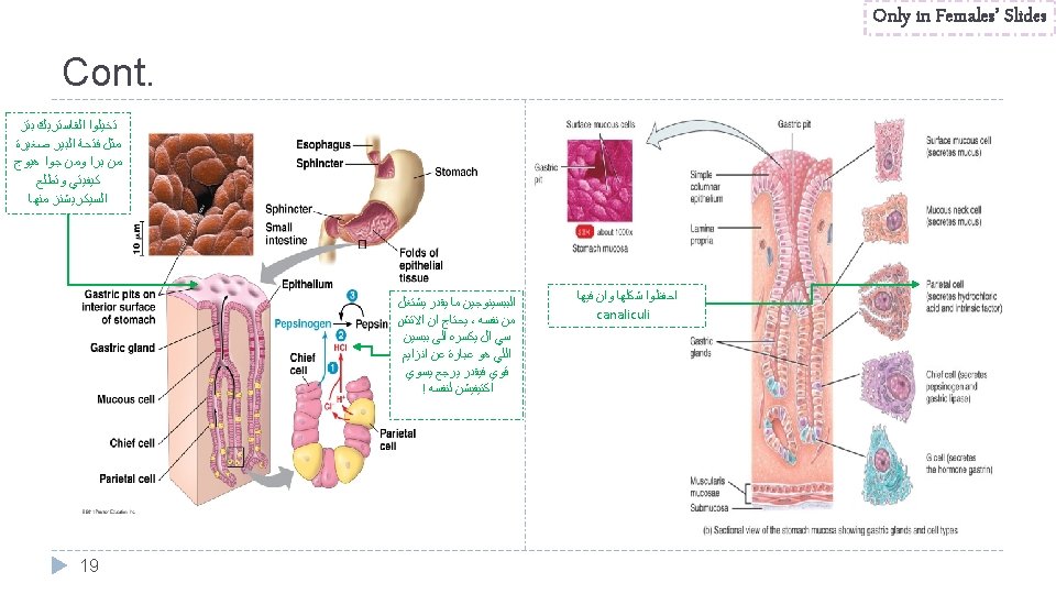
The Video here is very Important Gastric Secretion The stomach's mucosal lining, the glandular gastric mucosa, contains three main types of glands: 1. Cardiac glands 2. Oxyntic glands. 3. Pyloric glands They are tubular gland • - • They are composed of three types of cells: mucus neck cells, peptic (chief) cells, and parietal cells (oxyntic cells). These glands are the most abundant gastric glands, found in fundus and corpus. Secrete - Hydrochloric acid Pepsinogen Intrinsic factor Mucus Secretion of Bicarbonate from Pancreatic Cells 0: 35 17 Many G cells Location Located in body & fundus (In proximal 80%of stomach) Secrete - Mucus Protection Gastrin Pepsinogen This Video was included in the slides Location Located in the antrum (In the distal 20% of stomach)
Gastric Cells � Functionally different cell types compose glands: 1. Parietal cells (oxyntic cells) -most distinctive cells in stomach (HCl & intrinsic factor) (That’s why gastrectomy will cause Intrinsic factor deficiency then vit B-12 deficiency and therefore Anemia). The Parietal cells of stomach is the only source in GIT to pepsinogen and HCL 2. Chief cells (peptic cells), they are available in oxyntic glands & few in pyloric glands, pepsinogen. 3. Mucus neck cells: • HCO 3 - • Mucus The secreted pepsinogen is not active, that’s why it doesn’t destroy the gland itself. 4. G cells: gastrin (hormone) → increases HCl secretion. 5. D cells: somatostatin (antrum) → decreases HCl secretion. 6. enterochromaffin-like cell: histamine. 7. enteroendocrine cells. � Gastric mucosa has numerous openings called gastric pits. � Gastric glands empty into bottom of pits. 18
Only in Males’ Slides Oxyntic Gland � Structure of a Gastric Oxyntic Gland: � Parietal cells are the most distinctive cells in the stomach. Their structure is unique in that they have intracellular canaliculi as well as an abundance of mitochondria and ER. � This network consists of clefts and canals that are continuous with the lumen of the oxyntic gland. � HCl is secreted across the parietal cell microvillar membrane and flows out of the intracellular canaliculi into the oxyntic gland lumen. � The surface mucous cells line the entire surface of the gastric mucosa and the openings of the cardiac, pyloric, and oxyntic glands. � These cells secrete mucus and HCO 3 - to protect the gastric surface from the acidic environment of the stomach. � The distinguishing characteristic of a surface mucous cell is the presence of numerous mucus granules at its apex. 20
Gastric Cells & Gastric juice The Normal Locations of Gastric Cells: Only in Males’ Slides HCL The most important. The only source for it in the body is parietal cells. Pepsinogen Gastric juice � Electrolytes Intrinsic factor Mucus (mucus gel layer) 21
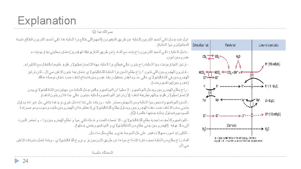
Only in Males’ Slides Hydrochloric acid secretion � Mechanism for secretion of hydrochloric acid: Water becomes dissociated into hydrogen ions and hydroxyl ions in the cell cytoplasm. Chloride ion is actively transported from the cytoplasm of the parietal cell into the lumen of the canaliculus and sodium ions are actively transported out of the canaliculus into the cytoplasm of the parietal cell. Carbon dioxide, either formed during metabolism in the cell or entering the cell from the blood, combines under the influence of carbonic anhydrase with the hydroxyl ions to form bicarbonate ions. These then diffuse out of the cell cytoplasm into the extracellular fluid in exchange for chloride ions that enter the cell 22 The hydrogen ions are then actively secreted into the canaliculus in exchange for potassium ions � CO 2 + H 2 O in the presence of carbonic anhydrase give> H 2 CO 3 -. � H 2 CO 3 - will break down to> HCO 2 + H+. � HCO 2> will be excreted into the blood in exchange with Cl, so the blood p. H around the stomach will increase (alkaline tide). � H+> will be secreted towards the lumen via K and H+ pump (exchanger) , which depends on ATP and the concentration of K in the lumen. � How do we get K? by 1. Na and K pump 2. K has the ability to leave the cell into the lumen actively, Cl also has the ability to leave the cell into the lumen actively. � To maintain the osmolarity water will follow to the lumen. � End results: H+ and Cl will be secreted into the lumen of the stomach. The Notes here is very Important
Cont. � Mechanism of HCl production: Only in Males’ Slides Only in Females’ Slides Depends on H/K ATPase Inhibited by: omeprazole H/K pump depends on [K]out [HCl] drives water into gastric content to maintain osmolality During gastric acid secretion: amount of HCO 3 - in blood = amount of HCl being secreted 23 Alkaline tide It means that the blood near parietal cells become alkaline ﺷﺮﺡ ﺍﻟﺼﻮﺭﺓ ﻓﻲ ﺍﻟﺴﻼﻳﺪ ﺍﻟﺠﺎﻱ � Postulated mechanism for secretion of hydrochloric acid. � The points labeled "P" indicate active pumps, and the dashed lines represent free diffusion and osmosis.
Only in Males’ Slides Neural and Hormonal Control � Gastric secretion is under neural and hormonal control. � Gastric acid secretion is mediated through neural and hormonal pathways. 1. Vagus nerve stimulation is the → neural effector. 2. Histamine and gastrin are the → hormonal effectors. � Parietal cells possess special histamine receptors, h 2 receptors, whose stimulation results in increased acid secretion. � Special neuroendocrine cells of the stomach, known as enterochromaffinlike (ECL) cells, are believed to be the source of this histamine. � They are located mostly in the acid-secreting regions of the stomach. � The mechanisms that stimulate the ecl cells to release histamine are poorly understood. � The effectiveness of cimetidine, (a h 2 blocker) in reducing acid secretion has indirectly demonstrated the importance of histamine as an effector of gastric acid secretion. � H 2 blockers are commonly used for the treatment of peptic ulcer disease or gastroesophageal reflux disease. 25
The Notes here is very Important Cont. � Vagus nerve (neural effector) either by: 1. Releasing ach (direct activation of parietal cells). 2. By releasing gastrin releasing peptide, GRP � Agents that stimulate and inhibit H+ secretion by gastric parietal cells. • • (indirect activation). � Gastrin (hormonal effector). � Enterochromaffin like cells release histamine. à Activates h 2 receptor (parietal cells) à Increases acid secretion. • Neural activation (direct). Hormonal activation (indirect), gastrin works on CCKB receptor because they are from the same family ”you have to know this its even important for the SMLE”. Gastrin releasing peptide is a neurotransmitter which is released in the presence of proteins or distention of the stomach. Histamine you have to know its H 2 receptor. There are different stimuli that activate parietal cells witch are: • Ach from vagus activates M 3 receptor. • Gastrin from G cells activates CCKB receptor. • Histamine from ECL cells activates H 2 receptor. These activators will stimulate H/K Pump and therefore, increased acid secretion. On the other hand, there are stimuli that inhibit parietal cells witch are: • Somatostatin from D cells. • Prostaglandins from arachidonic acid metabolism. These inhibitors will reduce c. AMP witch will inhibit H/K Pump and therefore, decreased acid secretion. 26
Only in Males’ Slides Rate of Secretion � � � At a low secretion rate, gastric juice contains: � high concentrations of Na+ and Cl- In gastric secretion, increased rate of secretion is � low concentrations of K+ and H+. better for digestion. Opposite to slivery glands , increased rate of secretion isn’t beneficial for digestion When the rate of secretion increases: � the concentration of Na+ decreases � whereas that of H+ increases significantly. � Also coupled with this increase in gastric secretion is an increase in Cl- concentration. To understand the changes in electrolyte composition of gastric juice at different secretion rates, remember that gastric juice is derived from the secretions of two major sources: � 1. parietal cells. 2. nonparietal cells. Secretion from nonparietal cells is probably constant; therefore, it is parietal secretion (HCl secretion) that contributes mainly to the changes in electrolyte composition with higher secretion rates. 27 -Low secretion rate (between meals) - high Na. Cl. -High secretion rate (after a meal)high HCl. -Always isotonic. Because of water secretion , Gastric secretion is always isotonic.
The stimulation of acid secretion resulting from the ingestion of food can be divided into three phases: the cephalic phase, the gastric phase, and the intestinal phase -involves the central nervous system. -Smelling, chewing, and swallowing food send impulses via the vagus nerves to the parietal and G cells in the stomach: 1 -The nerve endings release Ach, which directly stimulates acid secretion from parietal cells. 2 -The nerves also release gastrin-releasing peptide (GRP), → which stimulates G cells to release gastrin, → indirectly stimulating parietal cell acid secretion. Only in females’ Slides Only in Males’ Slides 1. The cephalic phase ( )ﺍﻟﺮﺋﻴﺴﻴﺔ ﺃﻮ ﺍﻟﻤﺮﻛﺰﻳﺔ 2. The gastric phase (The most important phase) 3. During the intestinal phase -is mainly a result of gastric distention and chemical agents such as digested proteins. 1 -Distention of the stomach stimulates mechanoreceptors, which stimulate the parietal cells directly through: A- short local (enteric) reflexes. B- long vago-vagal reflexes. 2 -Digested proteins in the stomach are also potent simulators of gastric acid secretion, an effect mediated through gastrin release. -Several other chemicals, such as alcohol and caffeine, stimulate gastric acid secretion through mechanisms that are not well understood. -protein digestion products in the duodenum stimulate gastric acid secretion through the action of the circulating amino acids on the parietal cells. -Distention of the small intestine, probably via the release of the hormone entero-oxyntin from intestinal endocrine cells, stimulates acid secretion. When there are proteins or distention within the intestines Inhibition of Acid Secretion Inhibitory hormones (Enterogastrones) Somatostatin (D-cells) in antrum 28 Secretin (S-cells) in duodenum Glucose-dependent insulinotropic peptide (GIP) in duodenum
Only in Males’ Slides Summary of the Phases (from slides) Cephalic phase (30%): So chewing gum increases HCl production, Because of Vagus. Smelling, Chewing and swallowing Stimulate 1 -parietal, 2 -G-Cells (via GRP) Gastric phase (60%): Firing of all the reflexes, mediators are: vagus, gastrin, histamine gastric distention proteins Intestinal phase (10%): By proteins and distention in releases entero-oxyntin hormone which increases HCl, its not mentioned in the schedule because it’s a local hormone. digested proteins 29 The percentage here is very Important
Only in Males’ Slidesc ﺍﺣﻔﻈﻮﺍ ﺍﻟﺠﺪﻭﻝ ﺯﻱ ﺍﺳﻤﻜﻢ Hormonal Control of Gastrointestinal Motility (GI Peptides) Hormone Gastrin Site of secretion Stimuli for secretion G cells of the antrum, • duodenum and • jejunum. • • Actions Protein Stimulates: gastric H+ secretion Distention of the and growth of gastric mucosa. stomach Vagal stimulation (GRP) Acid inhibits release I cells of the Cholecystokinin (CCK) duodenum, jejunum, and ileum. • • • Protein Fatty acids Acids Stimulates: pancreatic enzyme secretion, pancreatic HCO 3 secretion, gallbladder contraction, growth of the exocrine pancreas, and relaxation of the sphincter of oddi. Inhibits: gastric emptying. S cells of the duodenum, jejunum, and ileum • Acids and fat in the duodenum. Stimulates: pepsin secretion, pancreatic HCO 3 - secretion, biliary HCO 3 - secretion, and growth of the exocrine pancreas. Inhibits: gastric H+ secretion. • • • Protein Fatty acids Oral glucose Stimulates: insulin secretion from pancreatic β cells. Inhibits: gastric H+ secretion. Fat Acid Nerve Stimulates: • Gastric motility • Intestinal motility Secretin Glucose-dependent K cells of the insulinotropic peptide duodenum and (GIP) jejunum. Motilin 30 M cells of the duodenum and jejunum
Thank you!. ﺍﻋﻤﻞ ﻭ ﺃﻨﺖ ﺗﻌﻠﻢ ﺃﻦ ﺍﻟﻠﻪ ﻻ ﻳﻀﻴﻊ ﺃﺠﺮ ﻣﻦ ﺃﺤﺴﻦ ﻋﻤﻼ ، ﺍﻋﻤﻞ ﻟﺘﻤﺴﺢ ﺩﻣﻌﺔ ، ﺍﻋﻤﻞ ﻟﺘﺮﺳﻢ ﺑﺴﻤﺔ The Physiology 436 Team: Female Members: Male Members: Ghada Almazrou Hassan alshammari Team Leaders: Laila Mathkour Mohammad Alayed Nada Aldakheel Nouf Al. Amari Abdullah Alsaeed References: • • • 2017 -2018 Dr. Hana Alzamel’s Lecture. 2017 -2018 Dr. Mohammed Al Zoghaibi’s Lecture. Guyton and Hall Textbook of Medical Physiology (Thirteenth Edition. ) 31 . ﻓﺮﺩﻩ ﻟﻲ ﻭﻗﺖ ﺣﺎﺟﺘﻲ ﺇﻟﻴﻪ ﻳﺎ ﻣﻦ ﻻ ﺗﻀﻴﻊ ﻋﻨﺪﻩ ﺍﻟﻮﺩﺍﺋﻊ ، ﺍﻟﻠﻬﻢ ﺍﻧﻲ ﺍﺳﺘﻮﺩﻋﺘﻚ ﻣﺎ ﺣﻔﻈﺖ ﻭﻣﺎ ﻗﺮﺃﺖ ﻭﻣﺎ ﻓﻬﻤﺖ Contact us: