TESTIS SCROTUM SPERMATIC CORD MALE GENITAL SYSTEM REVIEW


- Slides: 44
TESTIS, SCROTUM & SPERMATIC CORD MALE GENITAL SYSTEM
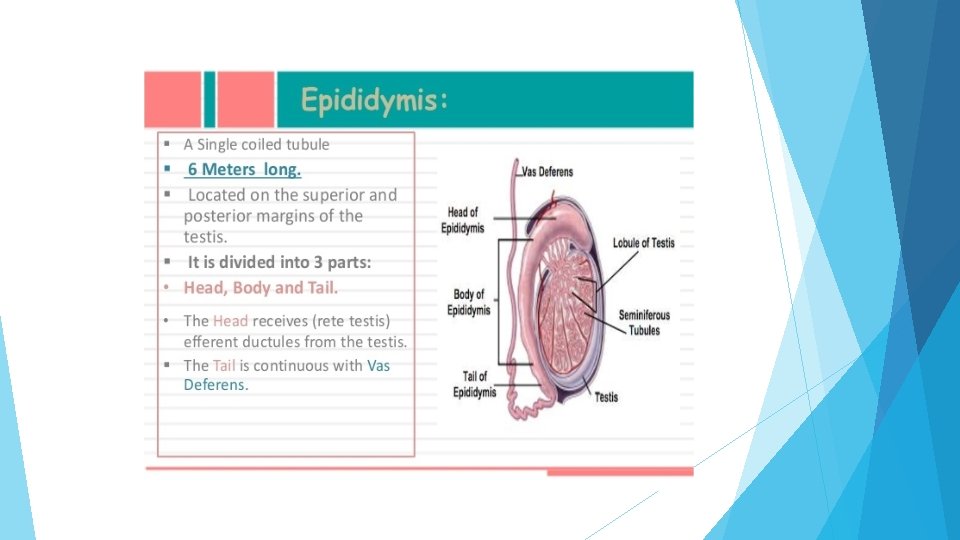
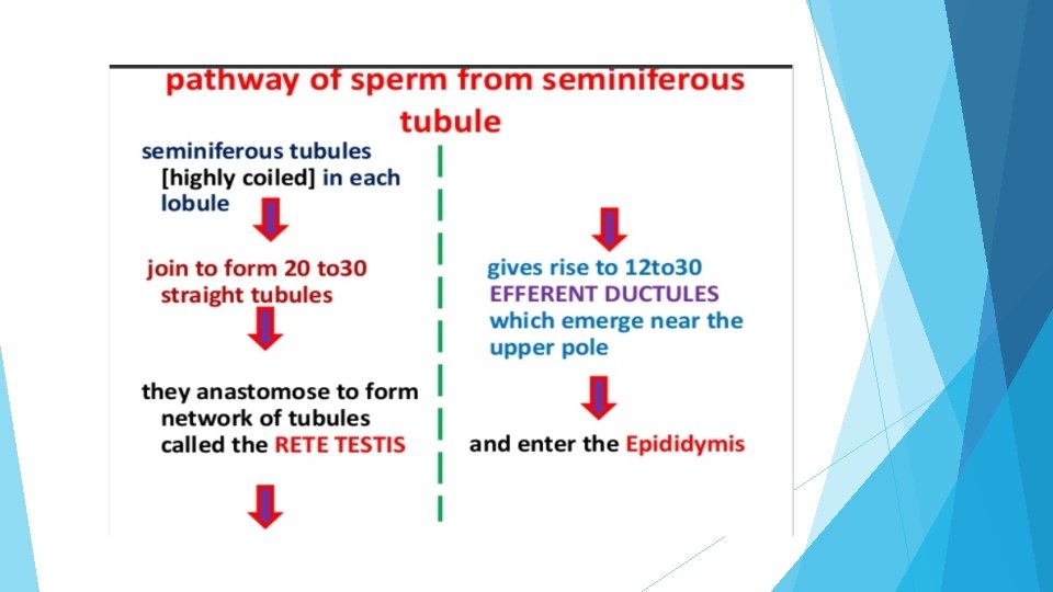
REVIEW OF MACROSCOPIC STRUCTURE OF TESTIS
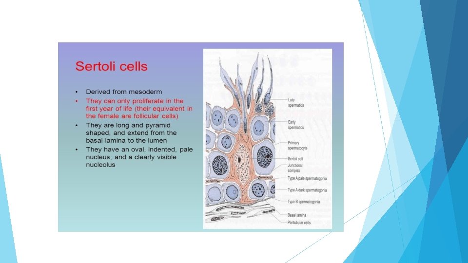
REVIEW OF MICROSCOPIC STRUCTURE OF TESTIS
ARRANGEMENTS OF CELLS INSIDE THE SEMINIFEROUS TUBULE
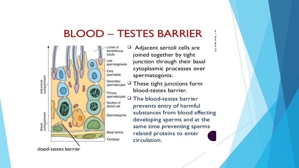
CONTENTS OF COMPARTMENTS WITHIN THE BLOOD-TESTIS BARRIER
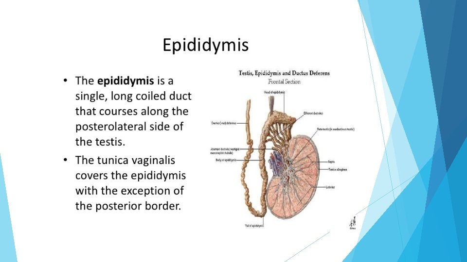
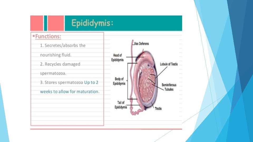
FUNCTIONS OF EPIDIDYMIS
HISTOLOGICAL STRUCTURE OF EPIDIDYMIS Epididymis is a long, convoluted tubule that is surrounded by connective tissue & a thin smooth muscle layer. Both cross section & longitudinal section of epididymis shows mature sperm within the lumen of epididymis. The lining pseudostratified columnar epithelium consists of tall columnar principal cells with long nonmotile stereocilia & small basal cells.
HISTOLOGICAL STRUCTURE OF EPIDIDYMIS
HISTOLOGICAL STRUCTURE OF EPIDIDYMIS
DIFFERENCE BETWEEN THE STRUCTURE OF TESTIS & EPIDIDYMIS
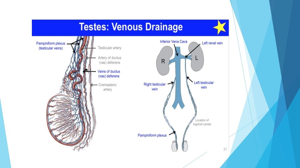
BLOOD SUPPLY OF TESTIS
BLOOD SUPPLY OF TESTIS
IMPORTANCE OF TESTICULAR BLOOD SUPPLY
HOW TESTICULAR VEIN IS FORMED About 15 to 20 veins appear from the posterior border of testis & epididymis & unite to form a pampiniform plexus which produce the bulk of spermatic cord. At superficial inguinal ring the plexus unite to form 4 veins & at the deep ring they further join to form 2 veins. Finally a single vein is formed in the posterior abdominal wall. Right testicular vein drain directly into inferior vena cava at an acute angle, left testicular vein drain into left renal vein almost at a right angle.
LYMPHATIC DRAINAGE & NERVE SUPPLY OF TESTIS
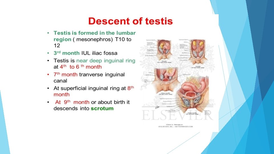
DESCENT OF TESTIS
FACTORS ASSOCIATED WITH THE DESCENT OF TESTIS
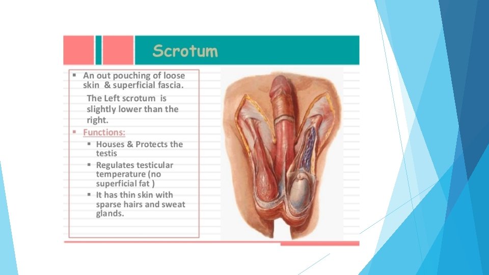
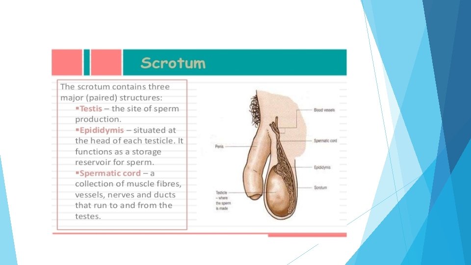
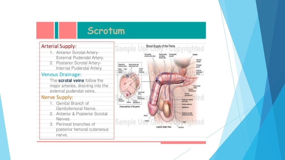
SCROTUM
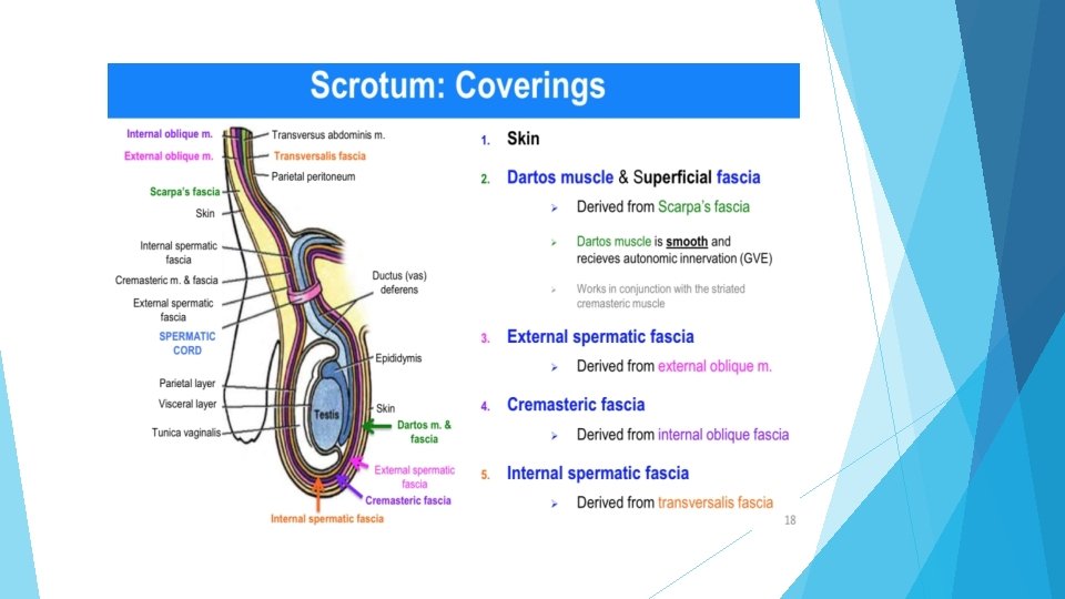
SCROTAL LAYERS
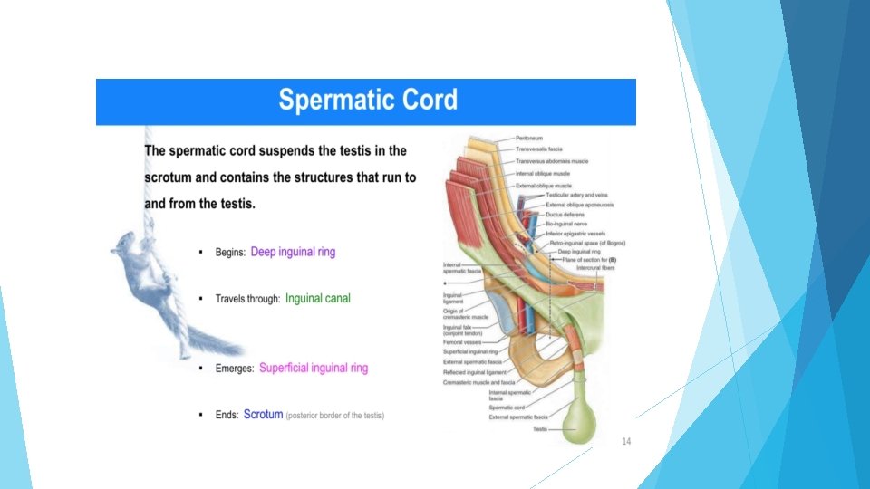
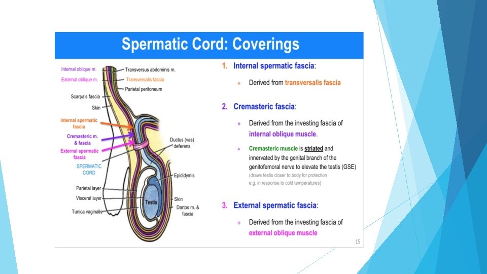
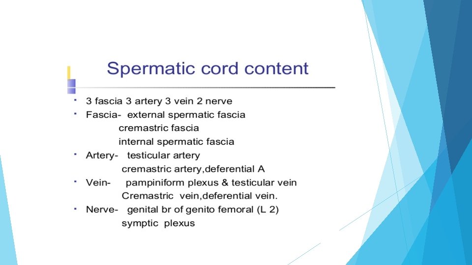
COVERINGS OF SPERMATIC CORD CONTINUE WITH SCROTAL LAYERS
FUNCTIONS OF SCROTAL MUSCLE
THANK YOU