Teresa Audesirk Gerald Audesirk Bruce E Byers Biology

Teresa Audesirk • Gerald Audesirk • Bruce E. Byers Biology: Life on Earth Eighth Edition Lecture for Chapter 5 Membrane Structure and Function Copyright © 2008 Pearson Prentice Hall, Inc.

Chapter 5 Outline • 5. 1 How Is the Structure of a Membrane Related to Its Function, p. 82 • 5. 2 How Do Substances Move Across Membranes? p. 85 • 5. 3 How Do Specialized Junctions Allow Cells to Connect and Communicate? p. 95

Section 5. 1 Outline • 5. 1 How is the Structure of a Membrane Related to Its Function? – The Plasma Membrane Isolates the Cell While Allowing Communication with Its Surroundings – Membranes Are “Fluid Mosaics” in Which Proteins Move Within Layers of Lipids – The Phospholipid Bilayer Is the Fluid Portion of the Membrane – A Mosaic of Proteins Is Embedded in the Membrane

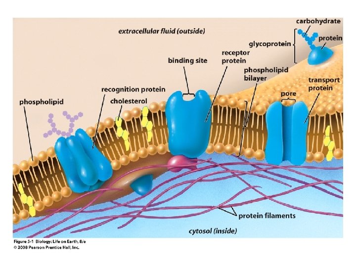
Plasma Membrane • Functions of the plasma membrane – Isolates the cell’s contents from environment – Regulates exchange of essential substances – Communicates with other cells – Creates attachments within and between other cells – Regulates biochemical reactions

Membranes Are “Fluid Mosaics” • Membranes are dynamic, ever-changing structures

Membranes Are “Fluid Mosaics” • “Fluid mosaic” model of a membrane proposed in 1972 – A lumpy, constantly shifting mosaic of “tiles” or proteins – Proteins float around in a sea of phospholipids

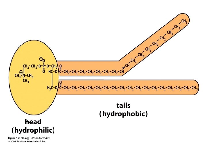
The Phospholipid Bilayer • Phospholipids are the basis of membrane structure – Polar, hydrophilic head – Two non-polar, hydrophobic tails

The Phospholipid Bilayer • The cell exterior and interior face watery environments

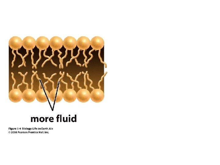
The Phospholipid Bilayer • Hydrophobic and hydrophilic interactions drive phospholipids into bilayers – – Double row of phospholipids Polar heads face outward and inward Non-polar tails mingle within the membrane Cholesterol in animal membranes keeps them flexible
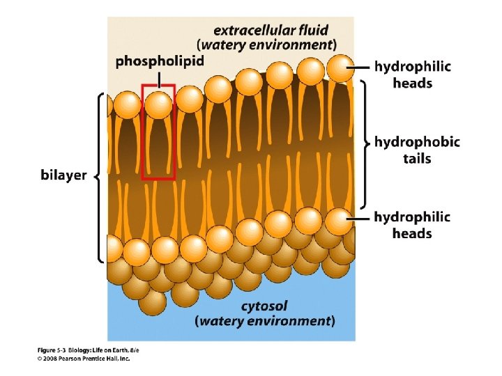

The Phospholipid Bilayer • Phospholipid bilayer is a flexible, fluid membrane to allow for cellular shape changes

The Phospholipid Bilayer • Individual phospholipid molecules are not bonded to one another • Some of the phospholipids have unsaturated fatty acids, whose double bonds introduce “kinks” into their “tails” • The above features make the membrane fluid


Membrane Proteins Form a Mosaic • Proteins are embedded in the phospholipid bilayer – Some proteins can float and drift – Other proteins are anchored by protein filaments in the cytoplasm – Many proteins have attached carbohydrates (glycoproteins)


Membrane Proteins Form a Mosaic • Categories of membrane proteins – – – Receptor Proteins Recognition Proteins Enzymatic Proteins Attachment Proteins Transport Proteins

Membrane Proteins Form a Mosaic • Receptor Proteins – Trigger cellular responses upon binding specific molecules, e. g. hormones • Recognition Proteins – Serve as identification tags on the surface of a cell
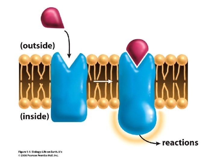

Membrane Proteins Form a Mosaic • Enzymes – Promote chemical reactions that synthesize or break apart biological molecules • Attachment Proteins – Anchor the cell membrane to inner cytoskeleton, to proteins outside the cell, and to other cells

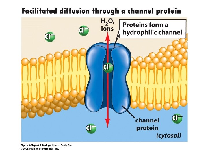
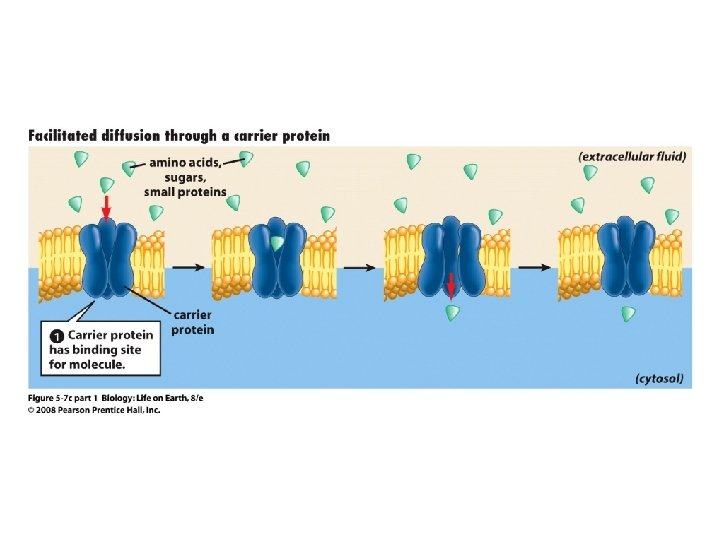
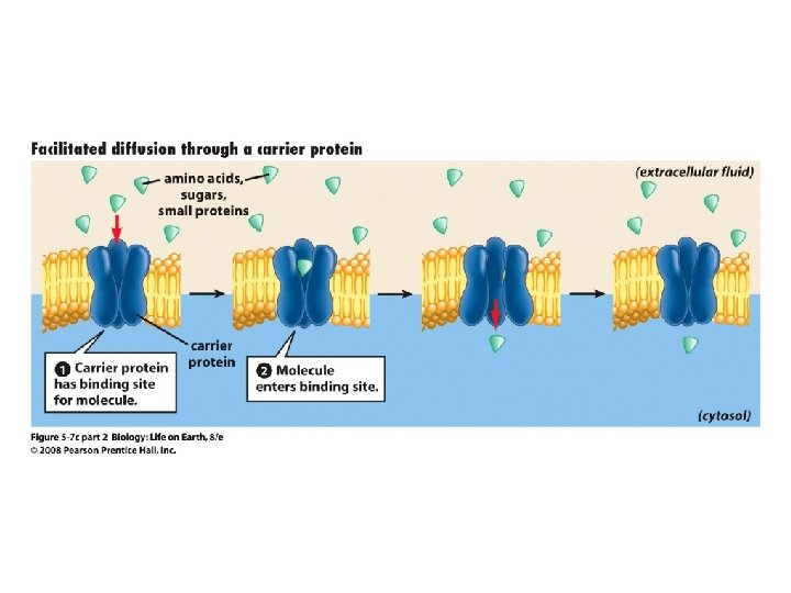
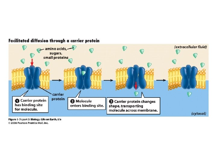
Membrane Proteins Form a Mosaic • Transport Proteins – Include channel and carrier proteins – Regulate import/export of hydrophilic molecules

Section 5. 2 Outline • 5. 2 How Do Substances Move Across Membranes? – Molecules in Fluids Move in Response to Gradients – Movement Across Membranes Occurs by Both Passive and Active Transport – Passive Transport Includes Simple Diffusion, Facilitated Diffusion, and Osmosis – Osmosis Plays an Important Role in Cells

Section 5. 2 Outline • 5. 2 How Do Substances Move Across Membranes? continued – Active Transport Uses Energy to Move Molecules Against Their Concentration Gradients – Cells Engulf Particles or Fluids by Endocytosis – Exocytosis Moves Material Out of the Cell – Exchange of Materials Across Membranes Influences Cell Size and Shape

Movement of Molecules in Fluids • Definitions relevant to substance movement – A fluid is a substance that can move or change shape in response to external forces – A solute is a substance that can be dissolved (dispersed as ions or molecules) in a solvent – A solvent is a fluid capable of dissolving a solute
 – The")
Movement of Molecules in Fluids • Definitions relevant to substance movement (continued) – The concentration of molecules is the number of them in a given volume unit – A gradient is a physical difference in temperature, pressure, charge, or concentration in two adjacent regions

Movement of Molecules in Fluids • Why molecules move from one place to another – Substances move in response to a concentration gradient • Molecules move from high to low concentration (diffusion) until dynamic equilibrium is reached
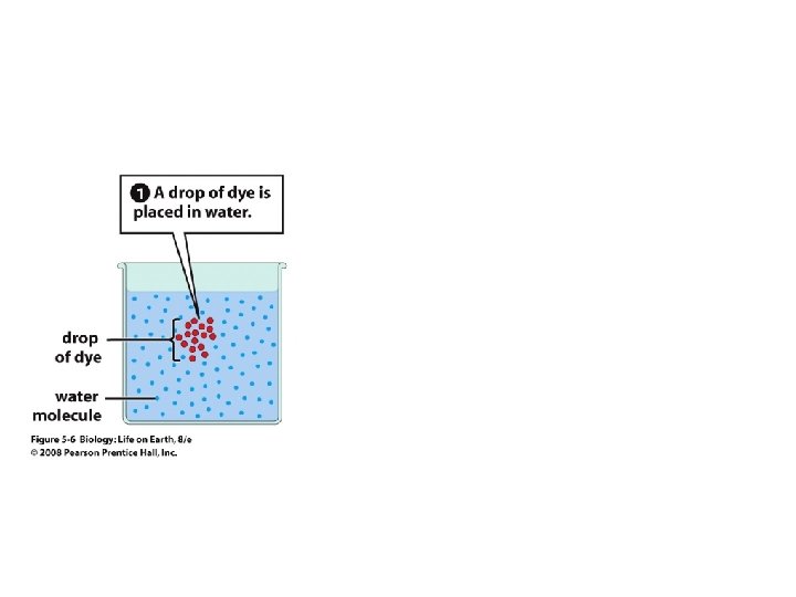
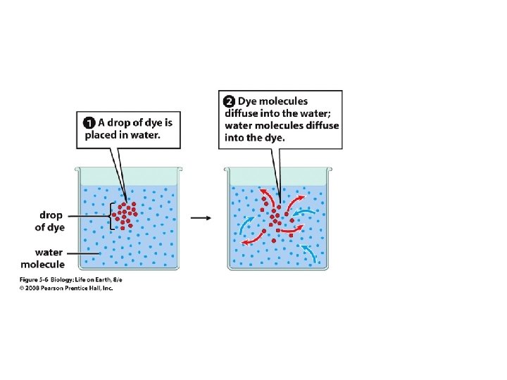
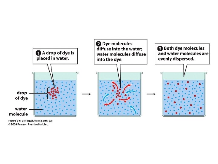

Movement of Molecules in Fluids • The greater the concentration gradient, the faster the rate of diffusion • Diffusion cannot move molecules rapidly over long distances

Movement Across Membranes • Concentration gradients of ions and molecules exist across the plasma membranes of all cells • There are two types of movement across the plasma membrane – Passive transport – Energy-requiring transport
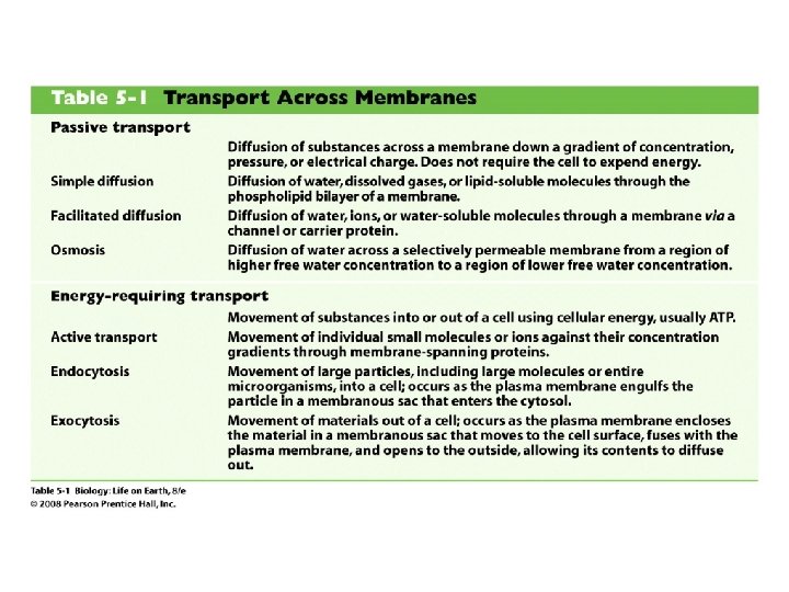

Movement Across Membranes • Passive transport – Substances move down their concentration gradients across a membrane – No energy is expended – Membrane proteins and phospholipids may limit which molecules can cross, but not the direction of movement

Movement Across Membranes • Energy-requiring transport – Substances are driven against their concentration gradients – Energy is expended

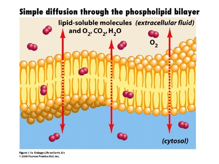
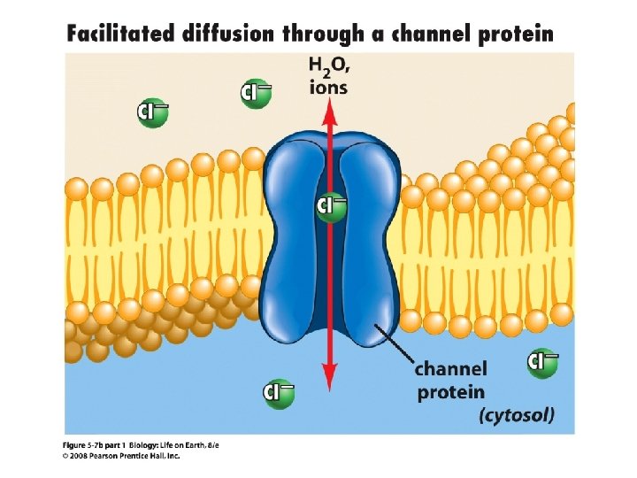
Passive Transport • Plasma membranes are selectively permeable – Different molecules move across at different locations and rates – A concentration gradient drives all three types of passive transport: simple diffusion, facilitated diffusion, and osmosis

Passive Transport • Simple diffusion – Lipid soluble molecules (e. g. vitamins A and E, gases) and very small molecules diffuse directly across the phospsholipid bilayer

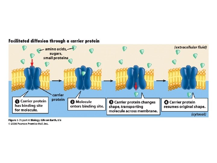
Passive Transport • Facilitated diffusion – Water soluble molecules like ions, amino acids, and sugars diffuse with the aid of channel and carrier transport proteins

Passive Transport • Osmosis – the special case of water diffusion – Water diffuses from high concentration (high purity) to low concentration (low purity) across a membrane – Dissolved substances reduce the concentration of free water molecules (and hence the purity of water) in a solution

Passive Transport • The flow of water across a membrane depends on the concentration of water in the internal or external solutions


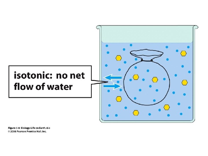
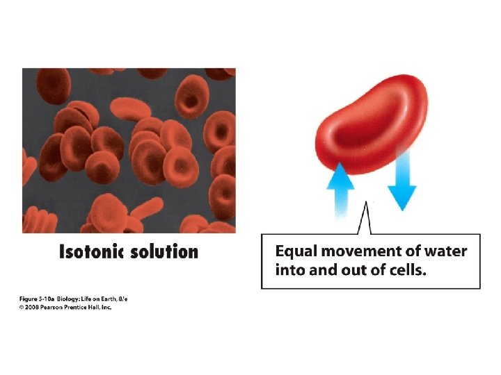
Passive Transport • Comparison terms for solutions on either side of a membrane – Isotonic solutions have equal concentrations of water and equal concentrations of dissolved substances • No net water movement occurs across the membrane
")
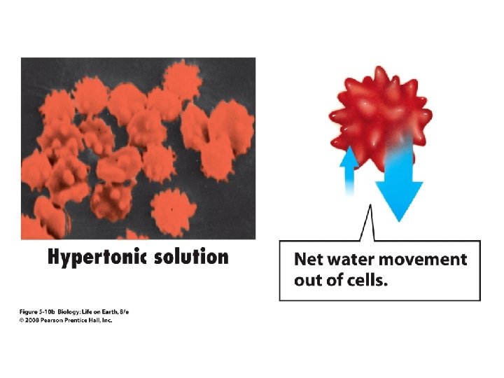
Passive Transport • Comparison terms for solutions on either side of a membrane (continued) – A hypertonic solution is one with lower water concentration or higher dissolved particle concentration • Water moves across a membrane towards the hypertonic solution
")
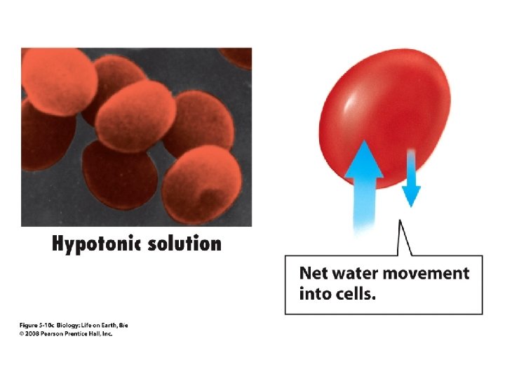
Passive Transport • Comparison terms for solutions on either side of a membrane (continued) – A hypotonic solution is one with higher water concentration or lower dissolved particle concentration • Water moves across a membrane away from the hypotonic solution

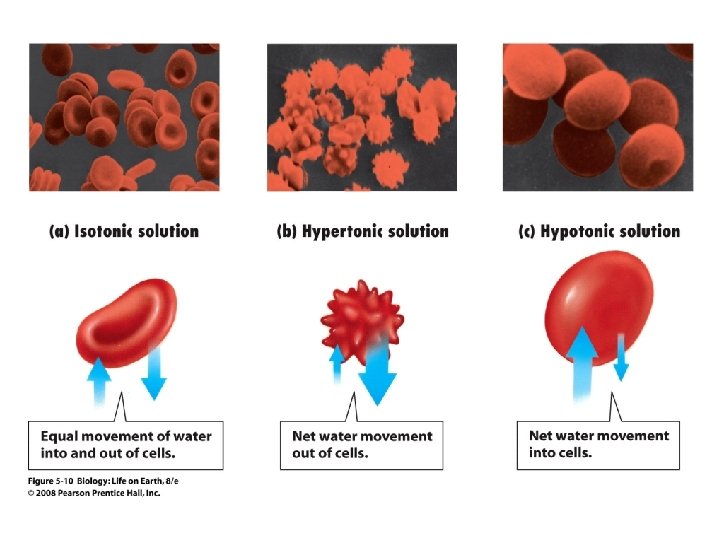
Passive Transport • The effects of osmosis are illustrated when red blood cells are placed in various solutions

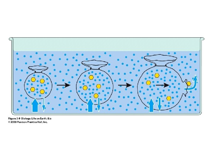
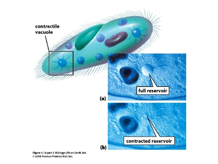
Passive Transport • Osmosis explains why fresh water protists have contractile vacuoles • Water leaks in continuously because the cytosol is hypertonic to fresh water • Salts are pumped into the vacuoles, making them hypertonic to the cytosol • Water follows by osmosis and is then expelled by contraction

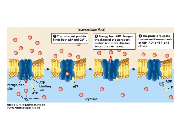
Active Transport • Cells need to move some substances against their concentration gradients

Active Transport • Active-transport membrane proteins move molecules across using ATP – Proteins span the entire membrane – Often have a molecule binding site and an ATP binding site – Often referred to as pumps

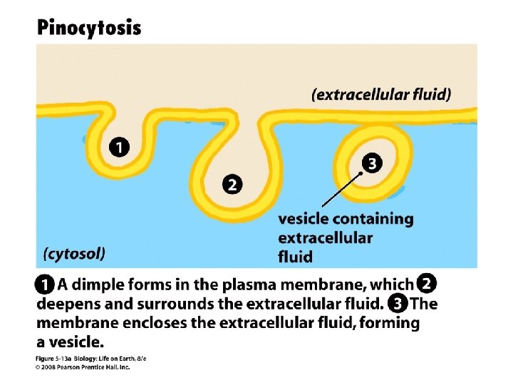
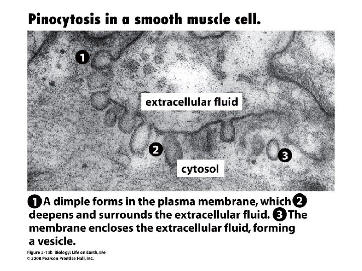
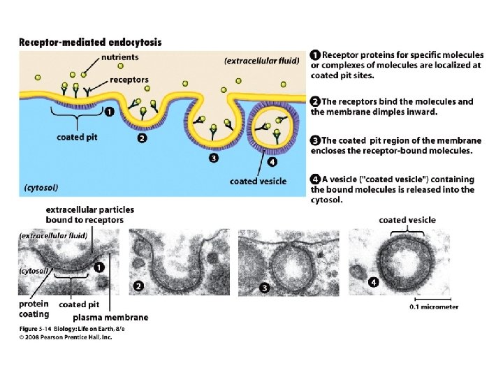
Endocytosis • Cells import large particles or substances via endocytosis

Endocytosis • Plasma membrane pinches off to form a vesicle in endocytosis – Types of endocytosis • Pinocytosis • Receptor-mediated endocytosis • Phagocytosis
 brings in droplet of extracellular")
Endocytosis • Types of endocytosis – Pinocytosis (“cell drinking”) brings in droplet of extracellular fluid

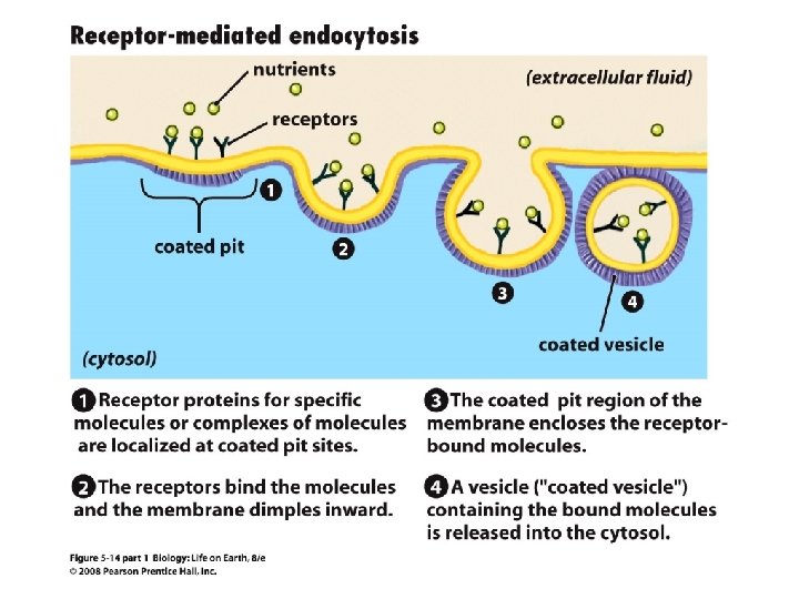
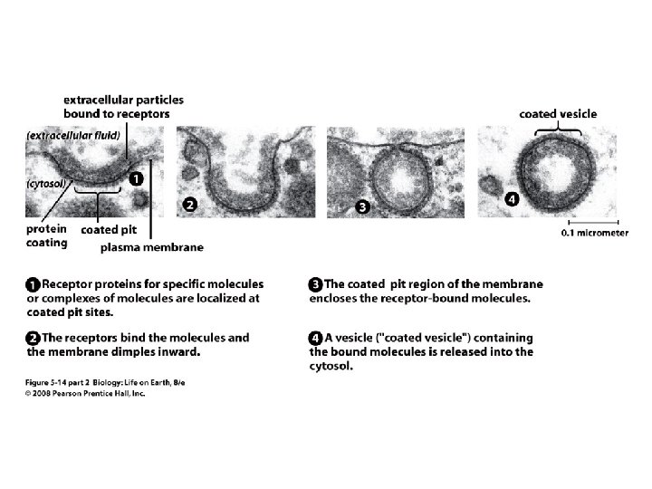
Endocytosis • Types of endocytosis – Receptor-mediated endocytosis moves specific molecules into the cell
 moves large particles or whole")
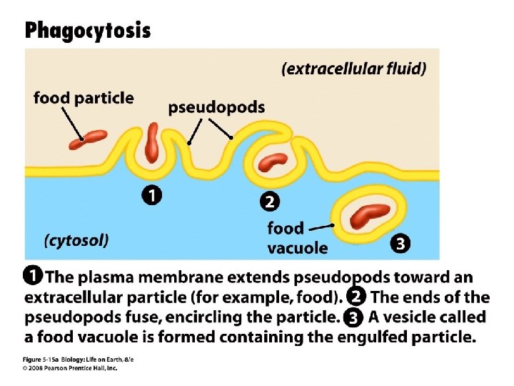
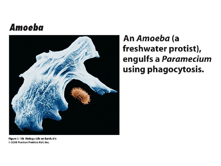
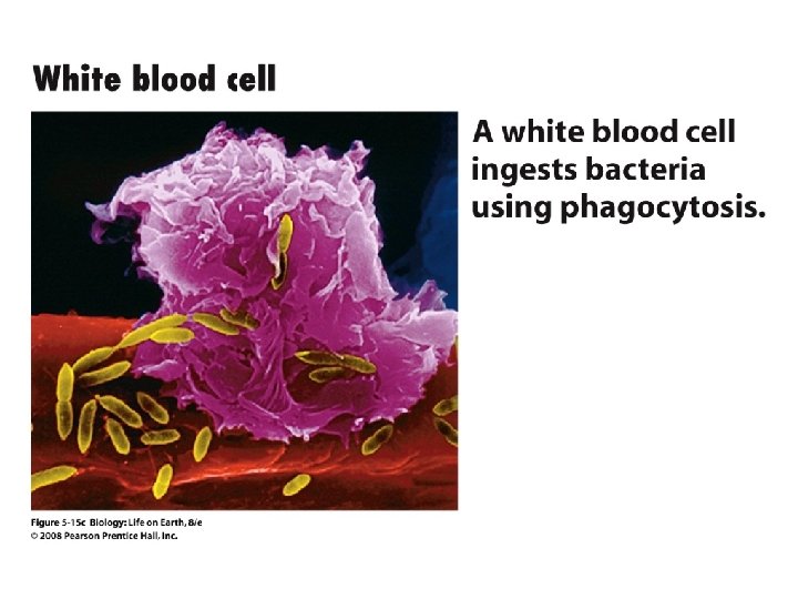
Endocytosis • Types of endocytosis – Phagocytosis (“cell eating”) moves large particles or whole organisms into the cell

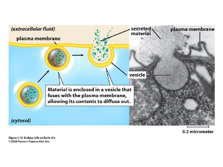
Exocytosis • Exocytosis – Vesicles join the membrane, dumping out contents in exocytosis

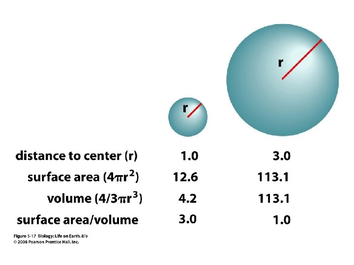
Cell Size and Shape • Exchange affects cell size and shape – As a spherical cell enlarges, its innermost parts get farther away from the plasma membrane – Also, its volume increases more rapidly than its surface area – A larger cell has a relatively smaller area of membrane for nutrition exchange than a small cell

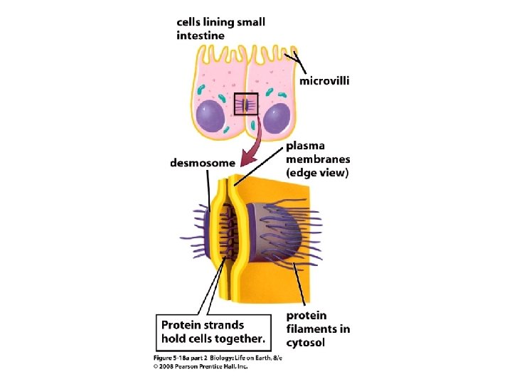
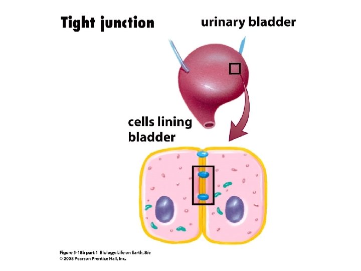
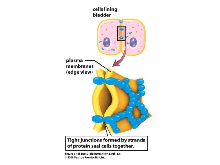
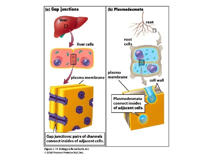
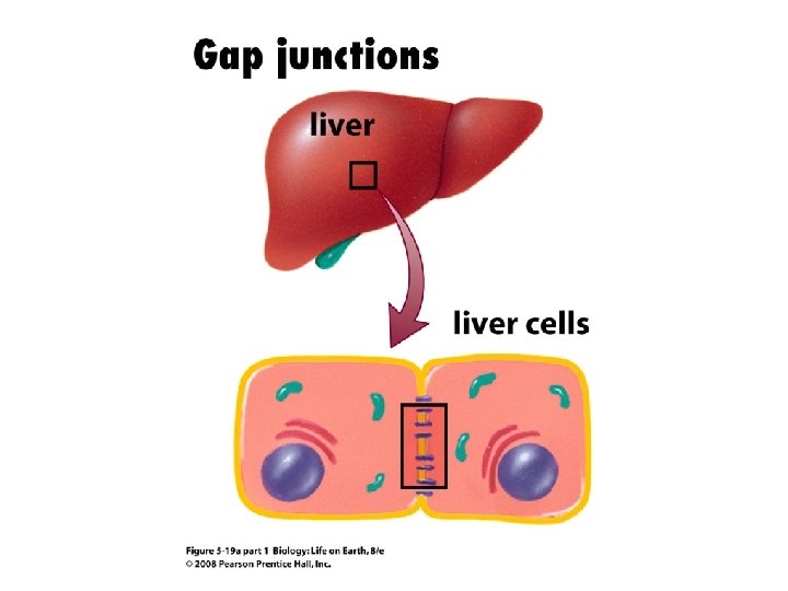
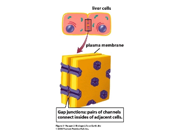
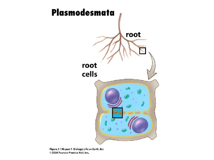
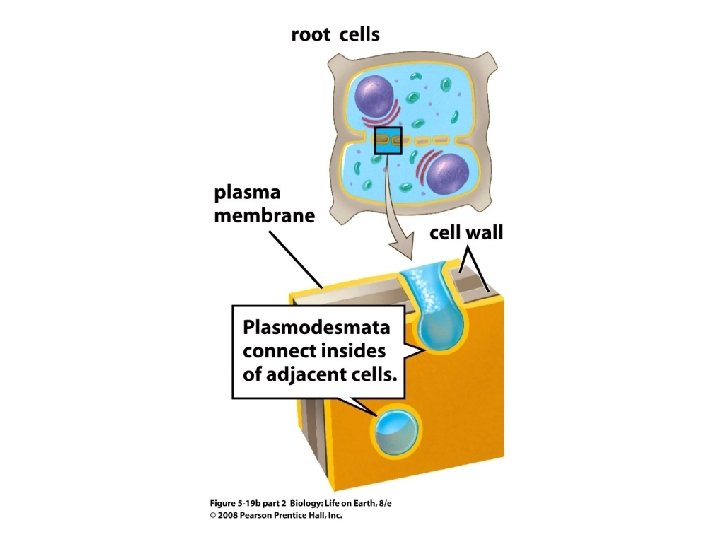
Section 5. 3 Outline • 5. 3 How Do Specialized Junctions Allow Cells to Connect and Communicate? – Desmosomes Attach Cells Together – Tight Junctions Make Cell Attachments Leakproof – Gap junctions and Plasmodesmata Allow Direct Communication Between Cells

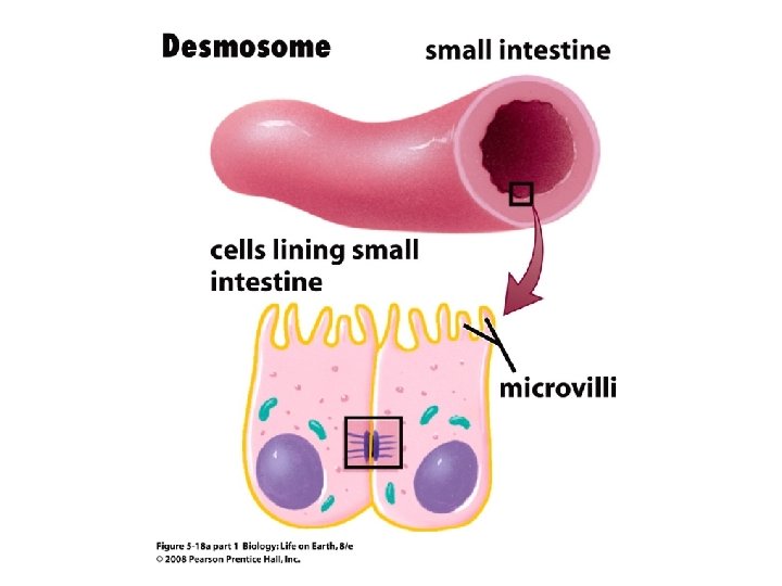
Desomosomes • Desmosomes attach cells together – Found where cells need to adhere tightly together under the stresses of movement (e. g. the skin)

Tight Junctions • Tight junctions make the cell leakproof – Found where tubes and sacs must hold contents without leaking (e. g. the urinary bladder)

Gap Junctions and Plasmodesmata • Gap junctions and plasmodesmata allow for communication – Cell-to-cell channels allowing for passage of hormones, nutrients, and ions in animal cells are gap junctions – Plant cells have cytoplasmic connections called plasmodesmata

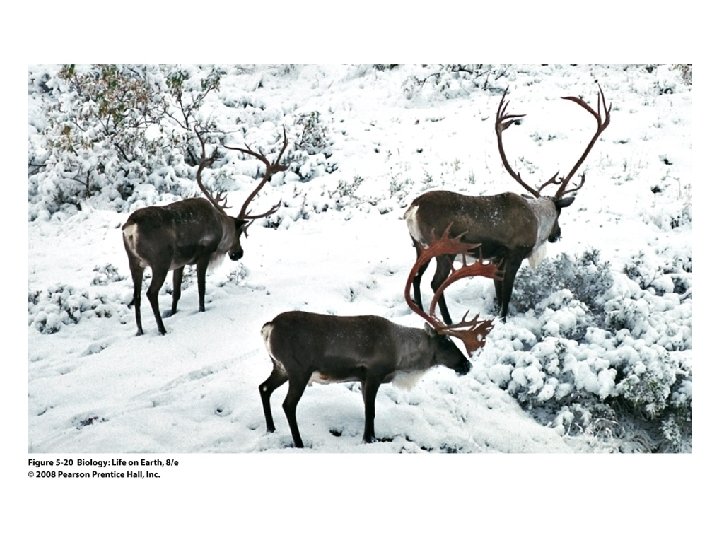
Caribous Legs and Membranes • Membrane function varies between organisms

Caribous Legs and Membranes • Plasma membrane phospholipids in caribous legs adapted for cold – Cold areas near hooves have more membrane unsaturated fatty acids to keep cell membranes fluid

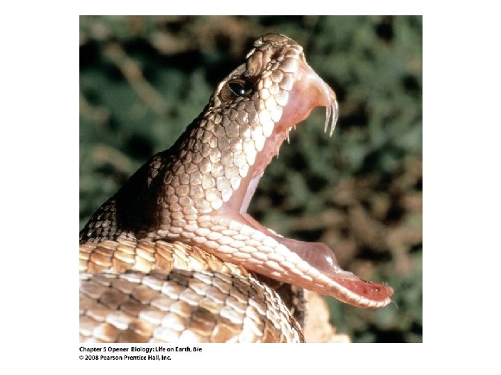
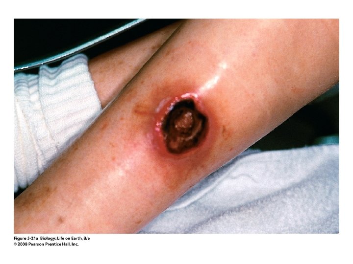
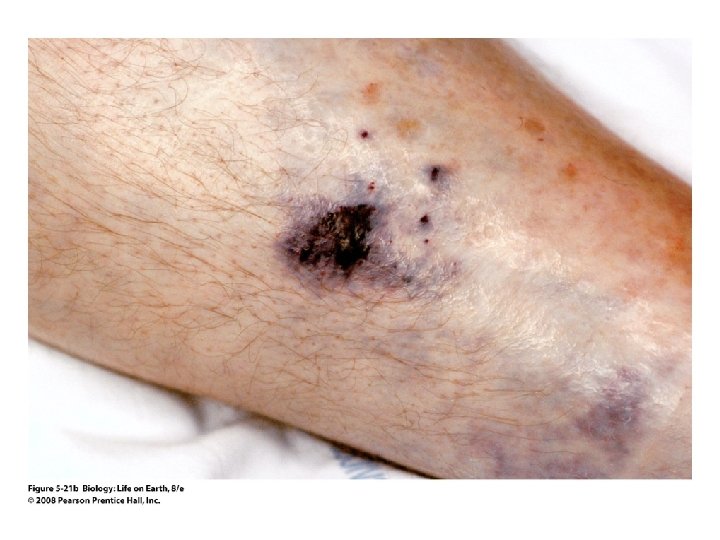
Viscous Venoms • Snake and spider venoms contain phospholipases, enzymes that break down phospholipids • Venoms attack cell membranes, causing cells to rupture and die • Cell death destroys tissue around bite
- Slides: 101