TERATOLOGY for pharmacist by Krisztina H Mink Semmelweis

TERATOLOGY for pharmacist by Krisztina H. -Minkó Semmelweis University, Faculty of Medicine, Department of Anatomy, Histology and Embyology

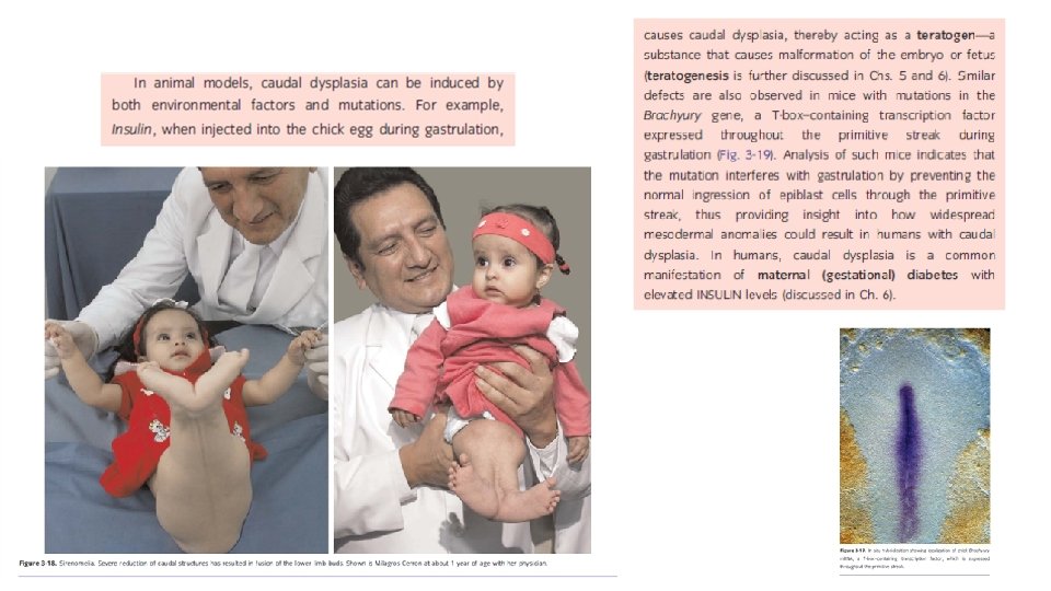
The initially flat three-layered embryonic disc undergoes morphogenesis to form a three-dimensional embryo with a tube-within-a-tube body plan and the beginnings of rudiments that will form all of the adult organs and systems. Morphogenesis results from differential growth. Differential growth is driven by a small number of fundamental cellular behaviors such as changes in cell shape, size, position, number, and adhesivity. If these behaviors are perturbed during embryogenesis, by a genetic mutation, environmental insult (i. e. , a teratogen), or a combination of the two, differential growth is abnormal and dysmorphogenesis results with the formation of a structural birth defect.

Dysmorphogenesis can result from both malformation Common examples, and deformation. include Down syndrome (trisomy 21) and 22 q 11. 2 deletion syndrome, two syndromes that result from genetic mutations. Malformations consist of primary morphologic defects Other syndromes can result from teratogen exposure. A common example is fetal alcohol syndrome, in an organ or body part resulting from abnormal also known as fetal alcohol spectrum disorder. developmental events that are directly involved in the development of that organ or body part. For example, failure of the neural groove to close results in a malformation called a neural tube defect. Similarly, failure of the digits to fully separate results in syndactyly, that is, fusion of the digits. Deformations consist of secondary morphologic defects that are imposed upon an organ or body part owing to mechanical forces; that is, deformations affect the development of an organ or body part indirectly. For example, if insufficient amniotic fluid forms (i. e. , oligohydramnios), deformation of the feet can occur due to mechanical constraints, resulting in club foot. Dysmorphogenesis can occur in an isolated organ or body part or can occur as a pattern of multiple primary malformations with a single cause. In the latter case, the condition is referred to as a syndrome.
 substances that are capable of causing a")
Teratogens are environmental (i. e. , nongenetic) substances that are capable of causing a birth defect when embryos or fetuses are exposed at critical times in development to sufficiently high doses (concentrations). The study of the role of environmental factors in disrupting development is known by the unfortunate name of teratology, which literally means the study of (developmental) monsters.

CONGENITAL MALFORMATION, TERATOLOGY, HISTORY • Teratology is the science that studies the causes, mechanisms, and patterns of abnormal development. teratology : development of „monsters” • 1941 Gregg (Australia) rubella infection of pregnants caused blindness, deafness , problems with heart • 1960 s years: Thalidomide scandal (phocomelia)

The first principle of teratology is that an embryonic structure is usually susceptible to teratogens only during specific critical sensitive periods, which usually correspond to periods of active differentiation and morphogenesis. Thus, a potent teratogen may have no effect on the development of an embryonic structure if it is administered before or after the critical period during which that structure is susceptible to its action. Because the major events of organogenesis take place during the first 8 weeks of development, that is the period during which the fetus is most vulnerable to teratogens.

A second principle of teratology is that an embryonic structure is susceptible to a critical dose of teratogen during its specific critical sensitive period. Thus, in teratologic studies a dose response curve is constructed for a suspected teratogen in which lowest dose has no effect and the highest dose is lethal to the embryo. A third principle of teratology is that susceptibility to a teratogen depends on the genetic constitution of the developing embryo or fetus. For example, if two embryos of the same age are exposed to the same dose of teratogen, one may develop severe cardiac malformations whereas the other may remain unaffected. The molecular basis for this difference in susceptibility might, for instance, be a genetic difference in the rate at which the enzyme systems of the two embryos detoxify the teratogen. Thus, there is a gene-environmental interaction underlying susceptibility to birth defects that varies from embryo to embryo.

• Incidence of abnormalities in Europe: 2 -3% of all live born children at the birth, -(congenital) • but in the first year: 4 -6%. Range of CONGENITAL ANOMALIES is very vide from the severe morfological defects till enzime defects (no morfological picture)
 genetic sources Developmental abnormalities")
CAUSES OF CONGENITAL ANOMALIES Envitonmental sources (teratogens) genetic sources Developmental abnormalities

Chromosome mutation 10% unknown Gene mutation 8% 50% Umwelt 7% multifactorial 25% ? ? : - age of the parents - different ethnical groups - familiar background (genetical) - seasons - social and geographical differences
")
AGE Incidence of Down syndrome is dependent on the age of mother (left figure) but the Apert syndrome is on the age of father (right blue line) Apert syndrome: turricephalus – tower head

RACE Incidence of cleft palate is different in different ethnical groups far-east > (2 x) caucasian > (2 x) afro-american

SEASON Anencephaly is abundant in the spring: it is caused by the deficit of the mother’s folic acid level. Given folic acid to the mothers cut down drastically the number of these fetuses. Anencephaly = cranioschisis – anterior part of the neural tube (neuroporus anterior) is not closed the brain will not be developed.

Frequency of anencephaly in different countries
: „to")
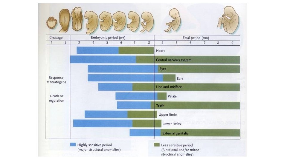
Critical periods during the pregnancy Pre-embryonic period: • 0 -3. embrionic weeks (EW): „to be or not to be”, the teratogen kills the whole embryo (abortion), or only some of the early cells and these cells will be substituted (no congen. malf. ) Embryonic peroid: • 3 -8. EW: most vulnerable period: time of the development of the most serious (major) anomalies Fetal period: • 8. EW: minor morphological or functional defects (e. g. mental retardation).

• The sensitivity of the different organs are different in the time: some organs are sensitive in early period (heart) the others are sensitive in later periods (genitals) and some of them are sensitive all along (nervous system). Diagram of sensitivity period
 :")
Different organs different sensitive periods Tetraciklin: - after the 120. embrionic day (ED) : milk-teeth and adult teeth become coloured -after ED 250: only the adult teeth become coloured Rubella infection: ED 0 - ED 60: cataracta (blindness )and heart problems ED 0 - ED 120: deafness https: //drmarthaszabolcs. com/2017/03/06/firstblog-post/#jp-carousel-140

Causes of CA genetic factors chromosome abnormalities Non-disjunction abnormal number of cromosome: aneuploidia monosomy trisomy abnormalities inside the chromosome reciprocal translocation deletion, duplication

Aneuploidia of the autosome chromosomes: Down syndrome: 21 trisomia symptomes: defects of the face, mental retardation, typical crease on the palm >35 age at pregnancy – higher risk Why these signs result from the trisomy is unknown

Aneuploidia of the autosome chromosomes: Down syndrome: 21 trisomia symptomes: defects of the face, mental retardation, typical crease on the palm >35 age at pregnancy – higher risk Why these signs result from the trisomy is unknown
")
Other trisomies: Trisomy of 13. Trisomy of 18. (rocker bottom feet)
: female but")
Aneuploidia of the sex chromosomes: Turner syndrome: 45 X, O (X monosomy): female but weak fenotype, steril Others: XXY: Klienefelter syndrome: male, but small testis, steril, long limbs XYY: seems to be normal male, high figure, but violent (choleric) behavior XXX: „super women”: seems to be normal feminin habit, but mental retardation is occure

abnormalities inside the chromosome reciprocal translocation deletion, duplication cause specific symptomes e. g. :
 but:")
Androgen insensitivity syndrome Here it is a nice man !!!!!! (he has testis) but: NO prostate, and any other male genital organs! NO ovary and female genital organs, only blind vagina It’s not rarely at photomodells. . . …

Andrej Pejic, the gender-fungible Australian male model
 , the")
Testicular feminization In this case the genotype is normal male ( XY) , the first steps of the sex determination is normal : the testis is developed, but the testosteron produced by the testis not able to act on the cells because the testosteron receptors are missing on the cells
 Syndaktilia: az ujjak összeolvadása. Ebben a humán esetben a")
Hox. D 13 pointmutation (polysyndactylia) Syndaktilia: az ujjak összeolvadása. Ebben a humán esetben a HOXD 13 gén pontmutációja a gén kieséséhez vezetett. Az üres csillagokkal jelölt képletek kézközépcsontok, a két kicsi csillag normálisan nem létező carpalis csontokat jelöl.
 phocomelia - heart")
Holt-Oram syndrome: -TBX 5 mutation -1: 100. 000 -limb bud (100%) phocomelia - heart defects (67%)
 2. drugs/chemicals 3.")
Causes of CA Environmental factors 1. biotic agents (bacteria, viruses, fungi) 2. drugs/chemicals 3. mechanical agents

1. Infectious Agents Teratogens • Rubella • German or Three-Day Measles • First trimester – most serious ED 0 - ED 60: cataracta (blindness )and heart problems ED 0 - ED 120: deafness • Cytomegalovirus (CMV) • Herpes Simplex Virus (HSV) • Varicella • Human Immunodeficiency Virus (HIV) • Congenital Syphilis • Toxoplasmosis

2. Mechanical Factors as Teratogens • Mechanical forces • • Restrict the mobility of the fetus Cause prolonged compression in an abnormal posture E. g. congenital dislocation of hip and clubfoot Malformed uterus

Mechanical Factors as Teratogens • Amniotic fluid • Absorb mechanical forces • Oligohydramnios • Significantly reduced fluid-quantity • Mechanically induced deformations of the limbs • Knee hyper-extends • Amniotic bands • Rings formed from amnion rupture • Local constriction during fetal growth • Intrauterine amputations
: causes problems in the brain development :")
3. DRUGS/CHEMICALS Etil-alcohol: Fetale Alcohol Syndrome (FAS): causes problems in the brain development : holoprosencephalia. Prosencephalon is the most anterior part of the neural tube : hemispheres, hypothalamus, hypophysis , eye developed from this part Thalidomid (Contergan): tranquillizier Many therapeutic drugs are known to be teratogenic; these include retinoids (vitamin A and analogs), the anticoagulant warfarin, the anticonvulsants valproic acid and phenytoin, and a number of chemotherapeutic agents used to treat cancer. Most teratogenic drugs exert their main effects during the embryonic period. Although, as stated above, most care must be exercised in administering certain anesthetics and other drugs even late in pregnancy or at term, because they may endanger the health of the fetus.

FAS: holoprosencephaly and craniofacial CA. Prosencephalon regulate not only the development of the brain but the head and face also B: cebocephalia C: no nose, hypotelorismus D: cyclopia

Some recreational drugs are also teratogenic; these include tobacco, alcohol, and cocaine. Cocaine, used by alarming numbers of pregnant women (the drug affected 300, 000 to 400, 000 newborns in 1990 in the United States), readily crosses the placenta and may cause addiction in the developing fetus. In some of the major cities of the United States, as many as 20% of babies are born to mothers who abuse cocaine. Unfortunately, fetal cocaine addiction may have permanent effects on the individual, although studies suggest that early intervention with intensive emotional and educational support in the first few years of life may be helpful. Two mechanisms have been proposed by which cocaine could cause preterm labor: cocaine, a potent constrictor of blood vessels, may cause abruption of the placental membranes (premature separation of the placenta from the uterus) by partly shutting off the flow of blood to the placenta; or as there is evidence that cocaine directly affects the contractility of the uterine myometrium (muscle layer), it perhaps makes the myometrium hypersensitive to signals that initiate labor. high frequency of preterm labor. https: //www. youtube. com/watch? v=QYICa. Ho 6 t. WQ
: This disorder affects 2 in 1000 live-born infants (Fig. 5")
Fetale Alcohol Syndrome (FAS): This disorder affects 2 in 1000 live-born infants (Fig. 5 -2). Consumption of amounts of alcohol as low as 80 g per day (i. e. , between two and three shots of a grain liquor such as rum) during the 1 st month of pregnancy can cause significant defects, and it has been suggested that even a single binge may be teratogenic. Common components of the disorder include defects of brain and face development, namely, microcephaly (small head), short palpebral fissures (eye openings), epicanthal folds (folds overeye lids), a low nasal bridge with a short nose, flat midface, minor external ear anomalies, and jaw anomalies including a thin upper lip with indistinct philtrum and micrognathia (small jaw). Chronic consumption of even quite small amounts of alcohol later in pregnancy can result in other, less-destructive effects, such as some degree of growth retardation and minor physical defects.
, often called small for gestational age (SGA), is a condition")
Intrauterine growth restriction (IUGR), often called small for gestational age (SGA), is a condition in which fetal growth in markedly retarded. IUGR carries a higher risk of perinatal mortality and morbidity, so IUGR is a life-threatening birth defect. A newborn is considered to be SGA if he/she weighs less than 2500 grams at term or falls below the 10 th percentile for gestational age. CAUSES: teratogen exposure such as congenital viral or bacterial infections, fetal chromosomal anomalies (e. g. , Down syndrome), maternal factors (such as preeclampsia, a condition affecting about 5% of pregnancies characterized by high blood pressure and protein in the urine), placental factors (such as placenta previa, or ‘‘low-lying’’ placenta, a condition in which the blastocyst implants near the uterine cervix and the placenta covers part of the opening of the cervix). involves the entire fetus
: tranquillizier thousands of pregnants used as a sleeping -pill")
Contergan scandal • Thalidomid (Contergan): tranquillizier thousands of pregnants used as a sleeping -pill in the 60’s • About 40 thousands person had got periferal nerve inflammation; • 8 -12 thousands baby born with phocomelia • among them 5 thousands grown up…

Thalidomid-case: phocomelia: the limbs are missing the diagram shows the amount of sold Contergan (Thalidomid), empty columns: in kilogramm!!!; stripped colums : number of phocomelia patient , in Germany between 1956 and 1964

In 1964 the drug was given to patients with leprosy as the last possibility for sleeping. And the drug dissolved the distress, gave good sleeping periods and mollified the leprosy. It seems to be very bid fair: it seems to be usable not only in leprosy but in some autoimmune deseases also Today we know that it has antiangiogenetic effect , that caused the problem in the developing embryo

Both maternal diabetes and maternal obesity during pregnancy constitute risk factors for birth defects of the fetus neural tube defects and heart defects

Maternal obesity (defined in the United States as a body mass index greater than 30 kg/meter squared) Is also a risk factor for birth defects. Fetuses born to obese women are 2 to 3. 5 times more likely than those born to average-weight women to have neural tube defects, heart defects, and omphalocele.

Thank You for attention!

References T. W. Sadler: Langman’s Medical Embryology, 7 th edition, 1995, Baltimore, Maryland, USA – képek Lectures of Dr. Ágnes Csáki and Dr. Nándor Nagy
- Slides: 46