Techniques used to study the brain in relation

Techniques used to study the brain in relation to behavior Pages 72 -77

Discuss the Use of Brain Imaging Technologies in Investigating / Biological Factors and Behavior l For a while, brain research was limited to studying victims of stroke or accident and using invasive methods such as autopsy to study their brains. ¡Comparing behavioral deviations observed in these people after the accident and abnormalities in brain structure discovered after their death led to some insights about the functions of certain brain areas. l This was how Broca’s and Wernicke’s areas were discovered. l It is an advantage to be able to use non-invasive methods to study the brain. ¡Such methods are called brain imaging techniques or neuroimaging

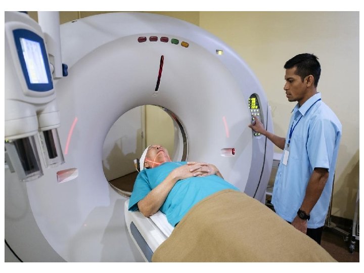
Discuss the Use of Brain Imaging Technologies in Investigating / Biological Factors and Behavior l Computerized axial tomography (CAT/CT) ¡Works on the principle of differential absorption of X -rays. ¡The strength of this technique is that it is a quick non-invasive method of studying brain structure l It has an advantage over standard X-rays because CAT records images of hard and soft tissues as well as blood vessels simultaneously. l Unlike some other techniques, CAT scans can be made for people who have implanted medical devices ¡The limitation is that CAT scans involve some level of radiation exposure
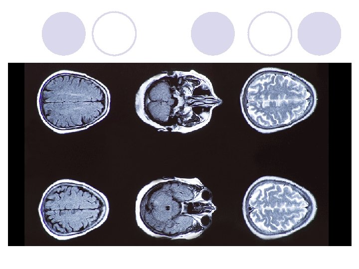

Discuss the Use of Brain Imaging Technologies in Investigating / Biological Factors and Behavior l Magnetic resonance imaging (MRI) ¡ MRI’s are often compared to CT scans because they have the same purpose: to produce a high-resolution three-dimensional image of the brain structure. l Unlike CT scans, MRI’s do not involve X-rays ¡ MRI’s are based on a principle that some atomic nuclei – in particular those of hydrogen atoms – can emit energy when placed in an external magnetic field. l When these pulses of energy are detected by the scanner, the relative distribution of hydrogen atoms in the brain can be mapped. l Hydrogen atoms exist naturally in the body, but their concentration in different types of tissue is different ¡ Advantages: l It allows non-exposure to radiation and, as a consequence, less risk of radiation-induced cancer l MRI has better resolution than a CAT scan. This makes it particularly useful for detecting abnormalities in soft tissue – such as the brain ¡ Disadvantages: l People with metal in their body cannot undergo the procedure (ex. cardiac pacemakers) because metal will attract to the magnetic field • Several deaths have been reported in patients with undisclosed metallic implants l Can be an issue for claustrophobic people because it requires being placed in a narrow tube for a long period of time (~ 45 min) l Lying still can be an issue for young children l Cost: MRI’s are more expensive than CAT scans l High resolution may lead to incidental findings. This may create anxiety and cause patients to seek unnecessary treatment

Discuss the Use of Brain Imaging Technologies in Investigating / Biological Factors and Behavior


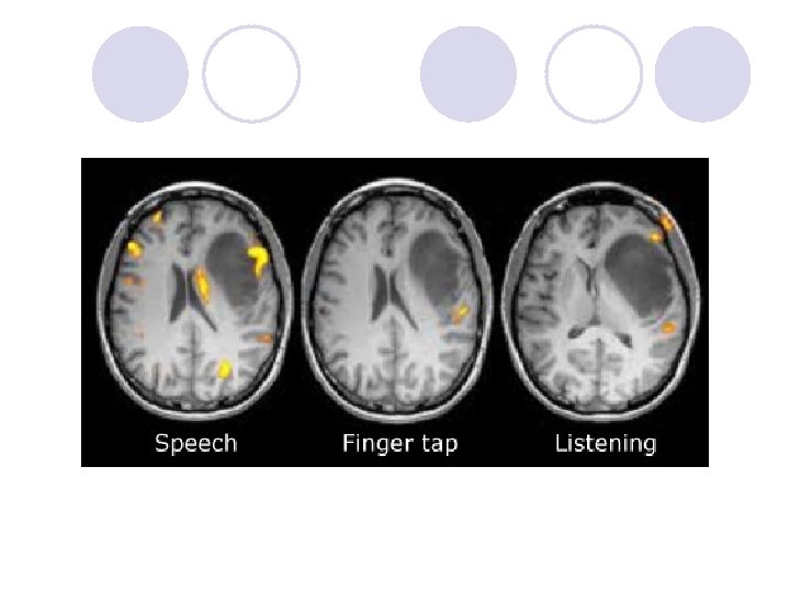
Discuss the Use of Brain Imaging Technologies in Investigating / Biological Factors and Behavior ¡ Functional magnetic resonance imaging (f. MRI) l While MRI and CAT scans are only able to reveal the structural features of the brain, f. MRI can also show the ongoing brain processes. l In a typical study, the subject is required to carry out some task in which peiods of activity are alternated with periods of rest. • The principle at work is that when a brain region is active during the performance of a task, the flow of oxygenated blood in that region increases. • The response of blood to rapidly changing magnetic fields differs depending on the flow and the level of oxygenation. l The signal that is analyzed by the f. MRI scanner to reconstruct brain activity is known as BOLD signal (blood-oxygen-level dependent). • There are other biomarkers as well, but BOLD is the most widely used. The flow of oxygenated blood directly correlates with the energy used by brain cells, and this directly corresponds to the level of activity in a specific brain region. l An f. MRI scan is characterized by spatial resolution and temporal resolution. • Spatial resolution: the ability to discriminate between nearby locations. Resolution is measured in VOXELS. A voxel is the smallest “brain particle” that we can see through a scanner. Typically, the size of a voxel that an f. MRI is able to operate with ranges from 1 to 5 mm. A voxel contains several million neurons and several billion synapses.

Discuss the Use of Brain Imaging Technologies in Investigating / Biological Factors and Behavior ¡ Temporal resolution: the smallest time period in which changes in brain activity can be registered. l Think of it as the rate at which snapshots of the brain are taken. l Currently, the temporal resolution achieved in f. MRI is about one second. l Advantages of f. MRI: ¡ It offers excellent spatial resolution (up to 1 -2 mm) ¡ unlike structural imaging techniques, it allows us to see brain processes. l Disadvantages of f. MRI: ¡ There is poor temporal resolution (about 1 second) when using f. MRI as compared to electromagnetic techniques such as an EEG (<1 millisecond) ¡ All the considerations that were relevant to MRI also apply to f. MRI: claustrophobia, cost, lengthy procedure and inability to use it with medical implants
 Scans l PET scans are very useful. ¡Like the f.")
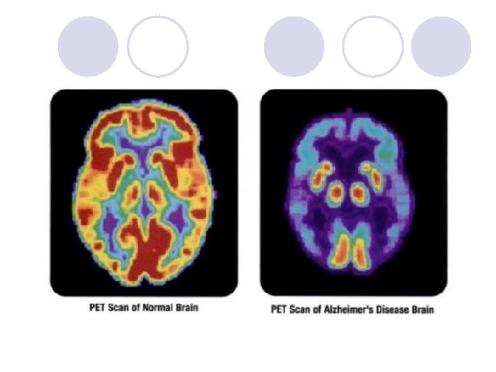
Positron Emission Tomography (PET) Scans l PET scans are very useful. ¡Like the f. MRI, PET scans uses blood flow as the indicator of brain activity. ¡It is done by injecting a radioactive substance into the blood and measuring the amount of brain activity. ¡PET scans have a decent spatial resolution of about 4 mm throughout the brain. However, its temporal resolution is only 30 -40 seconds, so quick processes are not easily detected.
, but they are used less")
PET Scans l Advantages ¡Good spatial resolution (4 mm), but they are used less and less these days given the existence of noninvasive alternatives (f. MRI) which do not require administration of a radioactive chemical. ¡PET is useful for detecting tumors and metastasis ¡It is often used in conjunction with MRI or CT. ¡It is often helpful in diagnosing causes of dementias ¡Scanners can be small – so small that a small PET scanner has been constructed that can be worn by a rat on its head like a hat

PET Scans l Disadvantages ¡The image displayed is not as clear as f. MRI. ¡Temporal Resolution (30 -40 seconds) ¡They are more difficult to use as compared to an FMRI ¡Radioactivity - It is only safe the first couple of times it is used ¡Discomfort – the injection of the tracer and the lengthy procedure (1 -2 hours)
 l Measures electric potentials generated by neural circuits. l When large groups")
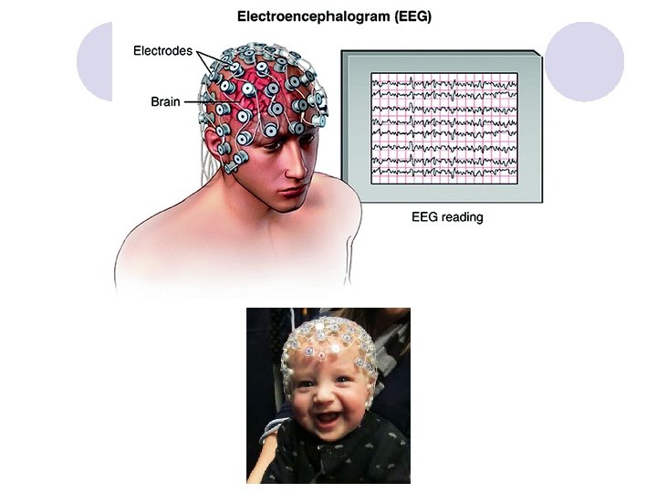
Electroencephalography (EEG) l Measures electric potentials generated by neural circuits. l When large groups of neurons fire synchronously, electric potentials generated by these impulses become detectable at the head surface. l Electrodes are attached to the scalp in predetermined points and pick up the changes in the electric potential of the scalp areas. l Used for sleep studies & epilepsy
 ¡Low-cost ¡Unlike PET and f. MRI,")
EEG l Advantages ¡Perfect temporal resolution (within milliseconds) ¡Low-cost ¡Unlike PET and f. MRI, EEG measures neural activity directly ¡Can be offered as a mobile service ¡EEG is silent ¡Completely non-invasive in comparison with other techniques

EEG l Disadvantages ¡Spatial resolution – EEG’s are not used to establish the origin location of the electrical signal. EEG measures brain activity “on the whole” ¡Not so good for detecting activity in subcortical areas. The farther away from the surface of the scalp, the weaker the signal ¡It takes considerable experience to interpret an EEG correctly because of a number of artifacts contribute to noise in the data (heartbeat, muscle movements, eye movements & blinks)
: used MRI")
The Use of Neuroimaging in Research Studies l Draganski et al (2004): used MRI to determine changes in brain structure in response to learning a simple juggling routine for three months l Freed et al (2001): used PET scans to study dopamine-producing cells in the brains of Parkinson’s disease patients l Fisher, Aron and Brown (2005): used f. MRI to study brain processes in response to looking at the picture of a loved one
- Slides: 20