Techniques in Electrophysiology What you are expected to


- Slides: 23

Techniques in Electrophysiology What you are expected to gain from this lecture: 1. Approaches 2. In-vivo vs. in-vitro preparations 3. Advantages & Pitfalls 4. Types of Measures

5 Common Ephys Approaches: 1. EEG 2. Extracellular/Local Field Potentials 3. Intracellular – Sharp Electrode 4. Patch-Clamp Configurations 5. Multi-Unit Array Recordings

EEGs Recording spontaneous brain (voltage volume conductance) activity from the scalp, described in rhythmic activity: Delta (<4 Hz), theta (4 -7 Hz), gamma (30100 Hz) Clinical Neuroscience: epilepsy, coma, tumors, stroke, focal brain damage, depth of anesthesia Coordinate cortical activity = high contribution Deep structure activity = low contribution Application to Cognitive Psychology: Evoked Potentials: time lock of EEG to presentation of stimuli Event Related Potentials: average of EEG over many trials of higher processing conditions (e. g. , memory, attention) N 1 or P 3 = coma recovery

Typical Slice/Culture Ephys Rig

Patch-Clamp Electrophysiology Apply positive pressure (2 -6 MΩ) Clear tissue as you move down Near cell membrane > ‘bubble’ Apply negative pressure > suction until 1 GΩ seal

4 Common Patch-Clamp Configurations Cell-Attached Whole-Cell Suction Pull Quickly Inside-Out Binding Site? Pull Slowly Outside-Out >1 GΩ seal – going ‘whole-cell’ does not compromise the seal: prevents leak current & extracellular buffer from entering the neuron

Perforated Patch Recording ~20 -30 min ~10 -15 min start Back-filling – nystatin, gramicidin, or amphotericin B (antibiotic/antifungal) – creates pores for select ions to pass Pros: Prevent dialysis of the intracellular contents & current run-down, used for hard to patch cells Cons: slow, high access resistance, weak membrane which leads to whole-cell configuration

Voltage vs. Current Clamp Voltage Clamp: holding the cell at a predetermined value (e. g. , -70 m. V) the amount of current (e. g. , m. A) required to maintain that value is recorded voltage-dependent K+ channels, spontaneous EPSCs Cons: Space Clamp (i. e. , inability to adequately maintain holding command in distal dendrites) & washout of cytosolic factors in whole-cell s. EPSC Somatic current injection producing AP firing Current Clamp: can be used to measure the ‘resting membrane potential’ current is injected into the cell to maintain a predetermined membrane potential (e. g. , -80 m. V) the injected current is constant and free fluctuations in the membrane potential are recorded AP waveform, plasticity of EPSPs, intrinsic excitability

Local Field Potentials - f. EPSPs SA = stimulus artifact A. Stimulation * = presynaptic fiber volley – presynaptic activity generated by stimulation B. f. EPSP = field excitatory postsynaptic potential PS = somatic population spike – coordinated spiking activity The initial slope of the f. EPSP (m. V/ ms) in the s. r. is a widely used measure in LTP studies PS A. B. SA * f. EPSP

Intracellular/Sharp Recording Intracellular recording – used ‘sharp’ glass electrodes with > 25 MΩ resistance (#1) records the change in membrane potential that the incoming current causes (#2) f. EPSP without a clear presynaptic fiber volley

Single Channel Recordings Cell-attached (CA), inside-out (IO), and outside-out (OO) patches Patch typically contains one or a few channels Measure channel open probability, open time at different voltages or in the presence of a test compound CA: stable (>20 GΩ seal), lowbackground but less control over holding potential IO: access to intracellular sites & signaling pathways, difficult to obtain, must replace bath solution from external to internal OO: repetitive & different doses, but less stable, disruption of cytoskeleton

Preparations 1. Acute slices 2. Organotypic cultures 3. Dissociated cultures 4. Cell Lines 5. In vivo

Acute Slices Widely used technique Usually from adolescent rodents, coronal sections Used the day they are made Best to do cardiac perfusion to maintain slice viability Buffer must be oxygenated and at the correct p. H/osmolarity Pros: treatments can be done in vivo, numerous brain regions can be prepared, slices are not too excitable, can combine ephys with confocal imaging, versatile (voltage or current clamp, fields, intracellular, plasticity, etc) Cons: difficult to get viable slices in adult rodents, confound of recordings in adolescents …translatation to adults, afferents are severed, there are changes in instrinsic excitability over the day of recording, bath application of drugs

Organotypic Slice Cultures Multiple brain regions (hpc, co-cultures) grown on porous membrane inserts Prepared from 2 -8 day old rodent pups Maintained for months Helios Gene Gun – can be used to load gold particles coated with c. DNA into cells on the day of culturing to change protein expression

Dissociated Cultures Typically prepared in low- or high-density from embryonic or <24 h old pups Hippocampal, Cortical, Striatal cultures are common Cultured Primary Dissociated Neurons Autaptic/Microisland Acutely Dissociated Neurons - the neurons preserve their dendritic structure proximal to the soma, maintain intact synaptic boutons, and are largely devoid of glial ensheathments.

Cultures Pros: Self-cleaning after insult during preparation, highly controllable experimental conditions, ease & success of growing & maintaining, can be used almost anytime, gene gun & lentiviral expression is easy, combine with imaging, focal drug application & whole-cell currents in dissociated neurons, glutamate uncaging/calcium transients in dendritic spines (dissociated neurons), versatile (current & voltage clamp, f. EPSPs, etc) Cons: Thin over time, loss of afferents (except hpc), developmental differences, contamination, highly excitable (transections), dissociated neurons don’t have intrinsic networks or glial cells, de novo expression of excitatory connections

Cell Lines HEK 293 Cells Xenopus oocytes PC-12 Adrenal Cells Pros: excellent for answering certain ? ’s Express select proteins Point mutation studies Model system for neuronal differentiation Cons: Non-mammalian , non-CNS cells Lack complete neuronal constituents (e. g. , signaling complexes)

In Vivo Recordings Performed under anesthesia or in freely-moving rodents Intra- & extra-cellular, whole-cell, single or multi-unit array recordings Network Properties: Can stimulate in one region and record in another (e. g. , m. PFC influence on NAc plasticity) Phase locking to brain rhythms (e. g. , m. PFC neurons & hippocampal theta)

In Vivo Recordings Lee et al. , 2006, Neuron, v 51, p 399

In Vivo Recordings

Multi-unit Array Recordings Pros: recording from an in vivo situation, network activity, population & single cell activity, phase locking of gamma & theta rhythms, correlation of neuronal or network activity with ongoing behavior, becoming more common Cons: Technically difficult, confound of anesthesia, application of mathematics to isolate data, probes are time-consuming to fabricate

Data, data AP: waveform, peak, half-width, AHP, frequency, back-propagating AP Subthreshold excitatory postsynaptic potentials: LTP, LTD Current-Voltage relationships: Mg unblock of NMDA receptors, shifts in voltage activation & inactivation curves Paired-pulse facilitation: second event that follows is up to 5 X as large due to increased probability of presynaptic vesicle release mini. EPSPs – recorded in presence of TTX: changes in amplitude: postsynaptic event changes in frequency: presynaptic release

Data, data Spike Sorting – used in multi-array recording to assign spikes to different neurons based on their spike properties Pharmacological & Electrical Isolation of distinct currents
 Insidan region jh
 Electrophysiology
 Fonction technique scooter
 Rain
Rain Good health is a choice agree or disagree
 If you think you can you can poem
 Tell me what you eat and i shall tell you what you are
 I will follow you follow you wherever
 Work immersion grading system
 Work immersion grading system
 When was abrasive water jet machining developed first
 Conditional expected value
 Value at risk formula
 Planning in nursing process
 Long run phillips curve
 Error expected identifier
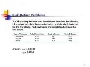
 How to calculate expected return
 Portfolio variance
 Research proposal methods
 Regularized risk minimization
 Expected value probability
 Expected value in probability
 Nusing care plan
 Expected value adalah

