
TARGETING OVARIAN CANCER WITH ANTIMISR 2 RADIOLABELED MABS
TARGETING OVARIAN CANCER WITH ANTI-MISR 2 RADIOLABELED MABS Emmanuel Deshayes, MD, Ph. D INSERM U 1194 Montpellier, France « Radiobiology and Targeted Radiotherapy Team » 30 April 2019 Erice, Italy
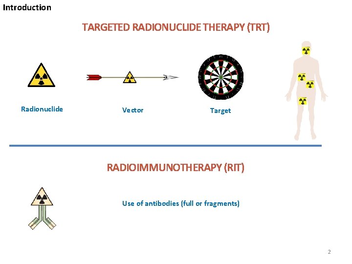
Introduction TARGETED RADIONUCLIDE THERAPY (TRT) Radionuclide Vector Target RADIOIMMUNOTHERAPY (RIT) Use of antibodies (full or fragments) 2
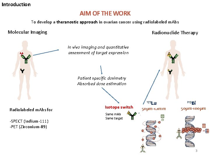
Introduction AIM OF THE WORK To develop a theranostic approach in ovarian cancer using radiolabeled m. Abs Molecular Imaging Radionuclide Therapy In vivo imaging and quantitative assessment of target expression Patient specific dosimetry Absorbed dose estimation Radiolabeled m. Abs for -SPECT (Indium-111) -PET (Zirconium-89) Isotope switch 177 Lu 50µm-1. 1 mm 213 Bi 50µm-100µm Same m. Ab Same target 3
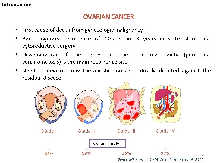
Introduction OVARIAN CANCER • First cause of death from gynecologic malignancy • Bad prognosis: recurrence of 70% within 3 years in spite of optimal cytoreductive surgery • Dissemination of the disease in the peritoneal cavity (peritoneal carcinomatosis) is the main recurrence site • Need to develop new theranostic tools specifically directed against the residual disease 5 years survival 4 Siegel, Miller et al. 2016, Reid, Permuth et al. 2017
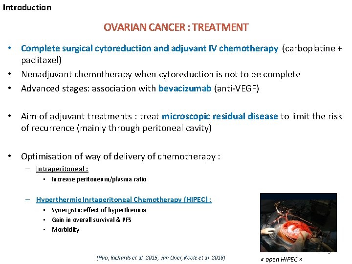
Introduction OVARIAN CANCER : TREATMENT • Complete surgical cytoreduction and adjuvant IV chemotherapy (carboplatine + paclitaxel) • Neoadjuvant chemotherapy when cytoreduction is not to be complete • Advanced stages: association with bevacizumab (anti-VEGF) • Aim of adjuvant treatments : treat microscopic residual disease to limit the risk of recurrence (mainly through peritoneal cavity) • Optimisation of way of delivery of chemotherapy : – Intraperitoneal : • Increase peritoneum/plasma ratio – Hyperthermic Inrtaperitoneal Chemotherapy (HIPEC) : • Synergistic effect of hyperthermia • Gain in overall survival & PFS • Morbidity (Huo, Richards et al. 2015, van Driel, Koole et al. 2018) « open HIPEC » 5
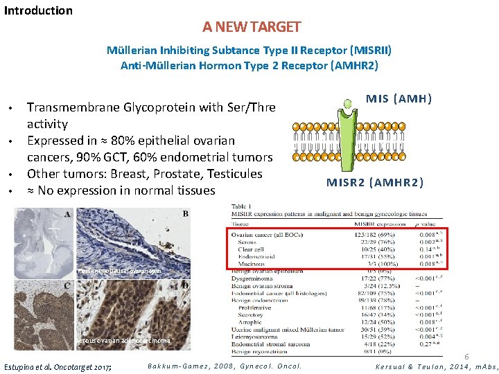
Introduction A NEW TARGET Müllerian Inhibiting Subtance Type II Receptor (MISRII) Anti-Müllerian Hormon Type 2 Receptor (AMHR 2) • • Transmembrane Glycoprotein with Ser/Thre activity Expressed in ≈ 80% epithelial ovarian cancers, 90% GCT, 60% endometrial tumors Other tumors: Breast, Prostate, Testicules ≈ No expression in normal tissues MIS (AMH) MISR 2 (AMHR 2) Post-menopausal ovarian epith Serous ovarian adenocarcinoma Estupina et al. Oncotarget 2017; Bakkum-Gamez, 2008, Gynecol. Oncol. 6 Kersual & Teulon, 2014, m. Abs,
Introduction ANTI-MISR 2 ANTIBODIES • • Initially developed by the team (Dr I. Teulon & Dr A. Pelegrin) Production via mouse ascites using lymphocyte hybridization Murine (16 F 12/12 G 4) or humanized (3 C 23 K) High affinity (> n. M) and specificity for human MISR 2 Internalization upon binding on MISR 2 Binds to MISRII independently from the natural ligand MIS In vivo anti-tumor efficacy (SC, IP) On going phase I clinical trial with 3 C 23 K (GM 102) « First In Human Safety, Pharmacokinetics and Anti-tumoral Activity of GM 102 in Gynecological advanced AMHR 2 + Cancers» (NCT 02978755). 7 Estupina et al. Oncotarget 2017; Kersual & Teulon, 2014, m. Abs
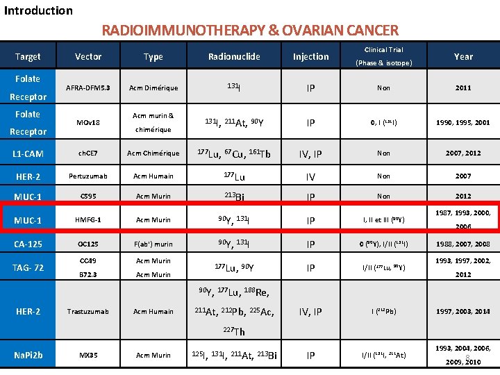
Introduction RADIOIMMUNOTHERAPY & OVARIAN CANCER Target Folate Receptor Clinical Trial Vector Type Radionuclide Injection AFRA-DFM 5. 3 Acm Dimérique 131 I IP Non 2011 131 I, 211 At, 90 Y IP 0, I (131 I) 1990, 1995, 2001 MOv 18 Acm murin & chimérique (Phase & isotope) Year L 1 -CAM ch. CE 7 Acm Chimérique 177 Lu, 67 Cu, 161 Tb IV, IP Non 2007, 2012 HER-2 Pertuzumab Acm Humain 177 Lu IV Non 2007 MUC-1 C 595 Acm Murin 213 Bi IP Non 2012 MUC-1 HMFG-1 Acm Murin 90 Y, 131 I IP I, II et III (90 Y) CA-125 OC 125 F(ab') murin 90 Y, 131 I IP 0 (90 Y), I/II (131 I) CC 49 Acm Murin B 72. 3 Acm Murin 177 Lu, 90 Y IP I/II (177 Lu, 90 Y) IV, IP I (212 Pb) IP I/II (131 I, 211 At) TAG- 72 1987, 1993, 2000, 2006 1988, 2007, 2008 1993, 1997, 2002, 2012 90 Y, 177 Lu, 188 Re, HER-2 Trastuzumab Acm Humain 211 At, 212 Pb, 225 Ac, 1997, 2003, 2014 227 Th Na. Pi 2 b MX 35 Acm Murin 125 I, 131 I, 211 At, 213 Bi 1993, 2004, 2006, 8 2009, 2010
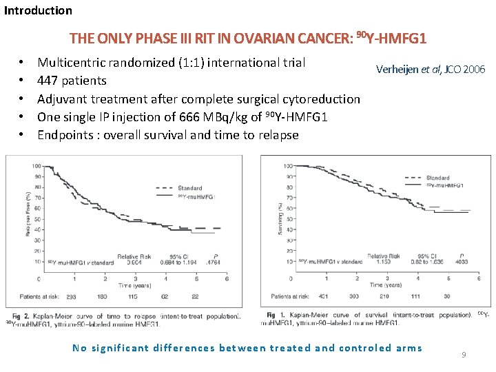
Introduction THE ONLY PHASE III RIT IN OVARIAN CANCER: 90 Y-HMFG 1 • • • Multicentric randomized (1: 1) international trial 447 patients Adjuvant treatment after complete surgical cytoreduction One single IP injection of 666 MBq/kg of 90 Y-HMFG 1 Endpoints : overall survival and time to relapse Verheijen et al, JCO 2006 No significant differences between treated and controled arms 9
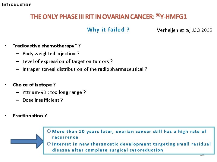
Introduction THE ONLY PHASE III RIT IN OVARIAN CANCER: 90 Y-HMFG 1 Why it failed ? • “radioactive chemotherapy” ? – Body weighted injection ? – Level of expression of target on tumors ? – Intraperitoneal distribution of the radiopharmaceutical ? • Choice of isotope ? – Yttrium-90 : too long range ? – Dose insufficient ? • Fractionation ? Verheijen et al, JCO 2006 More than 10 years later, ovarian cancer still has a high rate of recurrence Interest in new theranostic development targeting small residual disease after complete surgical cytoreduction 10
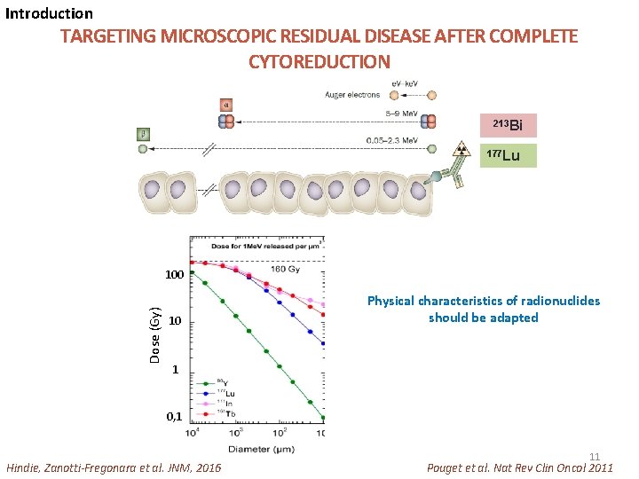
Introduction TARGETING MICROSCOPIC RESIDUAL DISEASE AFTER COMPLETE CYTOREDUCTION 213 Bi 177 Lu Dose (Gy) 100 10 Physical characteristics of radionuclides should be adapted 1 0, 1 Hindie, Zanotti-Fregonara et al. JNM, 2016 11 Pouget et al. Nat Rev Clin Oncol 2011
Objectives AIM OF THE WORK • To treat with radioimmunotherapy residual disease post cytoreductive surgery (small volume tumors : 1 -3 mm) by : • Developing new anti-MISR 2 radiopharmaceuticals • Find the best way of delivery • To develop some companion tools 12
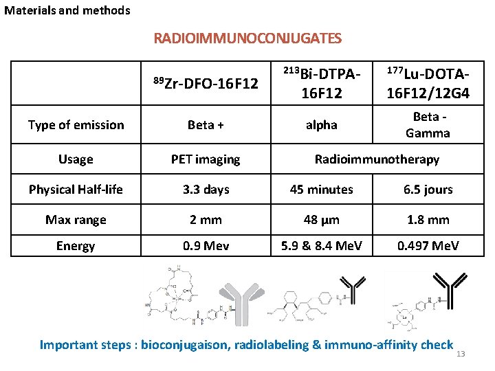
Materials and methods RADIOIMMUNOCONJUGATES 89 Zr-DFO-16 F 12 213 Bi-DTPA- 177 Lu-DOTA- 16 F 12/12 G 4 alpha Beta Gamma Type of emission Beta + Usage PET imaging Physical Half-life 3. 3 days 45 minutes 6. 5 jours Max range 2 mm 48 µm 1. 8 mm Energy 0. 9 Mev 5. 9 & 8. 4 Me. V 0. 497 Me. V Radioimmunotherapy Important steps : bioconjugaison, radiolabeling & immuno-affinity check 13
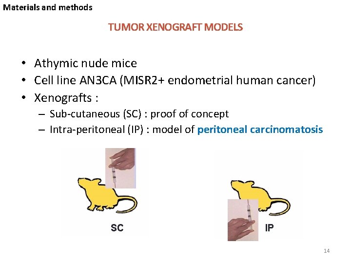
Materials and methods TUMOR XENOGRAFT MODELS • Athymic nude mice • Cell line AN 3 CA (MISR 2+ endometrial human cancer) • Xenografts : – Sub-cutaneous (SC) : proof of concept – Intra-peritoneal (IP) : model of peritoneal carcinomatosis SC IP 14
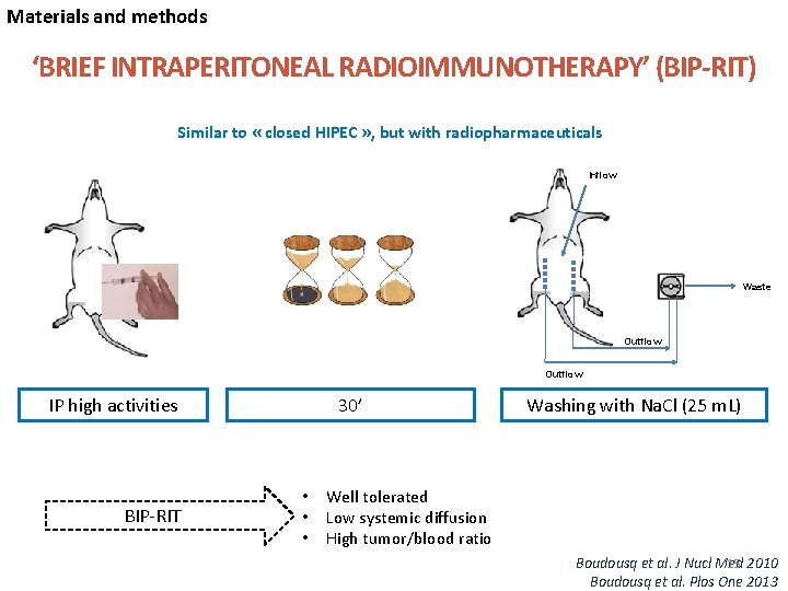
Materials and methods ‘BRIEF INTRAPERITONEAL RADIOIMMUNOTHERAPY’ (BIP-RIT) Similar to « closed HIPEC » , but with radiopharmaceuticals Inflow Waste Outflow 30’ IP high activities BIP-RIT • • • Washing with Na. Cl (25 m. L) Well tolerated Low systemic diffusion High tumor/blood ratio 15 Boudousq et al. J Nucl Med 2010 Boudousq et al. Plos One 2013
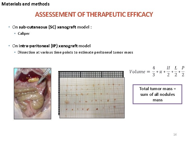
Materials and methods ASSESSEMENT OF THERAPEUTIC EFFICACY • On sub-cutaneous (SC) xenograft model : • Caliper • On intra-peritoneal (IP) xenograft model • Dissection at various time points to estimate peritoneal tumor mass Nodule 1 Total tumor mass = sum of all nodules mass 16
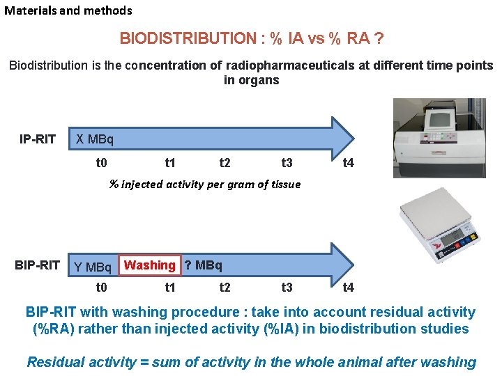
Materials and methods BIODISTRIBUTION : % IA vs % RA ? Biodistribution is the concentration of radiopharmaceuticals at different time points in organs IP-RIT X MBq t 0 t 1 t 2 t 3 t 4 % injected activity per gram of tissue BIP-RIT Y MBq Washing ? MBq t 0 t 1 t 2 t 3 t 4 BIP-RIT with washing procedure : take into account residual activity (%RA) rather than injected activity (%IA) in biodistribution studies 17 Residual activity = sum of activity in the whole animal after washing
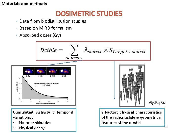
Materials and methods DOSIMETRIC STUDIES • Data from biodistribution studies • Based on MIRD formalism • Absorbed doses (Gy) Gy. Bq-1. s Cumulated Activity : temporal variations : • Pharmacokinetics • Physical decay S Factor: physical characteristics of the radionuclide & geometrical features of the model 18
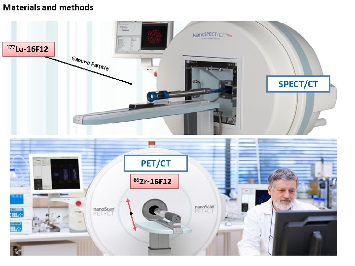
Materials and methods 177 Lu-16 F 12 Gam ma P artic le SPECT/CT PET/CT 89 Zr-16 F 12 19
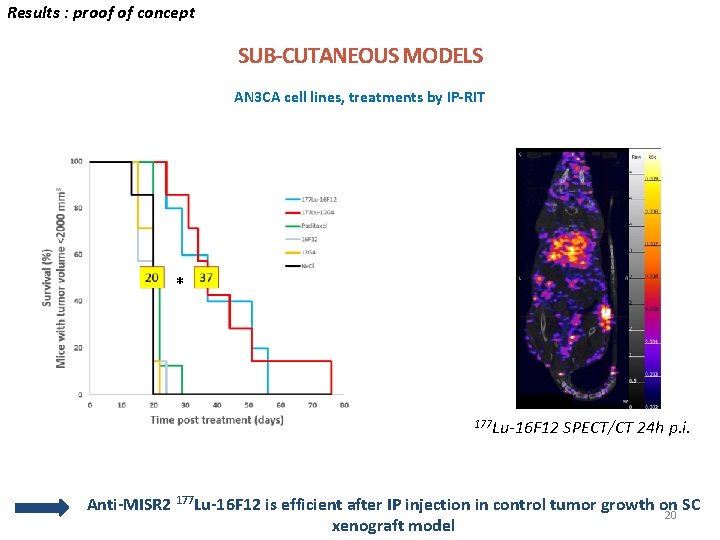
Results : proof of concept SUB-CUTANEOUS MODELS AN 3 CA cell lines, treatments by IP-RIT * 177 Lu-16 F 12 SPECT/CT 24 h p. i. Anti-MISR 2 177 Lu-16 F 12 is efficient after IP injection in control tumor growth on SC 20 xenograft model
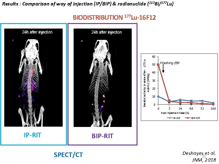
Results : Comparison of way of injection (IP/BIP) & radionuclide (213 Bi/177 Lu) BIODISTRIBUTION 177 Lu-16 F 12 Residul activity in mice after 177 Lua 16 F 12 (MBq) 60 Washing (BIP-RIT) 50 40 30 20 10 0 0 3 24 48 72 168 Post injection time (h) IP-RIT BIP-RIT SPECT/CT Deshayes et al. 21 JNM, 2018
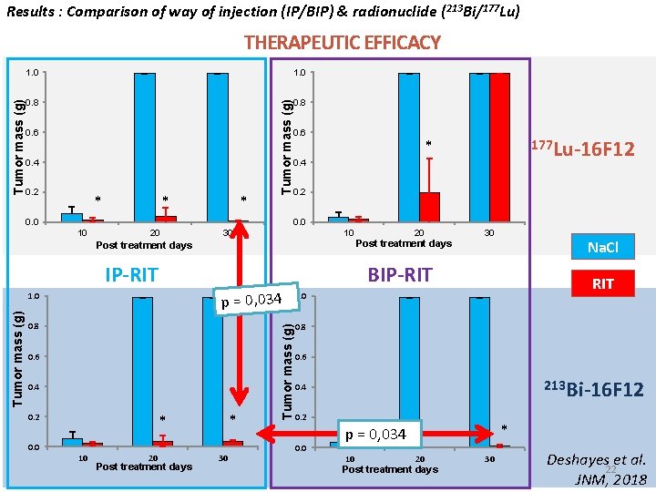
Results : Comparison of way of injection (IP/BIP) & radionuclide (213 Bi/177 Lu) 1. 0 0. 8 0. 6 0. 4 0. 2 * * * Tumor mass (g) THERAPEUTIC EFFICACY 0. 0 0. 6 0. 4 0. 2 0. 0 10 20 30 10 Post treatment days 0. 8 0. 6 0. 4 * * 0. 0 10 20 Post treatment days 30 Tumor mass (g) p = 0, 034 1. 0 0. 2 20 Post treatment days 30 Na. Cl BIP-RIT Tumor mass (g) 177 Lu-16 F 12 * RIT 1. 0 0. 8 0. 6 213 Bi-16 F 12 0. 4 0. 2 0. 0 * p = 0, 034 10 20 Post treatment days 30 Deshayes et al. 22 JNM, 2018
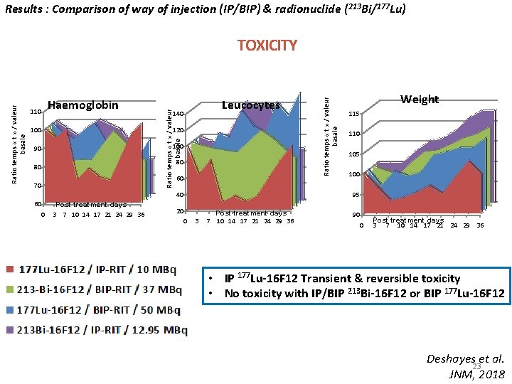
Results : Comparison of way of injection (IP/BIP) & radionuclide (213 Bi/177 Lu) 110 100 Leucocytes 140 120 100 90 80 80 60 70 40 60 0 Post treatment days 3 7 10 14 17 21 24 29 36 Ratio temps « t » / valeur basale Haemoglobin Ratio temps « t » / valeur basale TOXICITY Weight 115 110 105 100 95 20 0 3 Post treatment days 7 10 14 17 21 24 29 36 90 0 Post treatment days 3 7 10 14 17 21 24 29 36 • IP 177 Lu-16 F 12 Transient & reversible toxicity • No toxicity with IP/BIP 213 Bi-16 F 12 or BIP 177 Lu-16 F 12 Deshayes et al. 23 JNM, 2018
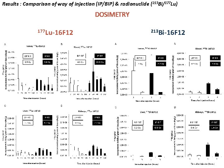
Results : Comparison of way of injection (IP/BIP) & radionuclide (213 Bi/177 Lu) DOSIMETRY 177 Lu-16 F 12 213 Bi-16 F 12 24
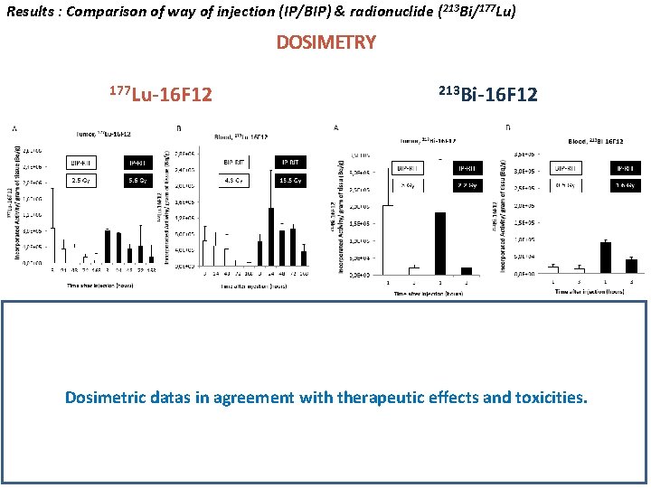
Results : Comparison of way of injection (IP/BIP) & radionuclide (213 Bi/177 Lu) DOSIMETRY 177 Lu-16 F 12 213 Bi-16 F 12 Dosimetric datas in agreement with therapeutic effects and toxicities. 25
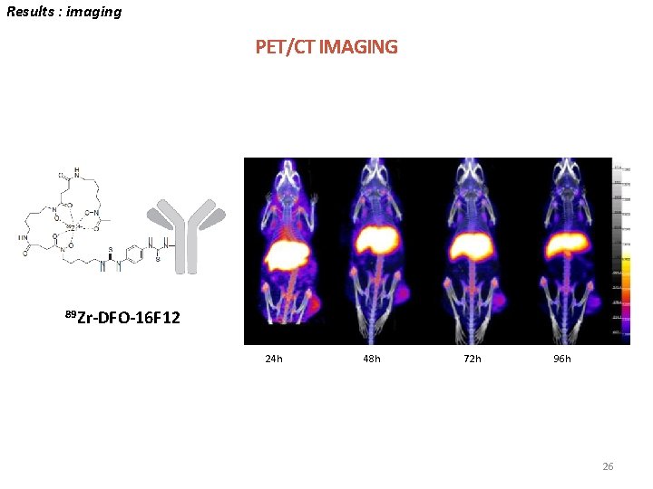
Results : imaging PET/CT IMAGING 89 Zr-DFO-16 F 12 24 h 48 h 72 h 96 h 26
CONCLUSION With anti-MISR 2 radiolabeled m. Abs : • BIP-RIT shows a favorable biodistribution and dosimetric profil whatever the type of isotope is used : 177 Lu or 213 Bi (low systemic diffusion and favorable tumor over blood ratio) • 213 Bi-16 F 12 appears well suited to target small residual disease of peritoneal carcinomatosis : – Therapeutic efficacy – No significant toxicity • Different results are concordant with absorbed doses • 89 Zr-16 F 12 may be suitable for theranostic applications
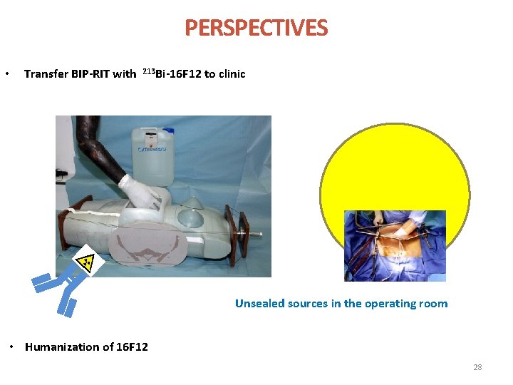
PERSPECTIVES • Transfer BIP-RIT with 213 Bi-16 F 12 to clinic Unsealed sources in the operating room • Humanization of 16 F 12 28
ACKNOWLEDGEMENTS Institut des Trans. Uraniens Karlsruhe, Germany Frank Bruchertseifer Alfred Morgenstern Biostatistique ICM/IRCM Marta Jarlier CRCINA Nantes, FRANCE Nicolas Chouin Radiobiology & TRT Team (U 1194) Nuclear medecine department Jean Pierre Pouget Isabelle Navarro-Teulon Catherine Lozza Vincent Boudousq Samuel Sevestre Alexandre Pichard Riad Ladjohouonlou Pierre Le Fur Muriel Busson ICM Val d’Aurelle Pierre Olivier Kotzki Marie-Claude Eberle Sophie Guillemard Lore Santoro Laurence Calas Cyril Fersing Barts Cancer Institute University of London, UK Roxana Kashani Joanna Koch Jane Sosabowski Julie Foster 29
- Slides: 29