Talkie Time and Recap Competencies Animals Process The


 Film Viewing https: //www. youtube. com/watch? v=UPrx. Qkjj. Ex. I Buzz Session")


 • Trabeculae – needle-like threads of spongy bone that")


 · A unit of bone ·")
 · Arranged")


: skin is intact • Open fracture (compound): skin is")


 Squamous Lambdoid Sutures")


 Mandible (1) Maxillae (2) Lacrimal (2) Inferior nasal conchae")


 • Smaller bodies • Larger spinal canal")
 • All articulate with ribs • Have heart-shaped")


 • Forms the posterior wall of pelvis •")


 = 1")


 • Fingers: •")


 • Also called the hip socket • Is the meeting")


")


 • Patella (kneecap) • Tibia and")
 Figure 8– 11")


 • Talus: • Calcaneus (heel bone): • transfers weight")


")


")


- Slides: 129
Talkie Time and Recap
Competencies: Animals Process: The SKELETAL SYSTEM_BIO 11/12 - IVa-h-1 • Give the functions of the bones • Describe the characteristics (composition) of the bones (types) • Identify the human bones of the body • Evaluate the Pathophysiology of bones
(15 mins) Film Viewing https: //www. youtube. com/watch? v=UPrx. Qkjj. Ex. I Buzz Session 1. What is the importance of Skeletal System? 2. What is it made of? Composition? 3. What disorders of bone that you know?
Anatomy of Skeletal System
SKELETAL SYSTEM • COMPOSED OF: -Bones -Cartilage -Joints -Ligaments
Functions of Skeletal System • SUPPORT: Hard framework that supports and anchors the soft organs of the body. • PROTECTION: Surrounds organs such as the brain and spinal cord. • MOVEMENT: Allows for muscle attachment therefore the bones are used as levers. • STORAGE: Minerals and lipids are stored within bone material. • BLOOD CELL FORMATION: The bone marrow is responsible for blood cell production.
Structure • Compact bone • Outer layer of bone, very hard and dense. • Organized in structural units called Haversian systems. • Matrix is composed of Ca salts (Ca carbonate and Ca phosphate) • Osteocytes – living bone cells that live in matrix. • Porous (Spongy) bone • Located in the ends of long bones. • Many spaces that are filled with red bone marrow which produces bone cells.
Structure • Spongy bone (cont’d) • Trabeculae – needle-like threads of spongy bone that surround the spaces. Add strength to this portion of the bone. • Cartilage • Matrix is a firm gel with chondrocytes suspended in the matrix.
Classification of Bones · Long bones · Typically longer than wide · Have a shaft with heads at both ends · Contain mostly compact bone • Examples: Femur, humerus Copyright © 2003 Pearson Education, Inc. publishing as Benjamin Cummings Slide 5. 4 a
Classification of Bones · Short bones · Generally cube-shape · Contain mostly spongy bone · Examples: Carpals, tarsals Copyright © 2003 Pearson Education, Inc. publishing as Benjamin Cummings Slide 5. 4 b
Classification of Bones on the Basis of Shape Figure 5. 1 Slide 5. 4 c
Classification of Bones · Flat bones · Thin and flattened · Usually curved · Thin layers of compact bone around a layer of spongy bone · Examples: Skull, ribs, sternum Copyright © 2003 Pearson Education, Inc. publishing as Benjamin Cummings Slide 5. 5 a
Classification of Bones · Irregular bones · Irregular shape · Do not fit into other bone classification categories · Example: Vertebrae and hip Copyright © 2003 Pearson Education, Inc. publishing as Benjamin Cummings Slide 5. 5 b
Microscopic Anatomy of Bone · Osteon (Haversian System) · A unit of bone · Central (Haversian) canal · Opening in the center of an osteon · Carries blood vessels and nerves · Perforating (Volkman’s) canal · Canal perpendicular to the central canal · Carries blood vessels and nerves Copyright © 2003 Pearson Education, Inc. publishing as Benjamin Cummings Slide 5. 10 a
Microscopic Anatomy of Bone · Lacunae · Cavities containing bone cells (osteocytes) · Arranged in coecentric rings · Lamellae · Rings around the central canal · Sites of lacunae Copyright © 2003 Pearson Education, Inc. publishing as Benjamin Cummings Figure 5. 3 Slide 5. 11 a
Microscopic Anatomy of Bone Figure 5. 3 Copyright © 2003 Pearson Education, Inc. publishing as Benjamin Cummings Slide 5. 10 b
Microscopic Anatomy of Bone · Canaliculi · Tiny canals · Radiate from the central canal to lacunae · Form a transport system Figure 5. 3 Copyright © 2003 Pearson Education, Inc. publishing as Benjamin Cummings Slide 5. 11 b
Changes in the Human Skeleton · In embryos, the skeleton is primarily hyaline cartilage · During development, much of this cartilage is replaced by bone · Cartilage remains in isolated areas · Bridge of the nose · Parts of ribs · Joints Copyright © 2003 Pearson Education, Inc. publishing as Benjamin Cummings Slide 5. 12
Bone Growth · Epiphyseal plates allow for growth of long bone during childhood · New cartilage is continuously formed · Older cartilage becomes ossified · Cartilage is broken down · Bone replaces cartilage Copyright © 2003 Pearson Education, Inc. publishing as Benjamin Cummings Slide 5. 13 a
Bone Growth · Bones are remodeled and lengthened until growth stops · Bones change shape by gravity &muscle pull · Bones grow in width through periostium Copyright © 2003 Pearson Education, Inc. publishing as Benjamin Cummings Slide 5. 13 b
Long Bone Formation and Growth Figure 5. 4 a Copyright © 2003 Pearson Education, Inc. publishing as Benjamin Cummings Slide 5. 14 a
Types of Bone Cells · Osteocytes · Mature bone cells · Osteoblasts · Bone-forming cells · Osteoclasts · Bone-destroying cells · Break down bone matrix for remodeling and release of calcium · Bone remodeling is a process by both osteoblasts and osteoclasts Copyright © 2003 Pearson Education, Inc. publishing as Benjamin Cummings Slide 5. 15
Fractures • Closed fracture (simple): skin is intact • Open fracture (compound): skin is open • Fracture reduction : 1 -closed reduction , no surgery is needed 2 -open reduction , surgery is needed
Repair of fracture • Healing time for simple fracture is 6 -8 weeks (longer in elderly people) • It occurs in FOUR major events • 1 -hematoma formation • 2 -fibrocartilage callus formation • 3 -bony callus formation • 4 -bone remodelling
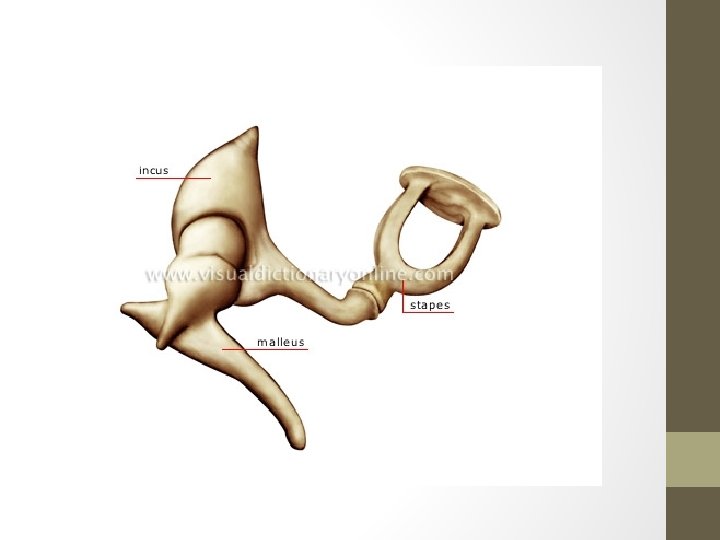
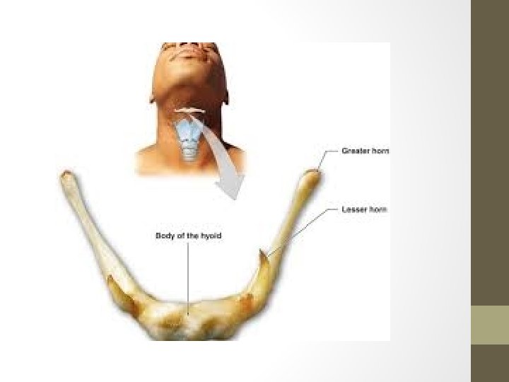
Skeletal system includes A. Axial division • Skull and associated bones • Auditory ossicles • Hyoid bones • Vertebral column • Thoracic cage(Ribs+ sternum) B. Appendicular -Pectoral girdle -Pelvic girdle division
The Axial Skeleton • Axial division • Skull and associated bones: • Auditory ossicles • Hyoid bones • Vertebral column • Thoracic cage • Ribs + sternum
The Skull and Associated Bones
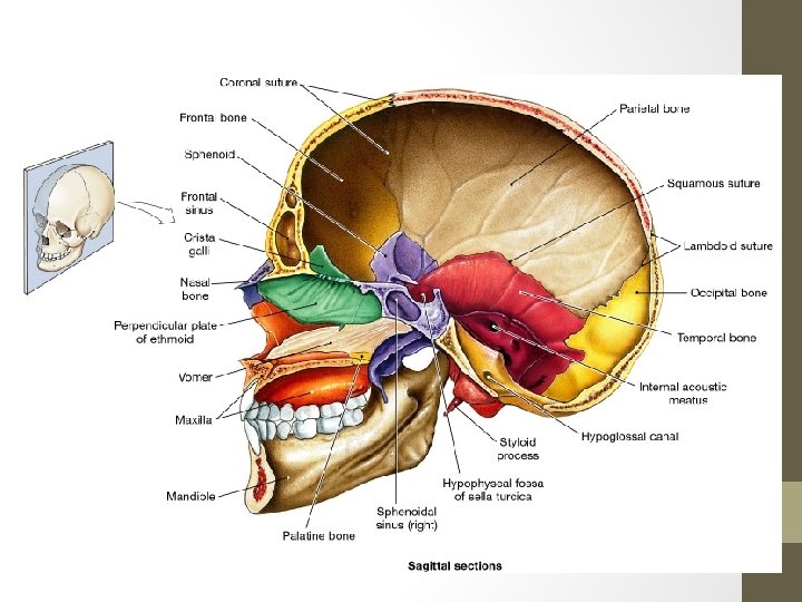
Sutures • Sutures – Immovable joints that join skull bones together • Form boundaries between skull bones • Four sutures: • Coronal – between parietal and frontal • Sagittal– between parietal bones • Lambdoid – between the parietal and occipital • Squamous – between the parietal and temporal ü Fontanels – usually ossify by 2 years of age
Sagittal Frontal (Coronal) Squamous Lambdoid Sutures
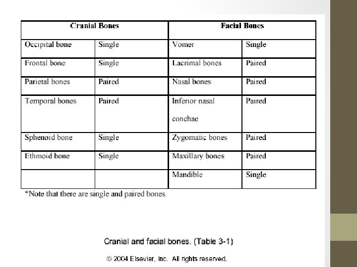
The Adult Skull • skull = 22 bones • cranium = 8 bones: frontal, occipital, 2 temporals, 2 parietals, sphenoid and ethmoid • facial bones = 14 bones: nasals, maxillae, zygomatics, mandible, lacrimals, palatines, inferior nasal conchae, vomer. • skull forms a larger cranial cavity -also forms the nasal cavity, the orbits, paranasal sinuses mandible and auditory ossicles are the only movable skull bones • cranial bones also: attach to membranes called meninges -stabilize positions of the brain, blood vessels -outer surface provides large areas for muscle attachment that move the head or provide facial expressions
Bones of the Cranium
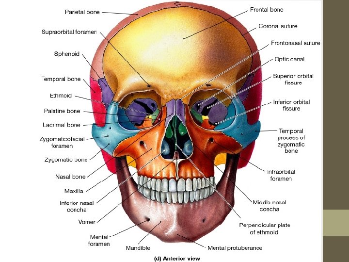
Frontal View
Frontal View
Parietal Frontal View
Temporal Frontal View
Nasal Frontal View
Vomer Frontal View
Zygoma Frontal View
Maxilla Frontal View
Mandible Frontal View
Frontal Parietal Temporal Nasal Vomer Maxilla Zygoma Mandible Frontal View
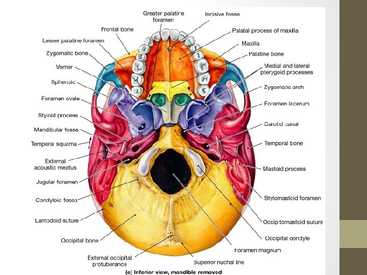
Lateral View
Frontal Lateral View
Parietal Lateral View
Temporal Lateral View
Nasal Lateral View
Zygoma Lateral View
Maxilla Lateral View
Mandible Lateral View
Occipital Lateral View
Mastoid Process Lateral View
External Auditory Meatus Lateral View
Parietal Frontal Sphenoid Temporal Occipital Mastoid Process Nasal Zygoma Maxilla Mandible External Auditory Meatus Lateral View
Figure 6. 4 Sectional Anatomy of the Skull, Part I
14 Facial Bones Nasal (2) Mandible (1) Maxillae (2) Lacrimal (2) Inferior nasal conchae (2) Zygomatic (2) Palatine (2) Vomer (1)
The Vertebral Column http: //www. wisc-online. com/objects/index. asp? obj. ID=AP 12104
Adult Vertebral Column • 26 vertebrae • 24 individual vertebrae • Sacrum • Coccyx • • • Seven cervical vertebrae Twelve thoracic vertebrae Five lumbar vertebrae Sacrum and coccyx are Fused together.
Typical Vertebrae • Body • weight bearing • Vertebral arch • pedicles • laminae • Vertebral foramen • Seven processes • 2 transverse • 1 spinous • 4 articular
Typical Cervical Vert. (C 3 -C 7) • Smaller bodies • Larger spinal canal • 1 st and 2 nd cervical vertebrae are unique • atlas & axis
Thoracic Vertebrae T 1 -T 12) • All articulate with ribs • Have heart-shaped bodies • Each side of the body bears demifacets for articulation with ribs
Thoracic Vertebrae • Allows rotation and prevents flexion and extension
Lumbar Vertebrae • Bodies are thick and strong • Allows flexion and extension – rotation prevented
Sacrum (S 1 – S 5) • Forms the posterior wall of pelvis • Formed from 5 fused vertebrae • Superior surface articulates with L 5 • Inferiorly articulates with coccyx
Sacrum Figure 7. 18 a, b
Coccyx • Is the “tailbone” • Formed from 3 – 5 fused vertebrae • Offers only slight support to pelvic organs
Bony Thorax • Forms the framework of the chest • Components of the bony thorax • Thoracic vertebrae – posteriorly • Ribs – laterally • Sternum and costal cartilage – anteriorly • Protects thoracic organs • Supports shoulder girdle and upper limbs • Provides attachment sites for muscles
The Bony Thorax Figure 7. 19 a
Sternum • Formed from three parts : • Manubrium – superior part • Articulates with medial end of clavicles • Body – bulk of sternum • Sides are articulate for costal cartilage of ribs 2– 7 • Xiphoid process – inferior end of sternum • Ossifies around age 40
Ribs • All ribs attach to vertebral column posteriorly • True ribs - superior seven pairs of ribs • Attach to sternum by costal cartilage • False ribs – inferior five pairs of ribs , attach indirectly to the sternum • floating ribs anteriorly. ribs 11– 12 are short and free
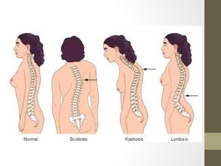
Disorders of the Axial Skeleton • Abnormal spinal curvatures • Scoliosis – an abnormal lateral curvature • Kyphosis – an exaggerated thoracic curvature • Lordosis – an inward lumbar curvature – “swayback” • Stenosis of the lumbar spine • A narrowing of the vertebral canal
The Appendicular Skeleton • Allows us to move and manipulate objects • Includes all bones other than axial skeleton, it includes: • the limbs (upper & lower limbs) • the supportive girdles (pectoral &pelvic girdles)
Appendicular Figure 8– 1
What are the bones of the pectoral girdle, their functions, and features?
The Pectoral Girdle • Also called the shoulder girdle • Connects the arms to the body • Positions the shoulders • Provides a base for arm movement
The Pectoral Girdle Figure 8– 2 a
The Pectoral Girdle • Consists of: • 2 clavicles • 2 scapulae • Connects with the axial skeleton only at the manubrium(claviculosternal joint)
The Clavicles Figure 8– 2 b, c
The Clavicles • Also called collarbones • Long, S-shaped bones • Originate at the manubrium (sternal end) • Articulate with the scapulae (acromial end)
The Scapulae • Also called shoulder blades • Broad, flat and triangular • Articulate with arms and collarbone
Anatomy of The scapula Figure 8– 3 a
What are the bones of the upper limbs, their functions, and features?
The Upper Limbs • Arms, forearms, wrists, and hands Note: arm (brachium) = 1 bone, the humerus
ANATOMY OF The Humerus Figure 8– 4
The Humerus • Also called the arm • The long, upper armbone • Articulates with the pectoral girdle
The Forearm Figure 8– 5
The Forearm • Also called the antebrachium • Consists of 2 long bones: • ulna (medial) • radius (lateral)
The Wrist Figure 8– 6
The Wrist • 8 carpal bones: • 4 proximal carpal bones • 4 distal carpal bones • allow wrist to bend and twist
Metacarpal Bones • The 5 long bones of the hand • Numbered I–V from lateral (thumb) to medial • Articulate with proximal phalanges
Phalanges of the Hands • Thumb: • 2 phalanges (proximal, distal) • Fingers: • 3 phalanges (proximal, middle, distal)
What are the bones of the pelvic girdle, their functions, and features?
The Pelvis • Consists of 2 ossa coxae, the sacrum, and the coccyx • Stabilized by ligaments of pelvic girdle, sacrum, and lumbar vertebrae
The Pelvic Girdle Figure 8– 7
The OSSA COXAE • Also called hipbones • Strong to bear body weight &stress of movement • Each is made up of 3 fused bones: • ilium (articulates with sacrum) • ischium • pubis
The Acetabulum (vinegar cup) • Also called the hip socket • Is the meeting point of the ilium, ischium, and pubis • Articulates with head of the femur (Hip joint))
The Pelvis Figure 8– 8
Divisions of the Pelvis Figure 8– 9
What are the structural and functional differences between the male and female pelvis?
Comparing the Male and Female Pelvis • Female pelvis: • smoother • lighter • less prominent muscle and ligament attachments
Comparing the Male and Female Pelvis Figure 8– 10
Pelvis Modifications for Childbearing • Enlarged pelvic outlet • Broad pubic angle (> 100°) • Less curvature of sacrum and coccyx • Wide, circular pelvic inlet • Broad, low pelvis • Ilia project laterally, not upwards
What are the bones of the lower limbs, their functions, and features?
The Lower Limbs • Functions: • weight bearing • motion Note: leg = lower leg; thigh = upper leg
Bones of the Lower Limbs • Femur (thigh) • Patella (kneecap) • Tibia and fibula (leg) • Tarsals (ankle) • Metatarsals (foot) • Phalanges (toes)
The Femur (longest, heaviest ) Figure 8– 11
The Patella • Also called the kneecap • Formed within tendon of quadriceps femoris
The Tibia and Fibula Figure 8– 13
The Tibia • • Also called the shinbone Supports body weight Larger than fibula Medial to fibula The Fibula • Attaches muscles of feet and toes • Smaller than tibia • Lateral to tibia
Bones of the Ankle (Tarsals) • Talus: • Calcaneus (heel bone): • transfers weight to ground • attaches Achilles tendon
The Ankle • Also called the tarsus: • consists of 7 tarsal bones Figure 8– 14 a
Feet: Metatarsal Bones • 5 long bones of foot • Numbered I–V, medial to lateral • Articulate with toes
Toes: Phalanges • Phalanges: • bones of the toes • Hallux: • big toe, 2 phalanges (distal, proximal) • Other 4 toes: • 3 phalanges (distal, medial,
Articulations (Joints)
Function • Holds bones together • Allows bones to move • All bones articulate with at least one other bone except the hyoid.
Classification of joints • Functional classification: focuses on the amount of movement (synarthrosis, amphiarthrosis and diarthrosis) • Structural classification: based on whether Fibrous, Cartilage or a joint cavity separates the bony regions at the joint. • As a general rule, fibrous joints are immovable and synovial joints are freely movable.
Types • Synarthroses • No movements • Primarily axial skeleton • Bones connected with fibrous tissue ligament • Examples: Skull sutures and distal Tibia/Fibula
Types • Amphiarthroses • Slightly movable • Axial skeleton • Connected by cartilage • Intervertebral joints, pubic symphysis
Types • Diarthroses – freely movable • Also called synovial (fluid filled joint cavity) • Primarily found in the limbs • Plane of movement depends on the joint
Disorders of joints • Dislocation: Bone is forced out of its position, Reduction is done by experts only • Sprain: excessive stretch on a ligament • Arthritis: inflammation of joints, may be -Acute: usually bacterial -Chronic: Rheumatoid , Osteoarthritis and Gouty arthritis END of Sk. system
Bone cancer
Application: Tracing of Bones